Full access
Articles
Published Online: 4 June 2021

A Longitudinal Study of Resting-State Connectivity and Response to Psychostimulant Treatment in ADHD

Abstract

Objective:

Psychostimulants are first-line pharmacological treatments for attention deficit hyperactivity disorder (ADHD), although symptom reduction varies widely between patients and these individual differences in treatment response are poorly understood. The authors sought to examine whether the resting-state functional connectivity within and between cingulo-opercular, striato-thalamic, and default mode networks was associated with treatment response to psychostimulant medication, and whether this relationship changed with development.

Methods:

Patients with ADHD (N=110; 196 observations; mean age at first observation, 10.83 years, SD=2.2) and typically developing control subjects (N=142; 330 observations; mean age at first observation, 10.49 years, SD=2.81) underwent functional neuroimaging on up to five occasions during development (age range, 6–17 years). For patients, symptoms were assessed on and off psychostimulant medication (methylphenidate-based treatments: N=132 observations, 67%; amphetamine-based treatments: N=64 observations, 33%) using the Diagnostic Interview for Children and Adolescents for parents. Linear mixed-effects models examined whether resting-state connectivity was associated with treatment response and its interaction with age. Comparisons with typically developing control subjects were performed to contextualize any significant associations.

Results:

Resting-state connectivity within the cingulo-opercular network was associated with a significant interaction between treatment response and age. Specifically, worse responses to treatment compared with better responses to treatment among patients and compared with typically developing control subjects were associated with an atypical increase in cingulo-opercular connectivity with increasing age from childhood to adolescence.

Conclusions:

This work delineates how resting-state connectivity may be associated over development with response to psychostimulants in ADHD. Functioning and development within the cingulo-opercular network may warrant further investigation as a contributor to differential response to psychostimulants.
Psychostimulants, including methylphenidate-based and amphetamine-based agents, are first-line pharmacological treatments for attention deficit hyperactivity disorder (ADHD) (13), one of the most prevalent disorders of childhood (4). While there are important differences in the actions of methylphenidate-based and amphetamine-based medications (5), both medication classes appear to increase dopamine and norepinephrine action, thus affecting facets of cognition pertinent to ADHD symptomatology (1, 5). Evidence from randomized double-blind placebo-controlled trials indicates that both subtypes of psychostimulants are efficacious relative to placebo in patients with ADHD (1, 2). Although meta-analytic findings suggest that effect sizes for treatment response relative to placebo are larger for amphetamine-based (d=−1.02) than methylphenidate-based agents (d=−0.78), the latter are often better tolerated in children and adolescents with ADHD, and, among patients who respond well to at least one psychostimulant, the largest portion (48%) respond equally well to both medication types. In light of this greater tolerability, methylphenidate-based agents are the most commonly prescribed ADHD medication in this population (1, 2).
Findings from in vivo neuroimaging studies converge on three sets of brain regions that are believed to be particularly relevant to the pathophysiology of ADHD and treatment response to psychostimulants (68). Several studies have investigated brain changes in response to acute doses of psychostimulant medication, reporting a normalization of cingulo-opercular and striato-thalamic hypoactivation and default mode deactivation during task-based functional MRI (fMRI) in patients with ADHD (6, 813). Abnormalities have also been reported in these networks at rest, although not without inconsistencies in the literature, perhaps in part due to methodological heterogeneity and small sample sizes (14, 15). However, effects of psychostimulants on resting-state connectivity and metabolism within these networks have also been reported (5, 1620; see reference 21 for a critical overview), as have effects on connectivity observed during task-based fMRI (22).
The literature has two major limitations. First, symptom reduction in response to psychostimulants varies widely between patients, with an estimated 10%−30% not benefiting adequately from treatment, and the scientific literature provides only limited insights on the neural correlates of treatment responsivity (1, 2). Although many neuroimaging studies have examined brain changes when patients are on versus off psychostimulant medication, little work has examined whether individual differences in treatment response are associated with differences in brain functioning, such as differences in brain functioning assessed while patients are off medication (8, 12). Since psychostimulants are believed to act via a modulation of functioning within cingulo-opercular, striato-thalamic, and default mode regions, it is plausible that treatment response could be associated with functioning within these regions (8). Second, ADHD is a developmental disorder, and the brain continues to develop throughout adolescence (2325), with cross-sectional comparisons indicating differences in children and adolescents relative to adults with the disorder (26), and longitudinal studies suggesting different neurodevelopmental trajectories in ADHD (27). However, most studies have used cross-sectional designs and did not examine potential relationships between neurodevelopmental change in brain networks and treatment response to psychostimulants.
In this study, 110 children and adolescents with ADHD underwent assessments of treatment response in tandem with functional neuroimaging scans up to five times during development. In addition, 142 typically developing control subjects underwent longitudinal neuroimaging. An accelerated longitudinal design was used, with the collection of multiple assessments per subject over time and subjects entering the study at different ages. This design allowed for a time-efficient coverage of the age range of interest, and it is well suited for the examination of developmental research questions such as whether treatment effects and their associations with brain functioning change with development (25).
In light of previous work showing changes in functional connectivity with development, as well as pharmaco-fMRI work implicating these networks in the actions of psychostimulants (8, 9), we examined whether resting-state connectivity within and between cingulo-opercular, striato-thalamic, and default mode networks would be associated with treatment response, as well as whether potential relationships changed with development. However, given that this is the first naturalistic study to examine possible associations between resting-state connectivity and long-term treatment response to psychostimulant medication, we did not form specific hypotheses regarding the direction of effects.

Methods

Participants

Patients 6–17 years of age (N=110) were selected from an ongoing longitudinal study taking place at the National Institutes of Health, the aim of which is to study the neural and genetic basis of ADHD symptoms using longitudinal data (27, 28). In this analysis, we included data collected from psychostimulant-treated patients with ADHD and typically developing control subjects (N=142) between February 2011 and October 2019. Patients with ADHD had to have good-quality neuroimaging data that was collected in tandem with an assessment of symptom response to psychostimulant medication. We defined typically developing control subjects as those with no more than three symptoms in total and no more than two symptoms of either inattention or hyperactivity/impulsivity at all time points. Principal exclusion criteria included IQ less than 80, neurologic disorders affecting brain structure, current substance dependence, and psychotic disorders.

Study Design

Patients and control subjects underwent symptom assessments and neuroimaging scans on up to five occasions during childhood and adolescence. A diagnosis of ADHD was established through a clinician-led interview with parents, using the Diagnostic Interview for Children and Adolescents–IV (29). This interview determines the number of symptoms of inattention and hyperactivity/impulsivity, with a range from zero to nine symptoms in each category. Establishing that the child had six or more symptoms of inattention and hyperactivity/impulsivity indicated a combined presentation, while six or more symptoms of inattention indicated a predominantly inattentive presentation, and six or more symptoms of hyperactivity/impulsivity indicated a predominantly hyperactive/impulsive presentation. Changes in symptom profiles with age were ascertained through the same interview at each time point: Parents provided reports on symptoms for the time window shortly after their child had taken psychostimulant medication. Parents also reported symptoms when their child was off medication, reflecting symptoms during days when the child had not taken medication (e.g., during weekends or during school holidays for some children), or in the late afternoon and evening for children who took psychostimulant medication with no breaks. Treatment response was thus defined as percent symptom reduction when rated on versus off medication for each patient at each time point. Patients did not take psychostimulant medication on the day of scanning. All psychostimulant daily dosages were converted to their oral methylphenidate-equivalent dosage (30, 31) and are expressed as methylphenidate-equivalents per kilogram of body weight per day.
Written informed consent was obtained from parents and/or legal guardians, and assent from children, as approved by the institutional review board of the National Human Genome Research Institute. Details regarding excluded data are provided in the online supplement.

Preprocessing

fMRI preprocessing was implemented in the fMRIPrep and xcpEngine packages (32, 33) (see the online supplement). We focused our analyses on three networks: the cingulo-opercular, striato-thalamic, and default mode networks. Full details regarding the rationale for choosing these networks, as well as details on region of interest definitions, are provided in the online supplement (see Table S1 and Figure S1).
The time courses from all regions of interest were correlated with each other to create a region-of-interest–to–region-of-interest connectivity matrix for each subject. Fisher’s r-to-z transformation was then applied to the Pearson correlation coefficients at each cell of the resultant matrices. We examined connectivity averaged within and between a priori defined cingulo-opercular, striato-thalamic, and default mode networks. Within-network connectivity was determined by averaging Fisher z-transformed correlation coefficients for intranetwork connections separately for each network, to create three within-network connectivity metrics (within cingulo-opercular, within striato-thalamic, and within default mode network). Between-network connectivity was calculated for each pairwise combination of networks (cingulo-opercular to striato-thalamic network, striato-thalamic to default mode network, and cingulo-opercular to default mode network) by averaging the Fisher z-transformed correlation coefficients of the relevant internetwork connections.
To determine which connections were driving network-level findings, exploratory follow-up edgewise analyses were performed at the level of individual region-of-interest–to–region-of-interest connections (see the online supplement).

Statistical Analysis

Participant characteristics and treatment response.

Analyses were conducted using linear mixed-effects models as implemented in the nlme package for R (http://www.r-project.org), while including a random term for each individual that was nested within a random term for each family, thereby accounting for both within-person and within-family dependence (34). This approach was used because our data contain a mixture of multiple observations per subject, measured at different and irregular intervals, as well as single observations per subject. Details on analyses conducted to examine relationships between demographic, clinical, and treatment response variables are provided in the online supplement.

fMRI analyses.

In model 1 we examined whether the development of resting-state connectivity varied as a function of treatment response to psychostimulant medication. We achieved this by regressing each connectivity metric against the interaction between treatment response (percent symptom reduction when patients were rated on versus off medication) and age while controlling for the main effects of age, sex, dosage, off-medication symptom severity, treatment response, and in-scanner motion.
To aid the interpretation of potential associations between the development of resting-state connectivity and treatment response to psychostimulant medication in the context of normative development, we next compared connectivity changes with age between “better” treatment responses in patients (i.e., a treatment response better than the mean), “worse” treatment responses in patients (i.e., a treatment response worse than the mean), and typically developing control subjects. In model 2 we thus regressed connectivity metrics against the interaction between treatment response group (better responses, worse responses, and typically developing control subjects) and age while controlling for the main effects of age, sex, treatment response group, and in-scanner motion. In model 3 we examined the effects of age on connectivity separately at each level of the group variable. Full model details are provided in the online supplement. In addition to examining treatment response–by–age interactions, we also examined the main effect of treatment response on brain functioning. We used the Benjamini-Hochberg method to correct for the number of within- and between-network metrics examined (35).

Results

Participant Characteristics and Treatment Response

The participants’ demographic and clinical characteristics are summarized in Table 1. There was a higher proportion of females in the typically developing group (61/142, 42.96%) compared with the ADHD group (28/110, 25.45%) (χ2=8.31, p=0.004), and sex was controlled for in all analyses. Patients were receiving methylphenidate-based medications during 132 observations (67.34%; mean dosage, 0.86 mg/kg per day of methylphenidate equivalents, range, 0.14–2.5) and amphetamine-based medications during 64 observations (32.65%; mean dosage, 0.82 mg/kg per day of methylphenidate equivalents, range, 0.23–2.14). A minority of participants switched between methylphenidate and amphetamine-based agents during the course of the study (N=7, 6.36%), and 69 participants (62.72%) always received methylphenidate and 34 (30.9%) always received amphetamine-based medications. Details on ADHD presentations are provided in the online supplement.
TABLE 1. Demographic and clinical characteristics of patients with ADHD and typically developing control subjects at each time point of the study
Characteristic or MeasureADHD Group (N=110)Typically Developing Group (N=142)Analysis
 N%N% 
Female2825.456142.96χ2=8.31, p=0.004
N     
 Time 1110100.00142100.00 
 Time 25449.098761.27 
 Time 31917.275337.32 
 Time 498.103323.24 
 Time 543.601510.56 
 MeanSDMeanSD 
Age (years)     
 Time 110.832.2010.492.81t=1.04, df=250, p=0.3
 Time 212.342.3511.892.54t=1.05, df=139, p=0.29
 Time 313.452.2713.152.45t=0.47, df=70, p=0.64
 Time 414.032.1214.072.14t=0.05, df=40, p=0.96
 Time 514.482.1915.161.40t=0.77, df=19, p=0.45
ADHD symptoms     
 Time 111.353.420.250.70t=37.69, df=250, p<0.001
 Time 210.203.430.240.61t=26.47, df=139, p<0.001
 Time 310.633.620.130.44t=20.95, df=70, p<0.001
 Time 410.112.710.180.64t=19.7, df=40, p<0.001
 Time 511.751.710.200.41t=25.37, df=19, p<0.001
ADHD treatment response (%)     
 Time 169.920.27   
 Time 267.790.24   
 Time 369.020.24   
 Time 470.100.24   
 Time 561.240.23   
Dosage (daily methylphenidate-equivalent mg/kg)     
 Time 10.820.46   
 Time 20.860.43   
 Time 30.880.36   
 Time 40.860.33   
 Time 50.880.20   
Treatment response remained stable with age (B=−0.009, t=−1.14, p=0.26, 95% CI=−0.03, 0.007) and was not associated with psychostimulant dosage (B=0.04, t=0.8, p=0.4, 95% CI=−0.05, 0.1), sex (B=0.01, t=0.27, p=0.8, 95% CI=−0.09, 0.1), off-medication symptoms (B=0.0008, t=0.13, p=0.85, 95% CI=−0.01, 0.01), or psychostimulant class (methylphenidate or amphetamine-based; B=0.02, t=0.45, p=0.65, 95% CI=−0.07, 0.1). Off-medication symptoms declined with age (B=−0.53, t=−6.3, p<0.001, 95% CI=−0.7, −0.3). Psychostimulant methylphenidate equivalent dosage was not associated with age (B=−0.02, t=−1.56, p=0.12, 95% CI=−0.04, 0.006), psychostimulant class (methylphenidate versus amphetamine; B=0.05, t=0.62, p=0.5, 95% CI=−0.1, 0.2), or off-medication symptoms (B=0.005, t=0.49, p=0.6, 95% CI=−0.02, 0.02).
Owing to the accelerated design, some participants had more scans than others available at the time of analysis (e.g., because of having started the study earlier). However, patients with fewer than three scans did not differ from those with three or more scans with regard to treatment response (B=0.02, t=0.45, p=0.7, 95% CI=−0.09, 0.1), off-medication symptoms (B=0.02, t=0.28, p=0.79, 95% CI=−1.5, 1.9), in-scanner motion (B=−0.05, t=−1.5, p=0.13, 95% CI=−0.1, 0.02), dosage (B=0.04, t=−0.36, p=0.73, 95% CI=−0.2, 0.3), or psychostimulant class (methylphenidate or amphetamine; χ2=0.06, p=0.8). The primary finding also remained significant when analyses were restricted to the first two or first three scans (see the online supplement). Thirteen of the 110 patients (11.81%) stopped taking psychostimulant medication permanently while part of the study. Four of them stopped treatment and were in remission (three or fewer off-medication symptoms); two patients, who were showing “worse” treatment responses (i.e., worse than the mean treatment response), also stopped medication. Finally, seven stopped medication even though they were showing “better” responses at their last assessment (i.e., better than the mean treatment response).

fMRI Results

Network results.

Model 1 showed that resting-state connectivity within the cingulo-opercular network was explained by a significant interaction between age and treatment response (B=−0.07, t=−3.4, adjusted p=0.006, 95% CI=−0.1, −0.03). No other within- or between-network metrics were significant even at an uncorrected threshold (p>0.1).
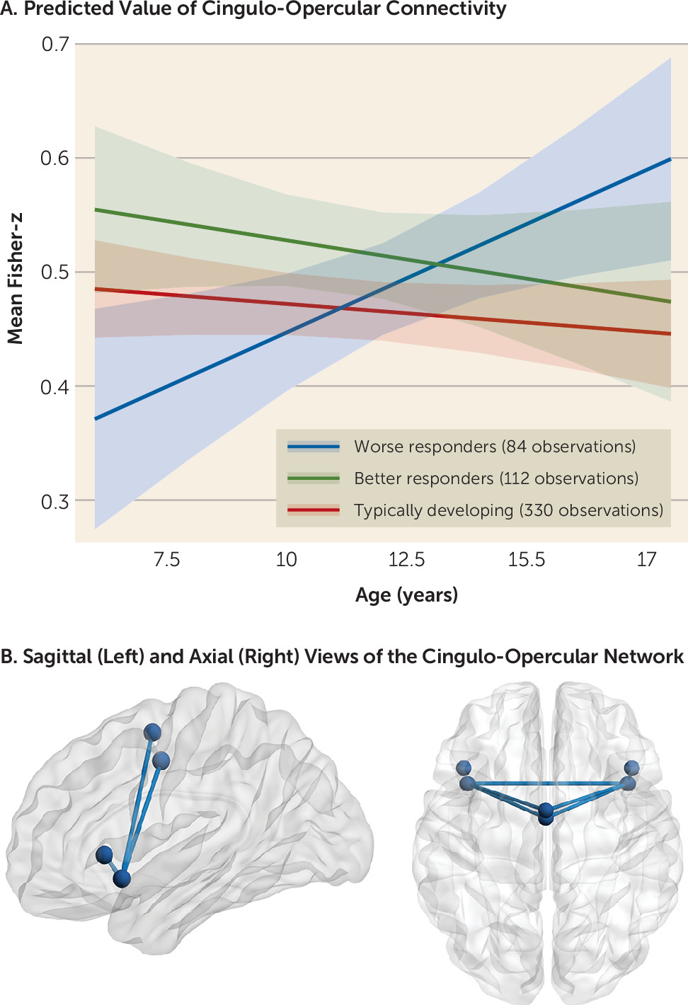
In model 2, a significant interaction between group and age on cingulo-opercular connectivity was found (F=4.86, df=2, 266, p=0.008). Specifically, age-related changes in connectivity that were associated with worse psychostimulant responses (i.e., a treatment response less than the mean; 84 observations from 63 participants; 25.4% female; mean age, 12.25 years) differed significantly from changes associated with better psychostimulant responses (i.e., a treatment response greater than the mean; 112 observations from 76 participants; 27.63% female; mean age, 11.33 years; B=−0.03, t=−2.79, p=0.006, 95% CI=−0.04, −0.008) and the typically developing control subjects (B=0.02, t=2.93, p=0.004, 95% CI=0.007, 0.04). In contrast, a better response to psychostimulant treatment was associated with age-related stability in cingulo-opercular connectivity that was similar to connectivity observed in the typically developing group (B=−0.003, t=−0.51, p=0.6, 95% CI=−0.02, 0.01).
Model 3 showed that worse responses to psychostimulant medication were associated with increased connectivity with increasing age (B=0.02, t=2.6, p=0.02, 95% CI=0.004, 0.04). Connectivity associated with better responses did not change with age (B=−0.007, t=−1.06, p=0.3, 95% CI=−0.02, 0.007), nor did connectivity change with age in the typically developing control subjects (B=−0.005, t=−1.49, p=0.14, 95% CI=−0.01, 0.001).
There were no significant main effects of treatment response or treatment response–by–age interactions for any other network metrics (all p values >0.1) (Figure 1). Sensitivity analyses and robustness checks, including those related to in-scanner motion, number of scans available, psychostimulant subtypes, symptom subscales, and comorbid disorders, are provided in the online supplement (see Table S2).
FIGURE 1. Effect of interaction of age and treatment response on cingulo-opercular connectivity in patients with ADHD and typically developing control subjectsa
a In panel A, the graph shows the predicted estimates derived from the linear mixed-effects model for the effect of the interaction of age and treatment response group (worse responders and better responders, as well as typically developing control subjects) on cingulo-opercular connectivity (F=4.86, df=2, 266, p=0.008). The y-axis represents the predicted mean of the Fisher z-transformed correlation coefficients for the cingulo-opercular network based on model estimates. Shading indicates 95% confidence intervals. In panel B, spheres represent nodes (regions of interest) for each of the cingulo-opercular regions, and lines represent cingulo-opercular edges (connections between nodes) that were significant at a nominal, uncorrected p-value threshold of 0.05 in the follow-up edgewise analysis.

Edgewise results.

No findings survived correction for multiple comparisons at the level of individual region-of-interest–to–region-of-interest connections. Connections that were significant at an uncorrected p threshold of 0.05 are presented in Table S3 in the online supplement, and, in line with the network-level findings, include multiple connections between regions of the cingulo-opercular network.

Discussion

In this study, we examined whether treatment response to psychostimulant medication was associated with resting-state connectivity within a set of cingulo-opercular, striato-thalamic, and default mode networks implicated in pathophysiological models of ADHD and in the therapeutic actions of psychostimulants, as well as whether the associations changed with development. We found that psychostimulant response showed overall stability with age. At a neural level, we found that cingulo-opercular functioning was associated with worse treatment responses in a way that changed with development.
We assessed symptoms on and off medication at each time point, meaning that we could separate symptom change due to psychostimulants from age-related symptom reduction. We found that response to psychostimulants remained stable with age in our observational study. This finding is in line with a recent double-blind randomized placebo-controlled treatment discontinuation trial in which methylphenidate discontinuation in children and adolescents who had undergone treatment for at least 2 years was associated with a worsening of ADHD symptoms, as compared with treatment continuation (36). The findings are also in line with pharmacoepidemiological work showing long-term associations between psychostimulants and decreases in injuries, accidents, and possibly substance abuse (see reference 37 for a critical review of this literature). Therefore, while causal inferences cannot be made given our observational design, our findings here are consistent with previous reports of stability in the efficacy of psychostimulant treatment with increasing age in children and adolescents with ADHD.
With regard to the brain, we found an association between psychostimulant treatment response and functional connectivity within the cingulo-opercular network. Better treatment responses were associated with a stable level of connectivity between nodes of this network during childhood and adolescence, whereas worse responses were associated with increased connectivity with increasing age. Moreover, typically developing children and adolescents showed age-related stability in cingulo-opercular connectivity, which tracked closely with the trajectory associated with better responses to psychostimulants. These findings are interesting in light of the literature reporting a normative segregation of networks during adolescence involving increasing within-network and decreasing between-network connectivity with age (23). Since network segregation has been proposed to be adaptive and associated with improvements in cognitive functioning with age, the present findings of increasing within-network cingulo-opercular connectivity only in the worse treatment responders might be deemed surprising (23). However, findings from longitudinal research on the development of within-cingulo-opercular-network connectivity have been mixed, and the fact that cingulo-opercular connectivity, at least as defined here, did not increase with age in the typically developing control subjects indicates that this increase associated with worse responses is atypical (38, 39). While the exact nature of the relationship between functional connectivity and cognitive and behavioral development is unknown, it is generally accepted that complex cognition and behaviors depend on a finely balanced interplay between multiple interconnected networks (23, 24). Consequently, the functioning of a given network is proposed to be highly dependent on the inputs and constraints provided by patterns of connectivity within and between other closely interdependent networks (23). Therefore, connectivity patterns associated with worse treatment responses might be interpreted as developmentally atypical and potentially out of line with ongoing structural, functional, and neurochemical development in other brain regions and networks, as well as with ongoing age-related changes in behavioral maturation and environmental demands (24).
That associations with treatment response were observed within the cingulo-opercular network is interesting in light of randomized placebo-controlled fMRI studies of acute methylphenidate effects in medication-naive boys with ADHD showing a normalization of functioning in this network (10, 11, 13, 22, 40), as well as improvements in task performance (11, 13, 40), with methylphenidate relative to placebo. Normalizing activation within the cingulo-opercular network therefore appears to be an important candidate mechanism of the acute effects of psychostimulant medication (6, 8, 11). Few studies have examined whether the pattern or degree of acute neural changes associated with taking psychostimulants depends on neural functioning as assessed when off medication (12). However, based on the present findings, the hypothesis emerges that psychostimulants may be better able to normalize recruitment within cingulo-opercular regions during cognitive demands when resting-state connectivity is already more in line with typical development. Alternatively, patients with already typical functioning in a network that undergoes modulation by psychostimulants may experience “better than typical” connectivity when medicated, which might allow them to compensate for their ADHD symptoms. No associations with treatment response were observed involving connectivity of the striato-thalamic and default mode networks, despite previous work showing changes in these networks in response to acute doses of psychostimulants (9, 10, 12, 13). However, in this study, participants underwent scanning during the temporary cessation of psychostimulant medication, and it is likely that treatment response also depends on changes in neural functioning that are induced by psychostimulant medication (12).
This study informs the understanding of the neural correlates of response to psychostimulants in a naturalistic setting. The findings suggest that characterization of developing functional connectivity may help us understand differences in psychostimulant responsivity. However, the study does not inform clinical decisions about the choice of psychostimulants, nor does it provide predictors of treatment response over time. Clinical translation of the findings depends not only on independent replication but also on the ability to characterize developing functional connectivity within a much shorter, clinically useful time frame than was used in this study. Furthermore, the costs of fMRI data need to be weighed against any gain in predictive power, particularly as psychostimulants act quickly, and adverse effects usually cease when the medication is stopped (1, 3). Finally, given the observational design, we cannot draw any conclusions about the causal direction of the relationship between treatment response and developing functional connectivity. Therefore, we argue that rather than setting the stage for use of fMRI in clinical decision making in the near future, the contribution of the present study is to further our understanding of potential biological processes associated with differences in treatment response to psychostimulant medication, which has been limited.
Further limitations of the present work include the fact that the data were collected as part of a naturalistic observational study, and therefore the sample was heterogeneous with regard to prescribed psychostimulant medications, duration of treatment, and other clinical and demographic variables. Relatedly, we included patients on methylphenidate and amphetamine-based agents, which have partly distinct mechanisms of action (5). However, we found that the relationship between cingulo-opercular connectivity, age, and treatment response did not vary significantly with psychostimulant class (see the online supplement). An additional limitation is that since patients were receiving long-term psychostimulant treatment, this may imply that they had been receiving at least some benefit from the drug. Therefore, the findings may not generalize to very poor treatment responders, who are more likely to cease taking the medication (2). In addition, ratings of ADHD symptoms were based on interviews with parents; therefore, they may have been biased by expectation effects on the part of parents, who were not blind to treatment. Moreover, we did not have objective data regarding treatment adherence and relied on parental report. Finally, as this analysis used data collected as part of a natural-history longitudinal cohort study, sample size was not determined according to a formal a priori power analysis. However, observed power estimates indicated that the study had adequate power to detect medium to large effect sizes for the interaction of interest in this study, in line with the detected effects (see the online supplement).
In summary, we provide the first longitudinal investigation of the relationship between brain connectivity and treatment response to psychostimulants in youths with ADHD. We report that worse responses to psychostimulant treatment may be tied to developmentally atypical functioning within a cingulo-opercular network that has been implicated in both ADHD and psychostimulant action. The findings therefore speak to a potential factor contributing to differences in response to psychostimulant medication.

Footnote

The authors report no financial relationships with commercial interests.

Supplementary Material

File (appi.ajp.2021.20091342.ds001.pdf)

References

1.
Cortese S: Pharmacologic treatment of attention deficit-hyperactivity disorder. N Engl J Med 2020; 383:1050–1056
2.
Cortese S, Adamo N, Del Giovane C, et al: Comparative efficacy and tolerability of medications for attention-deficit hyperactivity disorder in children, adolescents, and adults: a systematic review and network meta-analysis. Lancet Psychiatry 2018; 5:727–738
3.
Zuddas A, Banaschewski T, Coghill D, et al: ADHD treatment: psychostimulants, in Oxford Textbook of Attention Deficit Hyperactivity Disorder. Edited by Banaschewski T, Coghill D, Zuddas A. Oxford, UK, Oxford University Press, 2018, pp 379–392
4.
Polanczyk GV, Willcutt EG, Salum GA, et al: ADHD prevalence estimates across three decades: an updated systematic review and meta-regression analysis. Int J Epidemiol 2014; 43:434–442
5.
Faraone SV: The pharmacology of amphetamine and methylphenidate: relevance to the neurobiology of attention-deficit/hyperactivity disorder and other psychiatric comorbidities. Neurosci Biobehav Rev 2018; 87:255–270
6.
Rubia K: Cognitive neuroscience of attention deficit hyperactivity disorder (ADHD) and its clinical translation. Front Hum Neurosci 2018; 12:100
7.
Lukito S, Norman L, Carlisi C, et al: Comparative meta-analyses of brain structural and functional abnormalities during cognitive control in attention-deficit/hyperactivity disorder and autism spectrum disorder. Psychol Med 2020; 50:894–919
8.
Rubia K, Alegria AA, Cubillo AI, et al: Effects of stimulants on brain function in attention-deficit/hyperactivity disorder: a systematic review and meta-analysis. Biol Psychiatry 2014; 76:616–628
9.
Liddle EB, Hollis C, Batty MJ, et al: Task-related default mode network modulation and inhibitory control in ADHD: effects of motivation and methylphenidate. J Child Psychol Psychiatry 2011; 52:761–771
10.
Cubillo A, Smith AB, Barrett N, et al: Shared and drug-specific effects of atomoxetine and methylphenidate on inhibitory brain dysfunction in medication-naive ADHD boys. Cereb Cortex 2014; 24:174–185
11.
Kowalczyk OS, Cubillo AI, Smith A, et al: Methylphenidate and atomoxetine normalise fronto-parietal underactivation during sustained attention in ADHD adolescents. Eur Neuropsychopharmacol 2019; 29:1102–1116
12.
Peterson BS, Potenza MN, Wang Z, et al: An fMRI study of the effects of psychostimulants on default-mode processing during Stroop task performance in youths with ADHD. Am J Psychiatry 2009; 166:1286–1294
13.
Rubia K, Halari R, Mohammad A-M, et al: Methylphenidate normalizes frontocingulate underactivation during error processing in attention-deficit/hyperactivity disorder. Biol Psychiatry 2011; 70:255–262
14.
Sutcubasi B, Metin B, Kurban MK, et al: Resting-state network dysconnectivity in ADHD: a system-neuroscience-based meta-analysis. World J Biol Psychiatry 2020; 21:662–672
15.
Cortese S, Aoki YY, Itahashi T, et al: Systematic review and meta-analysis: resting-state functional magnetic resonance imaging studies of attention-deficit/hyperactivity disorder. J Am Acad Child Adolesc Psychiatry 2021; 60:61–75
16.
Yoo JH, Kim D, Choi J, et al: Treatment effect of methylphenidate on intrinsic functional brain network in medication-naïve ADHD children: a multivariate analysis. Brain Imaging Behav 2018; 12:518–531
17.
Silk TJ, Malpas C, Vance A, et al: The effect of single-dose methylphenidate on resting-state network functional connectivity in ADHD. Brain Imaging Behav 2017; 11:1422–1431
18.
An L, Cao X-H, Cao Q-J, et al: Methylphenidate normalizes resting-state brain dysfunction in boys with attention deficit hyperactivity disorder. Neuropsychopharmacology 2013; 38:1287–1295
19.
Battel L, Kieling RR, Kieling C, et al: Intrinsic brain connectivity following long-term treatment with methylphenidate in children with attention-deficit/hyperactivity disorder. J Child Adolesc Psychopharmacol 2016; 26:555–561
20.
Yang Z, Kelly C, Castellanos FX, et al: Neural correlates of symptom improvement following stimulant treatment in adults with attention-deficit/hyperactivity disorder. J Child Adolesc Psychopharmacol 2016; 26:527–536
21.
Pereira-Sanchez V, Franco AR, Vieira D, et al: Systematic review: medication effects on brain intrinsic functional connectivity in patients with attention-deficit/hyperactivity disorder. J Am Acad Child Adolesc Psychiatry 2021; 60:222–235
22.
Rubia K, Halari R, Cubillo A, et al: Methylphenidate normalises activation and functional connectivity deficits in attention and motivation networks in medication-naïve children with ADHD during a rewarded continuous performance task. Neuropharmacology 2009; 57:640–652
23.
Bassett DS, Xia CH, Satterthwaite TD: Understanding the emergence of neuropsychiatric disorders with network neuroscience. Biol Psychiatry Cogn Neurosci Neuroimaging 2018; 3:742–753
24.
Mareschal D, Johnson M, Sirois S, et al: Neuroconstructivism: How the Brain Constructs Cognition. Oxford, UK, Oxford University Press, 2007
25.
Shaw P, Gilliam M, Liverpool M, et al: Cortical development in typically developing children with symptoms of hyperactivity and impulsivity: support for a dimensional view of attention deficit hyperactivity disorder. Am J Psychiatry 2011; 168:143–151
26.
Hoogman M, Muetzel R, Guimaraes JP, et al: Brain imaging of the cortex in ADHD: a coordinated analysis of large-scale clinical and population-based samples. Am J Psychiatry 2019; 176:531–542
27.
Sudre G, Sharp W, Kundzicz P, et al: Predicting the course of ADHD symptoms through the integration of childhood genomic, neural, and cognitive features. Mol Psychiatry (Online ahead of print, November 10, 2020)
28.
Sudre G, Frederick J, Sharp W, et al: Mapping associations between polygenic risks for childhood neuropsychiatric disorders, symptoms of attention deficit hyperactivity disorder, cognition, and the brain. Mol Psychiatry 2020; 25:2482–2492
29.
Reich W: Diagnostic Interview for Children and Adolescents (DICA). J Am Acad Child Adolesc Psychiatry 2000; 39:59–66
31.
Nasky KM: stimulant dose conversion calculator. 2018. https://psychopharmacopeia.com/stimulant_conversion.php
32.
Ciric R, Rosen AFG, Erus G, et al: Mitigating head motion artifact in functional connectivity MRI. Nat Protoc 2018; 13:2801–2826
33.
Esteban O, Markiewicz CJ, Blair RW, et al: fMRIPrep: a robust preprocessing pipeline for functional MRI. Nat Methods 2019; 16:111–116
34.
Pinheiro J, Bates D: DebRoy S, et al: nlme: Linear and nonlinear mixed effects models. 2020. https://CRAN.R-project.org/package=nlme
35.
Benjamini Y, Hochberg Y: Controlling the false discovery rate: a practical and powerful approach to multiple testing. J R Stat Soc B 1995; 57:289–300
36.
Matthijssen AM, Dietrich A, Bierens M, et al: Continued benefits of methylphenidate in ADHD after 2 years in clinical practice: a randomized placebo-controlled discontinuation study. Am J Psychiatry 2019; 176:754–762
37.
Chang Z, Ghirardi L, Quinn PD, et al: Risks and benefits of attention-deficit/hyperactivity disorder medication on behavioral and neuropsychiatric outcomes: a qualitative review of pharmacoepidemiology studies using linked prescription databases. Biol Psychiatry 2019; 86:335–343
38.
Sylvester CM, Whalen DJ, Belden AC, et al: Shyness and trajectories of functional network connectivity over early adolescence. Child Dev 2018; 89:734–745
39.
Teeuw J, Brouwer RM, Guimarães JPOFT, et al: Genetic and environmental influences on functional connectivity within and between canonical cortical resting-state networks throughout adolescent development in boys and girls. Neuroimage 2019; 202:116073
40.
Rubia K, Halari R, Christakou A, et al: Impulsiveness as a timing disturbance: neurocognitive abnormalities in attention-deficit hyperactivity disorder during temporal processes and normalization with methylphenidate. Philos Trans R Soc Lond B Biol Sci 2009; 364:1919–1931

Information & Authors

Information

Published In

Go to American Journal of Psychiatry
American Journal of Psychiatry
Pages: 744 - 751
PubMed: 34086483

History

Received: 11 September 2020
Revision received: 25 December 2020
Revision received: 8 March 2021
Accepted: 22 March 2021
Published online: 4 June 2021
Published in print: August 01, 2021

Keywords

  1. Neurodevelopmental Disorders
  2. Cognitive Neuroscience
  3. Cingulo-Opercular Network
  4. Attention Deficit Hyperactivity Disorder (ADHD)
  5. Psychostimulants
  6. Resting-State Connectivity

Authors

Affiliations

Luke J. Norman, Ph.D. [email protected]
Section on Neurobehavioral and Clinical Research, Social and Behavioral Research Branch, National Human Genome Research Institute, NIH, Bethesda, Md. (Norman, Sudre, Bouyssi-Kobar, Sharp, Shaw); Office of the Clinical Director, NIMH, Bethesda, Md. (Sharp, Shaw)
Gustavo Sudre, Ph.D.
Section on Neurobehavioral and Clinical Research, Social and Behavioral Research Branch, National Human Genome Research Institute, NIH, Bethesda, Md. (Norman, Sudre, Bouyssi-Kobar, Sharp, Shaw); Office of the Clinical Director, NIMH, Bethesda, Md. (Sharp, Shaw)
Marine Bouyssi-Kobar, Ph.D.
Section on Neurobehavioral and Clinical Research, Social and Behavioral Research Branch, National Human Genome Research Institute, NIH, Bethesda, Md. (Norman, Sudre, Bouyssi-Kobar, Sharp, Shaw); Office of the Clinical Director, NIMH, Bethesda, Md. (Sharp, Shaw)
Wendy Sharp, M.S.W.
Section on Neurobehavioral and Clinical Research, Social and Behavioral Research Branch, National Human Genome Research Institute, NIH, Bethesda, Md. (Norman, Sudre, Bouyssi-Kobar, Sharp, Shaw); Office of the Clinical Director, NIMH, Bethesda, Md. (Sharp, Shaw)
Philip Shaw, M.D., Ph.D.
Section on Neurobehavioral and Clinical Research, Social and Behavioral Research Branch, National Human Genome Research Institute, NIH, Bethesda, Md. (Norman, Sudre, Bouyssi-Kobar, Sharp, Shaw); Office of the Clinical Director, NIMH, Bethesda, Md. (Sharp, Shaw)

Notes

Send correspondence to Dr. Norman ([email protected]).

Competing Interests

Supported by the Intramural Programs of NIMH and by the National Human Genome Research Institute (Z01-HG200378).

Metrics & Citations

Metrics

Citations

Export Citations

If you have the appropriate software installed, you can download article citation data to the citation manager of your choice. Simply select your manager software from the list below and click Download.

For more information or tips please see 'Downloading to a citation manager' in the Help menu.

Format
Citation style
Style
Copy to clipboard

There are no citations for this item

View Options

View options

PDF/ePub

View PDF/ePub

Get Access

Login options

Already a subscriber? Access your subscription through your login credentials or your institution for full access to this article.

Personal login Institutional Login Open Athens login
Purchase Options

Purchase this article to access the full text.

PPV Articles - American Journal of Psychiatry

PPV Articles - American Journal of Psychiatry

Not a subscriber?

Subscribe Now / Learn More

PsychiatryOnline subscription options offer access to the DSM-5-TR® library, books, journals, CME, and patient resources. This all-in-one virtual library provides psychiatrists and mental health professionals with key resources for diagnosis, treatment, research, and professional development.

Need more help? PsychiatryOnline Customer Service may be reached by emailing [email protected] or by calling 800-368-5777 (in the U.S.) or 703-907-7322 (outside the U.S.).

Media

Figures

Other

Tables

Share

Share

Share article link

Share