On the day before SPECT scanning and on 3 subsequent days, subjects received five drops of Lugol’s solution orally to reduce uptake of radioactive iodine into the thyroid. [
123I]β-CIT has been shown to bind with high affinity to dopamine and serotonin transporters
(31); in the brainstem, the radioactivity is specifically displaced by ligands binding to serotonin uptake sites, while in the striatum, the binding is displaced by ligands binding to dopamine transporters
(19, 20). Preparation of [
123I]β-CIT has been described previously
(32). Each subject received a dose of 222–259 MBq (6–7 mCi) of [
123I]β-CIT. Free concentrations of [
123I]β-CIT in blood plasma were assayed by thin-layer chromatography of plasma concentrated ultrafiltrates (30-kDa cutoff)
(33). A 60-minute SPECT scan was acquired at a mean of 22 hours (SD=1, range=21–26) after injection, when equilibrium at the dopamine and serotonin transporters is established
(19, 20). Specific displacement in the brainstem of the radioligand by citalopram has been demonstrated in humans over this time range
(20). During this time, smokers continued ad-lib use of nicotine to avoid effects of nicotine withdrawal; however, they were asked to abstain from smoking in the 60 minutes before SPECT scanning.
SPECT data were acquired with a CERASPECT gamma camera (Digital Scintigraphics, Waltham, Mass.) with a high-resolution (7.5 mm full width at half maximum) collimator in 120-projection step-and-shoot mode. The photopeak (143–175 keV) and two windows used for scatter correction (127–143 keV and 175–191 keV) were acquired. Reconstruction by back-projection with a 10th-order Butterworth filter (1-cm cutoff) generated an isotropic volume (1.67-mm voxels) of 64 128×128 transverse slices. The SPECT camera was calibrated before each scan session by imaging a 1-liter uniform flood phantom of known radioactivity similar to that observed in the brain (156 nCi/ml at 22 hours after injection).
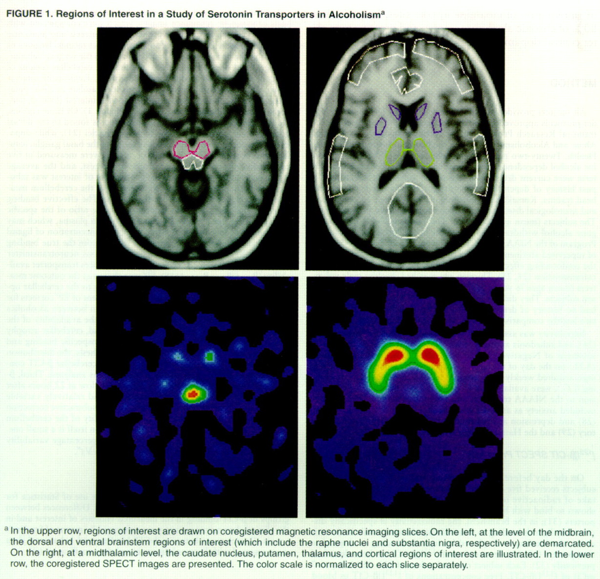
Individual regions of interest were drawn for each subject on the basis of magnetic resonance images, which were coregistered with the SPECT scans. Raters drawing regions of interest and analyzing SPECT data were blind to diagnosis and subject ratings. Regions of interest at the level of the midbrain demarcated the ventral (substantia nigra) and dorsal (raphe nuclei) brainstem; cerebellar regions of interest were drawn at the level of the pons. Regions of interest drawn at a midthalamic level demarcated the caudate nucleus, putamen, thalamus, and various cortical regions of interest (frontal, temporal, anterior cingulate, and occipital) (
figure 1). Of these regions, the highest density of serotonin transporters is found in the dorsal brainstem area, which contains the raphe nuclei
(21), while dopamine transporters are predominantly found in the basal ganglia (caudate and putamen)
(34). Regions of interest were measured in five consecutive slices, forming a volume of interest, and the average count per minute per milliliter in each volume of interest was tabulated and corrected for decay. Subtraction of the cerebellum measurement corrected for nonspecific binding. The effective binding potential (BP′=B
avail/K
d) was determined as the ratio of the specific binding to the free [
123I]β-CIT concentration in plasma, which may be assumed to be equal to the free synaptic concentration of ligand
(35). This effective binding potential differs from the true binding potential (BP=B
max/K
d) by virtue of endogenous neurotransmitter binding to transporter sites (B
endog), which reduces transporter availability (B
avail=B
max–B
endog)
(19). BP′ was chosen as the outcome measure instead of the ratio of the specific binding to the cerebellar uptake (V
3″)
(35) for three reasons. First, assessment of BP′ corrects for possible differences in radioligand metabolism between alcoholics and comparison subjects, which could affect the availability of the unmetabolized radioligand in the brain. Second, cerebellar atrophy in alcoholism can affect the assessment of nonspecific binding and thus systematically bias V
3″. To test this hypothesis, the distribution volume, V
2 (35), for the cerebellum (ratio of cerebellar β-CIT concentration to free concentration in plasma) was analyzed. Third, β-CIT retention in the cerebellum is exceedingly low at 22 hours after injection
(19, 20, 35); this leads to small and relatively variable SPECT measurements for the cerebellum. As a subtractive correction for nonspecific binding, the increased variability of the cerebellum data is of little consequence, since the correction itself is a small one. However, as a normalization divisor, the full percentage variability of the cerebellum data propagates directly into V
3″.