Full access
Regular Article
Published Online: 1 March 1999

Neuropsychological Functioning and MRI Signal Hyperintensities in Geriatric Depression

Publication: American Journal of Psychiatry

Abstract

OBJECTIVE: The purpose of this study was to examine the relationship between signal hyperintensities—a probable marker of underlying pathology—on T2-weighted magnetic resonance brain scans and neuropsychological test findings in elderly depressed and normal subjects. METHOD: Elderly subjects with a DSM-III-R diagnosis of major depression (N=41) and normal elderly comparison subjects (N=38) participated in a magnetic resonance imaging study (1.0-T) of signal hyperintensities in periventricular, deep white matter, and subcortical gray matter. Hard copies of scans were rated in random order by research psychiatrists blind to diagnosis; the modified Fazekas hyperintensity rating scale was used. Cognitive performance was independently assessed with a comprehensive neuropsychological battery. Clinical and demographic differences between groups were assessed by t tests and chi-square analysis. Relationships between neuropsychological performance and diagnosis and hyperintensities and their interaction were analyzed by using analysis of covariance, with adjustment for age and education. RESULTS: Elderly depressed subjects manifested poorer cognitive performance on several tests than normal comparison subjects. A significant interaction between hyperintensity location/severity and presence/absence of depression on cognitive performance was found: depressed patients with moderate-to-severe deep white matter hyperintensities demonstrated worse performance on general and delayed recall memory indices, executive functioning and language testing than depressed patients without such lesions and normal elderly subjects with or without deep white matter changes. CONCLUSIONS: Findings validate cognitive performance decrements in geriatric depression and suggest possible neuroanatomic vulnerabilities to developing particular neuropsychological dysfunction in depressed subjects. (Am J Psychiatry 1999; 156:438–444)
Recent investigations have postulated a relationship between signal hyperintensities on T2-weighted magnetic resonance scans and geriatric depression (15), although hyperintensities have also been reported in normal elderly subjects (58). The prevalence of these signal hyperintensities increases with age (5, 911), and they are more prominent in subjects with cerebrovascular disease risk factors (8, 1214) and, in some reports (5, 15), in subjects with dementia.
The clinical significance of signal hyperintensities and the relationship of these “lesions” to cognitive performance is unclear. While some reports have found a relationship between greater cognitive deficits in normal elderly individuals and more severe hyperintensity changes (1618), others have not (1922). Similarly, controversy exists regarding the significance of increased signal hyperintensities in cognitive disturbances in elderly depressed subjects (5, 20, 2325).
An extensive neuropsychological body of evidence in depression and geriatric depression demonstrates cognitive deficits that are reversible following successful somatic treatment of the depression (2630). This phenomenon, variably called “dementia syndrome of depression,” “depressive pseudodementia,” and “cognitive impairment of depression” (3133), may preferentially occur in elderly as compared to younger depressed subjects (33), although dissenting findings exist (26). The neuropsychological characteristics of late-life cognitive impairment of depression have been described as “subcortical” (34), in that the cognitive deficits observed resemble the deficits seen in neuropsychiatric conditions characterized by subcortical, rather than cortical, pathology. Furthermore, cognitive deficits in depression have been linked to subcortical-frontal lobe abnormalities (23, 35). Taken together, such data have contributed to a “subcortical dysfunction model” of learning and memory in affective illness (3638).
In geriatric patients, it is unclear whether cognitive impairment seen in depression is a consequence of depressive symptoms or is also mediated by underlying age-related brain changes that may play a role in the etiopathogenesis of the depression itself. While it has been suggested that hyperintensities present in depressed subjects may reflect a pathologic process that would also contribute to the impaired cognition seen in depression, the evidence for such a relationship remains inconclusive (24, 39, 40).
The purpose of this study was to extend examination of the relationship among signal hyperintensities, depression, and cognitive performance. Hyperintensity ratings in different brain locations and neuropsychological measures addressing different cognitive domains were evaluated in depressed and normal comparison subjects in whom dementia or a history of transient ischemic attack or stroke had been ruled out. We hypothesized that poorer cognitive performance would be associated with more severe hyperintensities.

METHOD

Subjects

Forty-one elderly depressed patients participated in the study. The patients were recruited from the Geriatric Psychiatry Service (inpatient, outpatient, and day hospital) at Hillside Hospital. Thirty-eight normal comparison subjects recruited from the community were solicited by means of advertisement in local newspapers or by word of mouth. All subjects received a Structured Clinical Interview for DSM-III-R (SCID) (41), were age 65 years or older, and were right-handed. Patients met the following inclusion criteria: DSM-III-R criteria for major depression, unipolar, as determined by the SCID and a score of 18 or greater on the 21-item Hamilton Depression Rating Scale (42).
Exclusion criteria for patients and comparison subjects included presence of a cardiac pacemaker, metallic clips, or other bodily metallic implants or artifacts (because of the magnetic resonance imaging [MRI] procedure); presence of significant acute medical illness or exacerbation of a chronic medical condition; presence of a neurodegenerative disorder, including Alzheimer’s disease or a related dementia (subjects did not meet DSM-III-R criteria for dementia); history of transient ischemic attack or stroke; and other past or current DSM-III-R diagnoses (for comparison subjects this included affective disorders). After complete description of the study to subjects, written informed consent was obtained.
The Clinical Global Impression for depression (43) and the Hamilton Depression Rating Scale (42) were administered to all subjects. Both psychiatric ratings were completed by an experienced geriatric psychologist (E.K.-G.) with demonstrated high interrater reliability (44). Other clinical and demographic data were assessed through use of a standardized format for information collection.

MRI Image Acquisition

Subjects were scanned in a 1.0-T whole-body MRI system (Siemens Magnetom). T2-weighted and proton density (intermediate) brain images were obtained in the axial plane. This series had a repetition time (TR) of 2500 msec and an echo delay time (TE) of 25 and 90 msec. The sequence produced 20 parallel sections in a 256×256 matrix with a 1.3 zoom factor. Axial images were 7 mm thick with a 0.7-mm gap between each section. A full coronal series was also obtained but was not used for the ratings.

MRI Scan Analysis

Hard copy images were printed for visual quantitative evaluation of signal hyperintensities. MRI scans of patients and comparison subjects were combined in a randomized order and evaluated under blind conditions by a research psychiatrist (K.R.R.K. or B.S.G.). Hyperintensities were assessed according to the modified Fazekas criteria (45). The modified Fazekas gradings follow an ascending degree of severity and frequency of hyperintensities (gradings=0–3) and rate them in three brain locations: periventricular region, deep white matter, and subcortical gray matter.
Interrater reliability for hyperintensity ratings were established by using intraclass correlation coefficients (periventricular hyperintensities ICC=0.86, deep white matter hyperintensities ICC=0.87, and subcortical gray matter lesions ICC=1.00).

Neuropsychological Measures

A neuropsychological test battery was administered to assess and quantify cognitive performance. On the basis of previous studies (24, 46, 47), selected tests were chosen from several domains for analyses. The battery consisted of tests representative of subcortical and cortical cognitive domains and included visuospatial functioning (block design of the WAIS-R [48]), attention/information processing (digit symbol of the WAIS-R [48], attention subscale of the Dementia Rating Scale [49]), language (Boston Naming Test [50]), executive functioning (initiation/perseveration subscale of the Dementia Rating Scale [49], Animal Naming Test [51], Controlled Oral Words Association Test [52]), and memory (Wechsler Memory Scale—Revised [53]). Both general memory and delayed recall memory index scores were derived according to the method of Woodard and Axelrod (54). The Dementia Rating Scale (49) was used as an overall measure of cognitive functioning. Because of occasional uncooperativeness, not all subjects completed every neuropsychological test.

Statistical Analysis

Clinical and demographic differences between depressed patients and comparison subjects were analyzed by using independent t tests and chi-square tests. To clarify the relationship among the regional distribution and severity of hyperintensities, neuropsychological function, and diagnosis of depression, the depressed and comparison subjects were dichotomized for the different brain region (periventricular, deep white matter, and subcortical gray matter) hyperintensity ratings into none/minimal (0–1 rating) and moderate/severe (2–3 rating). In this way each neuropsychological factor was then analyzed separately, with a main effect for group (depressed, comparison), a main effect for hyperintensity rating severity (in each brain area), and an interaction effect (group-by-hyperintensity ratings per area) being derived. Analysis of covariance (ANCOVA), with adjustment for the effects of age and years of education, was used to study both the main effects of group and hyperintensities, as well as their interactions, on relevant neuropsychological summary scores. Separate ANCOVAs were performed for periventricular, deep white matter, and subcortical gray matter regions. When significant interactions were observed, post hoc analyses of the cell means were performed. Because of the high number of ANCOVAs and increased chance of type II error, results were considered significant only if they met alpha criterion of 0.01. Two-tailed tests of probability were used to estimate statistical significance in all analyses.

RESULTS

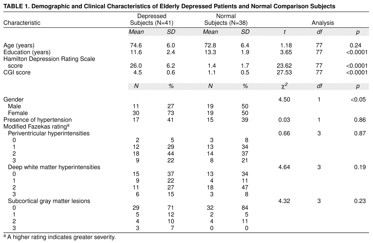
Age, sex distribution, years of education, depression ratings, and distribution of hyperintensity severity ratings are compared for depressed and normal subjects in table 1. Depressed patients were slightly, but nonsignificantly, older than normal comparison subjects. Although a higher proportion of men made up the comparison group, t test analyses revealed no significant differences between men and women on any of the neuropsychological test scores. Comparison subjects were also somewhat more educated than depressed patients. Scores on depression rating scales were, as expected, much higher for patients.
ANCOVA, which controlled for age and years of education, revealed a significant main effect of group on neuropsychological performance. Depressed patients performed worse on several cognitive tests, including general memory index (F=10.08, df=1, 73, p=0.002), block design (F=6.95, df=1, 61, p=0.01), and digit symbol (F=8.14, df=1, 50, p=0.006) and on the overall cognitive measure (the Dementia Rating Scale) (F=15.32, df=1, 72, p<0.0001). Hyperintensity severity as a main effect was not significant for any cognitive measures.
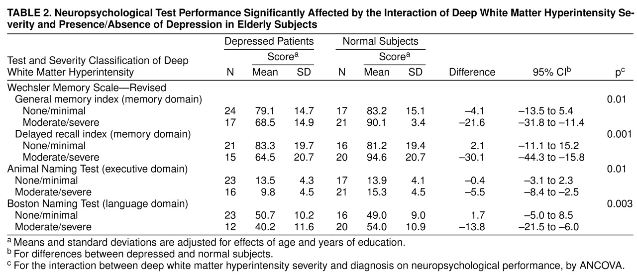
The interaction of group by hyperintensity location was examined to explore the influence of both factors on cognitive performance. Statistically significant differences were obtained as a function of group membership (depressed and comparison groups) and location of hyperintensities. The principal findings were that depressed subjects with moderate-to-severe hyperintensities in the deep white matter (N=17) performed significantly worse than depressed patients without such lesions (N=24) or comparison subjects with (N=21) and without (N=17) moderate-to-severe deep white matter hyperintensities on the following cognitive measures: general memory index (F=6.73, df=1, 73, p=0.01), delayed recall index (F=11.77, df=1, 66, p=0.001), Boston Naming Test (F=9.44, df=1, 65, p=0.003), and Animal Naming Test (F=6.84, df=1, 72, p=0.01). Table 2 presents age- and education-adjusted means for these tests. Ninety-five percent confidence intervals (CIs) for mean neuropsychological test differences between depressed and comparison subjects in both hyperintensity groups (i.e., none/minimal and moderate/severe) are presented. In contrast, no significant interaction effects were observed for group by hyperintensities in periventricular or subcortical gray matter areas.

DISCUSSION

The results of this study validate other reports that greater cognitive deficits exist in elderly depressed patients than in age-similar normal subjects and additionally suggest that deep white matter pathology may relate to some of these deficits. Poorer test performance in depressed patients than in comparison subjects occurred in several neuropsychological domains including attention/information, visuospatial, and memory processing, as well as overall cognitive functioning. The literature addressing cognitive dysfunction in depression ranges from studies reporting an absence of any cognitive changes (5557) to those reporting severe changes (36, 38, 58, >59). The results in the present study concur with studies reporting abnormalities in visuospatial functioning (24) and verbal (27, 60, 61) and visual (27, 62, 63) memory impairment in depressed as compared to normal subjects. Findings in the literature regarding attentional performance (64) and language processing (6568) are, however, more controversial. Not all studies of cognitive impairment in depression have examined a clear geriatric-only group. The findings in our study indicate that cognitive impairment in geriatric-only depressed patients cuts a fairly wide swath across multiple neuropsychological domains.
Evidence in prior reports is conflicting regarding the significance of MR signal hyperintensities on cognition in depressed and nondepressed geriatric populations. In normal elderly subjects, current findings reconcile with many studies that have failed to demonstrate a relationship between hyperintensities and cognitive performance (1922). On the other hand, other investigations have demonstrated a relationship between hyperintensities and impaired attention, visuospatial functioning, and speed of processing (1618). Some investigations of depressed patients have reported that patients with deep white matter lesions did not differ in terms of cognitive performance from those without such lesions (5, 47), whereas others have found an association between impairments in mental speed, executive functioning, and memory and presence of deep white matter (2325), periventricular (20), and subcortical gray matter (69) hyperintensities. This lack of consensus among studies may reflect differences in subject groups, cognitive tests administered, and MRI procedures/ratings; but it also likely is a consequence of the tremendous clinical heterogeneity of older depressed groups (70).
The present study differs from most other studies of elderly unipolar depressed subjects in that hyperintensities were rated in different brain regions (periventricular, deep white matter, subcortical gray matter) for the same patients, allowing simultaneous evaluation of the interaction of cognitive performance and distribution/severity of hyperintensities. In this study, less severe hyperintensities in all three brain regions in depressed and comparison subjects, and more severe hyperintensities in periventricular and subcortical gray regions in both groups, did not significantly influence cognitive performance. However, findings suggest that there is an interaction between the presence of depression and more severe signal hyperintensities in deep white matter that is associated with or hypothetically may mediate several, but not all, of the cognitive deficits observed in the depressed group. Of interest is that particular neuropsychological areas that were implicated (e.g., executive functioning, memory) are functions traditionally ascribed to subcortical/frontal brain systems (16, 36, 37) that have themselves been associated with an emerging neuroanatomy of depression (3, 39, 7173). We hypothesized that hyperintensities would be associated with cognitive impairment; however, only deep white matter changes were relevant in this study. Neuroanatomically, this suggests that interruptions between key cortical and subcortical gray matter regions/structures in the mood regulatory circuitry (i.e., in white matter tracts) may be more pertinent to cognitive deficits in geriatric depression than small, discrete lesions in the gray matter structures themselves (e.g., basal ganglia, thalamus), even though hyperintensities in both central gray and deep white matter regions have been linked to depression either as a neurobiological correlate or as a susceptibility factor (15, 15, 23, 69, 74, 75).
Furthermore, because more severe hyperintensities in deep white matter probably represent cerebrovascular disease changes histopathologically (76, 77), current data implicating moderate to severe—but not minimal—hyperintensities in the executive and memory function deficits seen in depressed subjects suggest that a threshold level of ischemic pathology in deep white matter may be necessary for these neuropsychological impairments to emerge during depression. Because similar deep white matter hyperintensity changes in elderly normal subjects were not significantly associated with any cognitive deficits, it appears that depression represents a vulnerability to or necessary cofactor for cognitive compromise associated with deep white matter hyperintensities.
Hyperintensities occur in deep white matter that contains tracts connecting subcortical and cortical brain structures such as the basal ganglia and frontal lobes in which depression state-dependent hypometabolism has been demonstrated in functional neuroimaging studies (78, 79). Furthermore, ascending neurotransmitter systems implicated in depression modulate these structures/circuits as well (3, 80, 81). As such, although deep white matter pathology itself may predispose an individual to depression, cognitive expression of deep white matter abnormalities may be contingent on a concurrent or “superimposed” state-dependent hypometabolism in similar or connected brain regions and/or a neurochemical disequilibrium associated with depression. In this way, more severe deep white matter changes may contribute to the so-called reversible cognitive impairment of depression (or dementia syndrome of depression or depressive pseudodementia) (3133). This notion thematically resonates with the finding of regional cerebral blood flow (CBF) abnormalities in depression with “additive” further regional CBF abnormalities in those depressed subjects with reversible cognitive impairment (82). Future studies that combine similar neuroimaging assessments with neuropsychological testing during depression and after recovery will help determine whether deep white matter changes in elderly depressed subjects are also associated with postrecovery evidence of persisting, but lesser, cognitive deficiencies—a phenomenon that has been demonstrated in a substantial proportion of remitted elderly depressed subjects who had experienced more evident cognitive difficulties when depressed (83).
Current findings suggest a possible brain substrate for some of the often reversible cognitive deficits encountered in elderly depressed patients. However, the MR hyperintensity rating methodology employed in this study scored hyperintensities regardless of where they occurred in the subcortical deep white matter (i.e., frontal, parietal, occipital, temporal regions). As such, further speculation on brain-behavior relationships are dependent on rating methods that more specifically localize hyperintensities in the brain. We are presently analyzing data based on more neuroanatomically specific ratings (74) in a different population of older depressed subjects and comparison subjects who also underwent an identical neuropsychological battery.
TABLE 1
TABLE 2

Footnote

Received Feb. 3, 1998; revision received July 23, 1998; accepted Aug. 6, 1998. From the Departments of Psychiatry and Radiology, Long Island Jewish Medical Center, Glen Oaks, N.Y., and Albert Einstein College of Medicine, Bronx, N.Y.; Department of Psychiatry, Duke University Medical Center, Durham, N.C.; and Department of Computer Information Systems and Decision Sciences, St. John’s University, Jamaica, N.Y. Address reprint requests to Dr. Kramer-Ginsberg, Hillside Hospital, Research Building, Long Island Jewish Medical Center, 75-59 263rd St., Glen Oaks, NY 11004; [email protected] (e-mail). Supported by NIMH grant MH-01098 (to Dr. Greenwald).

References

1.
Greenwald BS, Kramer-Ginsberg E, Krishnan KRR, Ashtari M, Aupperle PM, Patel M: MRI signal hyperintensities in geriatric depression. Am J Psychiatry 1996; 153:1212–1215
2.
O’Brien JT, Desmond P, Ames D, Schweitzer I, Harrigan S, Tress B: A magnetic resonance study imaging of white matter lesions in depression and Alzheimer’s disease. Br J Psychiatry 1996; 168:477–485
3.
Krishnan KRR: Neuroanatomic substrates of depression in the elderly. J Geriatr Psychiatr Neurol 1993; 6:39–58
4.
Coffey CE, Figiel GS, Djang WT, Weiner RD: Subcortical hyperintensity on magnetic resonance imaging: a comparison of normal and depressed elderly subjects. Am J Psychiatry 1990; 147:187–189
5.
Zubenko GS, Sullivan P, Nelson JP, Belle SH, Huff FJ, Wolf GL: Brain imaging abnormalities in mental disorder of late life. Arch Neurol 1990; 47:1107–1111
6.
Lindgren A, Roijer A, Rudling O, Norrving B, Larsson EM, Eskilsson J, Wallin L, Olsson B, Johansson BB: Cerebral lesions on magnetic resonance imaging, heart disease, and vascular risk factors in subjects without stroke: a population-based study. Stroke 1994; 25:929–934
7.
Schmidt R, Fazekas F, Offenbacher H, Dusek T, Zach E, Reinhart B, Grieshofer P, Freidl W, Eber B, Schumacher M, Koch M, Lechner H: Neuropsychological correlates of MRI white matter hyperintensities: a study of 150 normal volunteers. Neurology 1993; 43:2490–2494
8.
Meyer JS, Kawamura J, Terayama Y: White matter lesions in the elderly. J Neurol Sci 1992; 110:1–7
9.
Jernigan TL, Press GA, Hesselink JR: Methods for measuring brain morphologic features on magnetic resonance images: validation and normal aging. Arch Neurol 1990; 47:27–32
10.
Keretesz A, Black SE, Tokar G, Benke T, Carr T, Nicholson L: Periventricular and subcortical hyperintensities on magnetic resonance imaging. Arch Neurol 1988; 45:404–408
11.
Kirkpatrick JB, Hayman LA: White matter lesions in MR imaging of clinically healthy brains of elderly subjects: possible pathologic basis. Radiology 1987; 162:509–511
12.
Manolio TA, Kronmal RA, Burke GL, Poirier V, O’Leary DH, Gardin JM, Fried LP, Steinberg EP, Bryan RN: Magnetic resonance abnormalities and cardiovascular disease in older adults. Stroke 1994; 25:318–327
13.
Bots ML, Van Swieten JC, Breteler MMB, de-Jong PT, Van-Gijn J, Hofman A, Grobbee DE: Cerebral white matter lesions and atherosclerosis in the Rotterdam study. Lancet 1993; 341:1232–1237
14.
Awad IA, Spetzler RF, Hodak JA, Awad CA, Williams F, Carey R: Incidental lesions noted on magnetic resonance imaging of the brain: prevalence and clinical significance in various age groups. Neurosurgery 1987; 20:222–227
15.
Rabins PV, Pearlson GD, Aylward E, Kumar AJ, Dowell K: Cortical magnetic resonance imaging changes in elderly inpatients with major depression. Am J Psychiatry 1991; 148:617–620
16.
Boone KC, Miller BL, Lesser IM, Mehringer CM, Hill-Gutierrez E, Goldberg MA, Berman NG: Neuropsychological correlates of white-matter lesions in healthy elderly subjects. Arch Neurol 1992; 49:549–554
17.
Rao SM, Mittenberg W, Bernardin L, Haughton V, Leo GJ: Neuropsychological test findings in subjects with leuko-araiosis. Arch Neurol 1989; 46:40–44
18.
Steingart A, Hachinski VC, Lau C, Fox AJ, Lee D, Inzitari D, Merskey H, Diaz F, Cape R, Lee D, Inzitari D, Merskey H: Cognitive and neurologic findings in subjects with diffuse white matter lucencies on computed tomographic scan (leuko-araiosis). Arch Neurol 1987; 44:32–35
19.
Tupler LA, Doffey CE, Logue PE, Djang WT, Fagan SM: Neuropsychological importance of subcortical white matter hyperintensity. Arch Neurol 1992; 49:1248–1252
20.
Harrell LE, Duvall EF, Folks DG, Duke L, Bartolucci A, Conboy T, Callaway R, Kerns D: The relationship of high-intensity signals on magnetic resonance images to cognitive and psychiatric state in Alzheimer’s disease. Arch Neurol 1991; 48:1136–1140
21.
Schmidt R, Fazekas F, Offenbacher H, Lytwyn H, Blematl B, Niederkorn K, Horner S, Payer F, Freidl W: Magnetic resonance imaging white matter lesions and cognitive impairment in hypertensive individuals. Arch Neurol 1991; 48:417–420
22.
Hendrie HC, Farlow MR, Austrom MG, Edwards MK, Williams MA: Foci of increase T2 signal intensity on brain MR scans of healthy elderly subjects. AJNR Am J Neuroradiol 1989; 10:703–707
23.
Hickie I, Scott E, Mitchell P, Wilhelm K, Austin MP, Bennett B: Subcortical hyperintensities on magnetic resonance imaging: clinical correlates and prognostic significance in patients with severe depression. Biol Psychiatry 1995; 37:151–160
24.
Lesser IM, Boone KB, Mehringer CM, Wohl MA, Miller BL, Berman NG: Cognition and white matter hyperintensities in older depressed patients. Am J Psychiatry 1996; 153:1280–1287
25.
Lesser IM, Miller BL, Boone KB, Hill-Gutierrez E, Mehringer CM, Wong K, Mena I: Brain injury and cognitive function in late-onset depression. J Neuropsychiatry Clin Neurosci 1991; 3:33–40
26.
Tarbuck AF, Paykel ES: Effects of major depression on the cognitive function of younger and older subjects. Psychol Med 1995; 25:285–296
27.
Fromm D, Schopflocher D: Neuropsychological test performance in depressed patients before and after drug therapy. Biol Psychiatry 1984; 19:55–72
28.
Rabins PV, Merchant A, Nestadt G: Criteria for diagnosing reversible dementia caused by depression: validation by 2-year follow-up. Br J Psychiatry 1984; 144:488–492
29.
Cohen RM, Weingartner H, Smallberg SA, Pickar D, Murphy DL: Effort and cognition in depression. Arch Gen Psychiatry 1982; 39:593–597
30.
Sternberg DE, Jarvik ME: Memory functions in depression: improvement with antidepressant medication. Arch Gen Psychiatry 1976; 33:219–224
31.
Reifler BV: Mixed cognitive-affective disturbances in the elderly: a new classification. J Clin Psychiatry 1986; 47:354–356
32.
McAllister TW: Overview: pseudodementia. Am J Psychiatry 1983; 140:528–533
33.
Folstein MF, McHugh PR: Dementia syndrome of depression, in Alzheimer’s Disease: Senile Dementia and Related Disorders: Aging, vol 7. Edited by Katzman R, Terry RD, Bick KL. New York, Raven Press, 1978, pp 87–96
34.
Caine ED: Pseudodementia: current concepts and future directions. Arch Gen Psychiatry 1981; 38:1359–1364
35.
Cummings JL: The neuroanatomy of depression. J Clin Psychiatry 1993; 54:14–20
36.
Massman PJ, Butters NM, Delis DC: Some comparisons of the verbal learning deficits in Alzheimer dementia, Huntington disease and depression, in Dementia: Presentations, Differential Diagnosis, and Nosology. Edited by Emery VOB, Oxman TE. Baltimore, Johns Hopkins University Press, 1994, pp 232–248
37.
Massman PJ, Delis DC, Butters N, Dupont RM, Gillin JC: The subcortical dysfunction hypothesis of memory deficits in depression: neuropsychological validation in a subgroup of patients. J Clin Exp Neuropsychol 1992; 14:687–706
38.
Wolfe J, Granholm E, Butters N, Saunders E, Janowsky D: Verbal memory deficits associated with major affective disorders: a comparison of unipolar and bipolar patients. J Affect Disord 1987; 13:83–92
39.
Soares JC, Mann JJ: The anatomy of mood disorders: review of structural neuro-imaging studies. Biol Psychiatry 1997; 41:86–106
40.
Nussbaum PD: Pseudodementia: a slow death. Neuropsychol Rev 1994; 4:71–90
41.
Spitzer RL, Williams JBW, Gibbon M, First MB: Instruction Manual for the Structured Clinical Interview for DSM-III-R (SCID). New York, New York State Psychiatric Institute, Biometrics Research, 1988
42.
Hamilton M: A rating scale for depression. J Neurol Neurosurg Psychiatry 1960; 23:56–62
43.
Guy W (ed): ECDEU Assessment Manual for Psychopharmacology: Publication ADM 76-338. Washington, DC, US Department of Health, Education, and Welfare, 1976, pp 534–537
44.
Greenwald BS, Kramer-Ginsberg E: Age at onset of geriatric depression: relationship to clinical variables. J Affect Disord 1988; 15:61–68
45.
Krishnan KRR, Boyko OB, McDonald WM, Charles CH, MacFall JR, Tupler LA, Upchurch LW: Magnetic resonance morphometry: image-analysis methodology development for affective disorder. Depression 1993; 1:159–173
46.
Brettler MMB, Van Amerongen NM, Van Swieten JC, Claus JJ, Grobbe DE, Van Gijn J, Hofman A, Van Harskamp F: Cognitive correlates of ventricular enlargement and cerebral white matter lesions on magnetic resonance imaging. Stroke 1994; 25:1109–1115
47.
Dupont RM, Jernigan TL, Heindel W: Magnetic resonance imaging and mood disorders: localization of white matter and other subcortical abnormalities. Arch Gen Psychiatry 1995; 52:747–755
48.
Wechsler D: WAIS-R Manual. New York, Psychological Corp, 1981
49.
Mattis S: Dementia Rating Scale Professional Manual. Odessa, Fla, Psychological Assessment Resources, 1973
50.
Kaplan E, Goodglass H, Weintraub S: The Boston Naming Test, 2nd ed. Philadelphia, Lea & Febiger, 1983
51.
Goodglass H, Kaplan E: The Assessment of Aphasia and Related Disorders, 2nd ed. Philadelphia, Lee & Febiger, 1983
52.
Benton AL, Hamsher K: Multilingual Aphasia Examination Manual. Iowa City, University of Iowa, 1978
53.
Wechsler C: Wechsler Memory Scale, Revised. New York, Psychological Corp, 1987
54.
Woodard JL, Salthouse TA, Godsall RE, Green RC: Confirmatory factor analysis of the Mattis Dementia Rating Scale in patients with Alzheimer’s disease. Psychol Assessment 1996; 8:85–91
55.
Williams JM, Little MM, Scates S, Blockman N: Memory complaints and abilities among depressed older adults. J Consult Clin Psychol 1987; 55:595–598
56.
Niederehe G, Camp CJ: Signal detection analysis of recognition memory in depressed elderly. Exp Aging Res 1985; 11:207–213
57.
Popkin SJ, Callagher D, Thompson LW, Moore M: Memory complaint and performance in normal and depressed older adults. Exp Aging Res 1982; 8:141–145
58.
Siegfried K: Cognitive symptoms in late-life depression and their treatments. J Affect Disord Suppl 1985; 1:S33–S40
59.
Savard RJ, Rey AC, Post RM: Halstead-Reitan Category Test in bipolar and unipolar affective disorders. J Nerv Ment Dis 1980; 168:297–304
60.
Kopelman MD: Clnical test of memory. Br J Psychiatry 1986; 148:517–525
61.
La Rue A, D’Elia LF, Clark EO, Spar JE, Jarvik LF: Clinical tests of memory in dementia, depression, and healthy aging. Psychol Aging 1986; 1:69–77
62.
Richards PM, Ruff RM: Motivational effects on neuropsychological functioning: comparison of depressed versus non-depressed individuals. J Consult Clin Psychol 1989; 57:396–402
63.
Piersma HL: Wechsler Memory Scale performance in gero­psychiatric patients. J Clin Psychol 1986; 42:323–327
64.
Breslow R, Kocsis J, Belkin B: Memory deficits in depression: evidence utilizing the Wechsler Memory Scale. Percept Mot Skills 1980; 51:541–542
65.
Alexander MP, Naeser MA, Palumbo CL: Correlations of subcortical CT lesion sites and aphasia profiles. Brain 1987; 110:961–991
66.
Butters N, Wolfe J, Martone M, Granholm E, Cermak LS: Memory disorders associated with Huntington’s disease: verbal recall, verbal recognition, and procedural memory. Neuropsychologia 1985; 23:729–743
67.
von Cramon D, Hebel N: Disorders of learning and memory in focal cerebral tissue lesions. Fortschr Neurol Psychiatr 1989; 57:544–550
68.
Luria AR, Majovski LV: Basic approaches used in American and Soviet clinical neuropsychology. Am Psychol 1977; 32:959–968
69.
Coffey CE, Figiel GS, Djang WT, Saunders WB, Weiner RDl: White matter hyperintensity on magnetic resonance imaging: clinical and neuroanatomic correlates in the depressed elderly. J Neuropsychiatry 1989; 2:135–144
70.
Caine ED, Lyness JM, King DA: Reconsidering depression in the elderly. Am J Geriatr Psychiatry 1993; 1:4–20
71.
Salloway S, Cummings J: Subcortical disease and neuropsychiatric illness. J Neuropsychiatry Clin Neurosci 1994; 6:93–99
72.
Weinberger DR: A connectionist approach to the prefrontal cortex. J Neuropsychiatry Clin Neurosci 1993; 5:241–253
73.
Gupta SR, Naheedy MH, Young JC, Ghobrial M, Rubino FA, Hindo W: Periventricular white matter changes and dementia: clinical, neuropsychological, radiological, and pathological correlation. Arch Neurol 1988; 45:637–641
74.
Greenwald BS, Kramer-Ginsberg E, Krishnan KR, Ashtari M, Auerbach C, Patel M: Neuroanatomical localization of magnetic resonance imaging signal hyperintensities in geriatric depression. Stroke 1998; 29:613–617
75.
Coffey CE, Wilkinson WE, Weiner RD, Parashos IA, Djang WT, Webb MC, Figiel GS, Spritzer CE: Quantitative cerebral anatomy in depression. Arch Gen Psychiatry 1993; 50:7–16
76.
Braffman BH, Zimmerman RA, Trojanowski JQ, Gonatas NK, Hickey WK, Schlaepfer WW: Brain MR: Pathological correlation with gross and histopathological change, II: hyperintense white-matter foci in elderly. Am J Neuroradiol 1988; 9:629–636
77.
Fazekas F, Kleimert R, Offenbacher H, Schmidt R, Kleinert G, Payer F, Radner H, Lechner H: Pathologic correlates of incidental MRI white matter hyperintensities. Neurology 1993; 43:1683–1689
78.
Baxter LR, Schwartz JM, Phelps ME, Mazziotta JC, Guze BH, Selin CE, Gerner RH, Sumida RM: Reduction of prefrontal cortex glucose metabolism common to three types of depression. Arch Gen Psychiatry 1989; 46:243–250
79.
Baxter LR, Phelps ME, Mazziotta JC, Schwartz JM, Gerner RH, Selin CE, Sumida RM: Cerebral metabolic rates for glucose in mood disorders: studies with PET and fluorodeoxyglucose F18. Arch Gen Psychiatry 1985; 42:441–447
80.
Nolte J, Angevine JB Jr: The Human Brain in Photographs and Diagrams. St. Louis, Mosby-Year Books, 1995
81.
Brown SL, Steinberg RL, van Praag HM: The pathogenesis of depression: reconsideration of neurotransmitter data, in Handbook of Depression and Anxiety: A Biological Approach. Edited by den Boer JA, Ad Sitsen JM. New York, Marcel Dekker, 1994, pp 317-347
82.
Dolan RJ, Bench CJ, Brown RG, Scott LC, Friston KJ, Frackowiak RSJ: Regional cerebral blood flow abnormalities in depressed patients with cognitive impairment. J Neurol Neurosurg Psychiatry 1992; 55:768–773
83.
Abas MA, Sahakian BJ, Levy R: Neuropsychological deficits and CT scan changes in elderly depressives. Psychol Med 1990; 20:507–520

Information & Authors

Information

Published In

Go to American Journal of Psychiatry
American Journal of Psychiatry
Pages: 438 - 444
PubMed: 10080561

History

Published online: 1 March 1999
Published in print: March 1999

Authors

Affiliations

Elisse Kramer-Ginsberg, Ph.D.
Blaine S. Greenwald, M.D.
K. Ranga Rama Krishnan, M.D.
Bruce Christiansen, Ph.D.

Metrics & Citations

Metrics

Citations

Export Citations

If you have the appropriate software installed, you can download article citation data to the citation manager of your choice. Simply select your manager software from the list below and click Download.

For more information or tips please see 'Downloading to a citation manager' in the Help menu.

Format
Citation style
Style
Copy to clipboard

There are no citations for this item

View Options

View options

PDF/ePub

View PDF/ePub

Get Access

Login options

Already a subscriber? Access your subscription through your login credentials or your institution for full access to this article.

Personal login Institutional Login Open Athens login
Purchase Options

Purchase this article to access the full text.

PPV Articles - American Journal of Psychiatry

PPV Articles - American Journal of Psychiatry

Not a subscriber?

Subscribe Now / Learn More

PsychiatryOnline subscription options offer access to the DSM-5-TR® library, books, journals, CME, and patient resources. This all-in-one virtual library provides psychiatrists and mental health professionals with key resources for diagnosis, treatment, research, and professional development.

Need more help? PsychiatryOnline Customer Service may be reached by emailing [email protected] or by calling 800-368-5777 (in the U.S.) or 703-907-7322 (outside the U.S.).

Media

Figures

Other

Tables

Share

Share

Share article link

Share