The American Psychiatric Association (APA) has updated its Privacy Policy and Terms of Use, including with new information specifically addressed to individuals in the European Economic Area. As described in the Privacy Policy and Terms of Use, this website utilizes cookies, including for the purpose of offering an optimal online experience and services tailored to your preferences.

Please read the entire Privacy Policy and Terms of Use. By closing this message, browsing this website, continuing the navigation, or otherwise continuing to use the APA's websites, you confirm that you understand and accept the terms of the Privacy Policy and Terms of Use, including the utilization of cookies.

×
History of PsychiatryFull Access

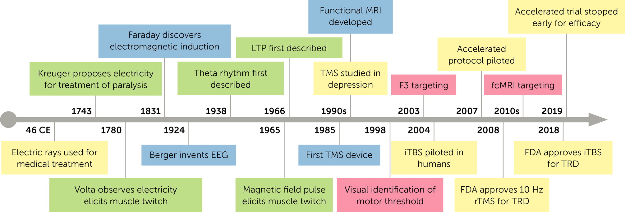
History and Future Directions of rTMS for Treatment of Depressive Disorders

The development of transcranial magnetic stimulation (TMS) offers an illustrative example of how a breadth of expertise and technology can come together to transform the treatment of depression. This article outlines the history of this development.

rTMS Origins: Biophysics

Michael Faraday’s discovery of electromagnetic induction in 1831 is the foundational breakthrough in physics that eventually enabled TMS, just over 150 years later. The pioneering work in electromagnetism by Michael Faraday and his student James Clerk Maxwell (coincidentally born in 1831) enabled physicists and then physiologists to study the electrophysiological effects of magnetic fields (1, 2). Pulsed magnetic fields were first shown to elicit twitches in skeletal muscles in animal models and humans in 1965 (3). Two decades later, the first device to generate cortical activity with the use of a pulsed magnetic field was developed by the medical physicist Anthony Barker and his team (4). This first TMS device delivered one pulse every 3 seconds and demonstrated that stimulation of the motor cortex elicited movements of the contralateral body.

In the decades to follow, improvements in the engineering of TMS coils, including improvements in pulse parameters, cooling technology, and dual coil configurations, would enable the transformative application of this technology in both neurophysiological and clinical research (5). These technological developments offered researchers a wide range of stimulation parameters to investigate, including stimulation dose, frequency, rhythm, and target. Repetitive TMS (rTMS) became a focus of therapeutic intervention, because rTMS was shown to produce a lasting increase or decrease in focal cortical activity. Whether rTMS increased or decreased cortical activity depended on the specific pulse parameters used (6). “High-frequency” stimulation, in the range of 5 Hz–20 Hz, became widely considered to increase activity in the cortical region stimulated.

Clinical Debut

From early in the 1990s, on the basis of brain lesion studies and functional neuroimaging studies, dysfunction of the left dorsolateral prefrontal cortex (L-dlPFC) was associated with depression. Soon thereafter, rTMS researchers focused on the L-dlPFC as a target site in the treatment of major depressive disorder (as defined by DSM-IV at that time) (7, 8). This line of research led to the landmark double-blind multisite study by O’Reardon et al. (9) that began recruitment in 2004, in which patients for whom pharmacotherapy for major depressive disorder had failed received either high-frequency rTMS over the L-dlPFC or sham treatment. rTMS produced significantly greater response and remission rates. This study was published in 2007, and in the following year, the Food and Drug Administration (FDA) approved the first rTMS protocol for patients whose depression failed to respond to at least one adequate trial of pharmacotherapy (10): daily rTMS treatment sessions, five times weekly for 4–6 weeks, with pulses delivered at 10 Hz, for a total of 3,000 pulses to the L-dlPFC over 37.5 minutes (9, 11).

rTMS at the Bench: Long-Term Potentiation

Although the clinical efficacy of rTMS has been demonstrated, the exact mechanism by which rTMS results in lasting changes in cortical activity and symptom relief is still not fully defined. However, long-term potentiation (LTP) of neuronal activity in key brain regions is suspected to play a central role (6, 12). LTP is considered to be foundational to many forms of learning and memory and is one of the major mechanisms of synaptic plasticity. In brief, LTP involves a lasting increased efficiency of signal transmission across synapses in response to particular patterns of stimulation, such as specific forms of rTMS (13). LTP is mediated through complex changes in pre- and postsynaptic molecular machinery, including cell surface receptors, neurotransmitter release, and cytoarchitecture (13). LTP reflects the principle that “cells that fire together, wire together,” a paraphrase from the visionary book, The Organization of Behavior (1949) by Donald Hebb, the so-called “father of neuropsychology.” Evidence supporting the existence of LTP was first detailed in the mid-1960s (14). Around this time, the prominent psychiatrist Eric Kandel was also advancing the molecular study of memory formation, including the study of LTP. Dr. Kandel would go on to share the Nobel Prize in Physiology or Medicine in 2000 for this work.

Bench to Bedside: LTP to Theta Burst

An understanding of LTP would later enable the development of theta burst stimulation, a more efficient stimulus pattern for LTP and, in turn, a more efficient rTMS pulse pattern. However, the origins of the term “theta” started earlier, with the development of electroencephalography (EEG). In 1924, the psychiatrist Hans Berger recorded the first human EEG (15). Dr. Berger categorized brain wave patterns by frequency ranges, and he named the alpha (8 Hz–13 Hz) and beta (13 Hz–30 Hz) ranges. Naming by Greek letters would resume by order of discovery, and the theta range (4 Hz–8 Hz) would be identified decades later (16). Although scalp EEG allowed for the noninvasive study of human cortical electrical activity, concurrent neurophysiological studies in animal models also discovered strong bursts of hippocampal neuronal activity in frequency ranges similar to theta, and thus these hippocampal rhythms were similarly named. Although the link between cortical and hippocampal theta rhythms is not fully understood, the hippocampus is thought to play an important role in generating theta rhythms in widespread cortical regions (17). In the 1970s, in vivo microelectrode recording from rat brains demonstrated that hippocampal neurons would fire a burst of action potentials in a theta-range frequency during exploratory behavior (18). This prompted hypotheses about the role of theta bursts in learning and memory, and by the early 1990s, it was shown that LTP in hippocampal synapses was maximally facilitated by a burst of electrode stimulation in the theta-range frequency (19).

While O’Reardon and colleagues (9) were leading the clinical trial that led to the initial FDA approval of rTMS for the treatment of depression, rTMS researchers who were initiating preclinical trials of rTMS pulse patterns modeled the endogenous theta rhythms seen in mammalian brains. The first pilot study of theta burst stimulation (TBS) using rTMS in healthy volunteers was published in 2004 and promised a great boost to potentiating the efficiency of rTMS (20). Additional refinement favored an intermittent TBS (iTBS)—that is, TBS with pauses in between brief trains of stimulation. By 2013, recruitment was started for a randomized noninferiority multisite trial in Canada, comparing iTBS to the then-conventional 10-Hz rTMS over the L-dlPFC for treatment-resistant major depressive disorder (21). Although iTBS delivered a fifth of the total number of pulses in less than a tenth of the time, results published in 2018 showed iTBS to be noninferior, with similar tolerability. Both treatment arms achieved a 50% symptom reduction in about 50% of patients and remission in 30%. That same year, the FDA approved an iTBS protocol for treatment-resistant major depressive disorder, with a schedule similar to the previous 10-Hz protocol but delivering rapid bursts of pulses at a 5-Hz theta-like rate, for a total of 600 pulses to the L-dlPFC in just over 3 minutes.

Treatment Personalization: Dose and Target

Individual differences in scalp-cortex distance and cortical cytoarchitecture affect the cortical response to TMS (22). The motor threshold (MT) emerged as a concept that allowed TMS machine output to be personalized. The MT is the individualized minimum “dose” of TMS machine output (as a percentage of its maximum output) needed to elicit a contralateral motor response. In 1998, it was shown that visualizing movements of the contralateral hand effectively approximated electromyographic measures of the MT, simplifying the procedure even further (23). Visual determination of the MT continues to be a widely used method in TMS clinics and research, because it allows for relatively quick determination of the MT without the use of additional diagnostic tools (such as electromyographic detection of the MT or EEG over target frontal cortical regions) (24). However, it is important to recognize that the use of the MT involves the key assumption that the response threshold of other cortical regions, such as the dlPFC, is sufficiently similar to the response threshold of the motor cortex (24).

The 2007 study protocol by O’Reardon and colleagues (9) targeted the dlPFC by using a “5-cm rule,” in which the coil would be placed 5 cm anterior to the site of stimulation that elicited motor movements in the right thumb. However, by 2003, the EEG F3 electrode placement in the international 10-20 system was identified as a more reliable method to target the dlPFC (25). Further heuristics for coil positioning and improvements on this system would follow. As might be expected, neuroimaging-guided targeting would be proposed, with research protocols involving structural imaging techniques promptly giving way to the application of functional imaging techniques. By the early 2010s, rTMS studies began to use functional connectivity MRI (fcMRI), a functional neuroimaging technique that quantifies the association of activity between different brain regions and allows for the mapping of neural networks. fcMRI studies have suggested that stimulating the specific region of the dlPFC that is most anticorrelated with activity in the subgenual anterior cingulate cortex, a region previously implicated as hyperactive in depression, might further optimize treatment response rates (26).

Contemporary Developments and Future Directions

Accelerated TMS protocols with five to ten sessions in a single day were shown to be successful as early as 2010 (27), and the rapidly improved efficiency of iTBS greatly facilitated accelerated treatment schedules. By 2017, recruitment began for a double-blind randomized sham-controlled trial that combined an accelerated course of 50 treatment sessions over just 5 days with individualized targeting guided by fcMRI (28). Additionally, each of the treatment sessions were run for 10 minutes rather than 3 minutes, resulting in a total of 90,000 pulses delivered in just 5 days. At a planned interim analysis, the trial was stopped early for efficacy: 50% symptom reduction and remission were achieved in 71% and 57% of participants in the treatment arm, respectively, compared with 13% and 0% of those in the control group. Although a nonsignificant trend for symptom relapse was noted over 4 weeks of follow-up, a 50% symptom reduction and remission were maintained in 64% and 43% of those in the treatment arm, respectively. Such accelerated schedules are especially appealing for future studies in inpatient settings and where rapid treatment of depression is needed for acute management of suicidality. On the other hand, most trials have thus far focused on the index course of treatment and have had limited follow-up. Future research and development will also likely turn to characterizing the role for maintenance treatment.

Whereas convulsive therapy has generally been reserved for the most severe cases of depression, the emerging accessibility and effectiveness of rTMS may position it as an important complement to both psychotherapy and pharmacotherapy in the treatment of depression. The history thus far of rTMS development, from foundational theories to the latest in accelerated and personalized iTBS protocols (summarized in Figure 1), offers an inspiring reminder of how incremental interdisciplinary developments can come together to produce leaps in medical treatment. This trajectory also highlights the importance of having psychiatrists invested across the full spectrum of translational research, from bench science and technological development to clinical trials and back.

FIGURE 1.

FIGURE 1. Key advances in the history of repetitive transcranial magnetic stimulation (rTMS)a

aSubjects of development are grouped by color: clinical observations and treatment are yellow; neurophysiological studies, green; physics and engineering, blue; and treatment personalization, red. Timeline not to scale. EEG=electroencephalography; fcMRI=functional connectivity MRI; iTBS=intermittent theta burst stimulation; LTP=long-term potentiation; TRD=treatment-resistant depression. For additional details, see references 1, 2, 3, 10, 26.

Dr. Levit is a third-year resident in the Department of Psychiatry, University of British Columbia, Vancouver.
References

1. Fitzgerald PB, Daskalakis ZJ: Repetitive Transcranial Magnetic Stimulation Treatment for Depressive Disorders. Berlin, Springer, 2013 CrossrefGoogle Scholar

2. McWhirter L, Carson A, Stone J: The body electric: a long view of electrical therapy for functional neurological disorders. Brain 2015; 138:1113–1120 CrossrefGoogle Scholar

3. Walsh V, Pascual-Leone A, Kosslyn SM: Transcranial Magnetic Stimulation: A Neurochronometrics of Mind. Cambridge, MA, MIT Press, 2003 Google Scholar

4. Barker AT, Jalinous R, Freeston IL: Non-invasive magnetic stimulation of human motor cortex. Lancet 1985; 325:1106–1107 CrossrefGoogle Scholar

5. Goetz SM, Deng Z-D: The development and modelling of devices and paradigms for transcranial magnetic stimulation. Int Rev Psychiatry 2017; 29:115–145 CrossrefGoogle Scholar

6. Klomjai W, Katz R, Lackmy-Vallée A: Basic principles of transcranial magnetic stimulation (TMS) and repetitive TMS (rTMS). Ann Phys Rehabil Med 2015; 58:208–213 CrossrefGoogle Scholar

7. George MS, Wassermann EM, Williams WA, et al.: Daily repetitive transcranial magnetic stimulation (rTMS) improves mood in depression. Neuroreport 1995; 6:1853–1856 CrossrefGoogle Scholar

8. Pascual-Leone A, Rubio B, Pallardó F, et al.: Rapid-rate transcranial magnetic stimulation of left dorsolateral prefrontal cortex in drug-resistant depression. Lancet 1996; 348:233–237 CrossrefGoogle Scholar

9. O’Reardon JP, Solvason HB, Janicak PG, et al.: Efficacy and safety of transcranial magnetic stimulation in the acute treatment of major depression: a multisite randomized controlled trial. Biol Psychiatry 2007; 62:1208–1216 CrossrefGoogle Scholar

10. Cohen SL, Bikson M, Badran BW, et al.: A visual and narrative timeline of US FDA milestones for transcranial magnetic stimulation (TMS) devices. Brain Stimul 2022; 15:73–75 CrossrefGoogle Scholar

11. Yan J: FDA approves new option to treat major depression. Psychiatr News 2008; 43:2–17 CrossrefGoogle Scholar

12. Chervyakov AV, Chernyavsky AY, Sinitsyn DO, et al.: Possible mechanisms underlying the therapeutic effects of transcranial magnetic stimulation. Front Hum Neurosci 2015; 9:303 CrossrefGoogle Scholar

13. Hayashi Y: Molecular mechanism of hippocampal long-term potentiation: towards multiscale understanding of learning and memory. Neurosci Res 2022; 175:3–15 CrossrefGoogle Scholar

14. Lømo T: The discovery of long-term potentiation. Philos Trans R Soc B Biol Sci 2003; 358:617–620 CrossrefGoogle Scholar

15. Haas LF: Hans Berger (1873–1941), Richard Caton (1842–1926), and electroencephalography. J Neurol Neurosurg Psychiatry 2003; 74:9 CrossrefGoogle Scholar

16. Ray WJ, Slobounov S: Fundamentals of EEG methodology in concussion research; in Foundations of Sports-Related Brain Injuries. Edited by Slobounov S, W Sebastianelli. Berlin, Springer, 2006 CrossrefGoogle Scholar

17. Nuñez A, Buño W: The theta rhythm of the hippocampus: from neuronal and circuit mechanisms to behavior. Front Cell Neurosci 2021; 15:31 CrossrefGoogle Scholar

18. Hill AJ: First occurrence of hippocampal spatial firing in a new environment. Exp Neurol 1978; 62:282–297 CrossrefGoogle Scholar

19. Capocchi G, Zampolini M, Larson J: Theta burst stimulation is optimal for induction of LTP at both apical and basal dendritic synapses on hippocampal CA1 neurons. Brain Res 1992; 591:332–336 CrossrefGoogle Scholar

20. Huang Y-Z, Rothwell JC: The effect of short-duration bursts of high-frequency, low-intensity transcranial magnetic stimulation on the human motor cortex. Clin Neurophysiol 2004; 115:1069–1075 CrossrefGoogle Scholar

21. Blumberger DM, Vila-Rodriguez F, Thorpe KE, et al.: Effectiveness of theta burst versus high-frequency repetitive transcranial magnetic stimulation in patients with depression (THREE-D): a randomised non-inferiority trial. Lancet 2018; 391:1683–1692 CrossrefGoogle Scholar

22. Stokes MG, Chambers CD, Gould IC, et al.: Simple metric for scaling motor threshold based on scalp-cortex distance: application to studies using transcranial magnetic stimulation. J Neurophysiol 2005; 94:4520–4527 CrossrefGoogle Scholar

23. Pridmore S, Fernandes Filho JA, Nahas Z, et al.: Motor threshold in transcranial magnetic stimulation: a comparison of a neurophysiological method and a visualization of movement method. J ECT 1998; 14:25–27 CrossrefGoogle Scholar

24. Westin GG, Bassi BD, Lisanby SH, et al.: Determination of motor threshold using visual observation overestimates transcranial magnetic stimulation dosage: safety implications. Clin Neurophysiol 2014; 125:142–147 CrossrefGoogle Scholar

25. Herwig U, Satrapi P, Schönfeldt-Lecuona C: Using the international 10-20 EEG system for positioning of transcranial magnetic stimulation. Brain Topogr 2003; 16:95–99 CrossrefGoogle Scholar

26. Fox MD, Halko MA, Eldaief MC, et al.: Measuring and manipulating brain connectivity with resting state functional connectivity magnetic resonance imaging (fcMRI) and transcranial magnetic stimulation (TMS). Neuroimage 2012; 62:2232–2243 CrossrefGoogle Scholar

27. Holtzheimer PE, McDonald WM, Mufti M, et al.: Accelerated repetitive transcranial magnetic stimulation for treatment-resistant depression. Depress Anxiety 2010; 27:960–963 CrossrefGoogle Scholar

28. Cole EJ, Phillips AL, Bentzley BS, et al.: Stanford Neuromodulation Therapy (SNT): a double-blind randomized controlled trial. Am J Psychiatry 2022; 179:132–141 LinkGoogle Scholar