We examined 24 healthy subjects without any history or signs of neurologic or psychiatric disease (12 men, mean age=57.1 years, SD=12.5, range=36–72; and 12 women, mean age=55.0, SD=10.6, range=33–74, three of whom were under the age of natural menopause
[9]) by using a GE Advance PET scanner (General Electric Medical Systems, Milwaukee) in high-sensitivity three-dimensional mode
(10). We used [
11C]FLB 457
(7,
11) as tracer, prepared essentially as described by Lundkvist et al.
(12). The mean injected level of radioactivity and the mean amount of [
11C]FLB 457 used were 224 MBq (SD=39) (6.05 mCi, SD=1.05) and 1.56 μg (SD=0.54) for men, and 218 MBq (SD=37) (5.89 mCi, SD=1.00) and 1.61 μg (SD=0.5) for women, respectively. All subjects gave their written informed consent after procedures had been fully explained.
The radioactivity was measured in a consecutive series of 16 frames (3 × 60 seconds, 4 × 180 seconds, 9 × 360 seconds) with a total duration of 69 minutes. To rule out structural lesions and to provide anatomical reference, we obtained a magnetic resonance imaging (MRI) brain scan for all subjects by using a 1.5-T Siemens Magnetom (Erlangen, Germany). With the MRI as reference
(13), regions of interest were delineated for eight brain regions outside the striatum in each hemisphere. Cortical trace regions of interest (0.5–1 × 2–5 cm) were delineated on three to five consecutive MRI planes, and the surface extension of the Brodmann’s areas was guided by anatomical landmarks provided by an anatomical brain atlas
(14). Regions of interest were copied from MRI planes to the corresponding PET planes. Regions of interest were not delineated for the striatum, since the duration of [
11C]FLB 457 PET examination is too short to obtain equilibrium conditions and a reliable determination of the density of D
2-like receptors in this region
(11). The binding potential (B
max/K
d) was calculated by using the cerebellum as a reference tissue in the full reference tissue model developed for analysis of reversible radioligand binding to receptors. This method has recently been cross-validated for [
11C]FLB 457
(8) with good reproducibility
(15). For the statistical analysis, the regions were grouped into three major areas: the frontal cortex, the temporal cortex, and the thalamus. The frontal cortex included the anterior cingulate (Brodmann’s areas 24 and 32), the prefrontal (areas 9 and 10) and the dorsolateral prefrontal cortices (area 46). The temporal cortex included the medial and lateral temporal cortices, and the thalamus included the medial and lateral thalamus. The mean volumes of the three combined regions for all subjects were: 6840 mm
3 (SD=1200) for the frontal cortex, 5000 mm
3 (SD=980) for the temporal cortex, and 2190 mm
3 (SD=350) for the thalamus.
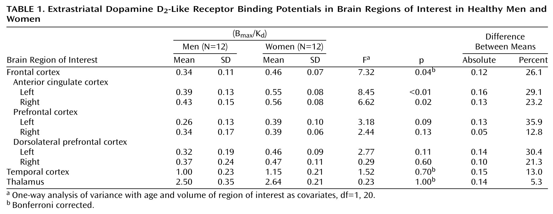
The binding potentials in men and women were compared by using one-way analysis of variance, with age and volume of the region of interest as covariates. To control for the effect of three comparisons, Bonferroni corrections were used. Subregions were studied if the result in the Bonferroni-corrected test was significant. Differences in the mean level of corresponding measurements on opposite hemispheres were tested by using a matched pairs t test. P values less than 0.05 were interpreted as statistically significant.