It has been suggested that posttraumatic stress disorder (PTSD) has not only long-lasting psychological but also long-lasting biological morbid effects. A 50-year prospective study demonstrated that combat exposure predicted early death and chronic illnesses by the age of 65
(1). A link between exposure to severe stress and later physical diseases has also been mentioned
(2), but there is no biological evidence for this link as yet. A series of studies revealed several biological dysfunctions in patients with current PTSD, such as low levels of 24-hour urinary cortisol excretion
(3), high natural killer (NK) cell activity
(4) and high CD4 and CD8 T cell numbers
(5,
6). To our knowledge, however, no study has focused on chronic biological effects among subjects who had PTSD in the past but who are now in remission. This study investigates the association between a past history of PTSD and any present immune dysfunction; our goal was to detect any long-lasting morbid effects of this disorder.
Method
We administered the Japanese versions of the Events Check List
(7) and the Impact of Event Scale—Revised
(8) to 1,550 male workers randomly recruited from a medium-sized Japanese industrial company. Written informed consent was obtained from all subjects. Past exposure to traumatic events was examined through the Events Check List, which comprises 15 categories such as natural disasters, violence, bullying, etc. Men who had been exposed to such events were screened for traumatic symptoms by using the Japanese version of the Impact of Event Scale—Revised
(8), which has demonstrated 100% sensitivity for subjects with current and lifetime diagnosis of PTSD when a cutoff point of 24 was applied. Participants with a score greater than 24 were interviewed with the Diagnostic Interview Schedule for DSM-IV
(9) to diagnose current and past PTSD and other axis I disorders.
Exclusion criteria included a previous history of any axis I disorder other than PTSD, use of any psychotropic medication within 2 months before this study, and any physical illness that needed treatment.
We recruited as many comparison subjects as possible from participants who had been exposed to trauma but whose Impact of Event Scale—Revised scores were below 25. The comparison subjects were matched for age and smoking because these factors affect immunity.
Blood samples were collected in heparinized tubes (Becton-Dickinson, Franklin Lakes, N.J.) at 10:00 a.m. and stored at room temperature for no longer than 4 hours before the assays. To determine WBC subset counts, the total numbers of WBC and leukocyte differential counts were determined with a Coulter counter (Beckman Coulter, Inc., Fullerton, Calif.). Lymphocyte subsets were determined by flow cytometry analysis (EPICS XL, Beckman Coulter, Inc.) according to standard methods. Enumeration by flow cytometry included the following cells: T cells (CD3/fluorescein isothiocyanate [FITC]), B cells (CD19/phycoerythrin [PE]), two types of T cell subsets (CD4/FITC, CD8/PE), and NK cells (CD3/phycoerythrin-Texas red [ECD], CD16/FITC, and CD56/PE). All antibodies were purchased from Beckman Coulter, Inc.
For determination of interferon γ (IFN-γ) and interleukin-4 (IL-4), a whole blood assay was applied
(10). Aliquots of 50 μl of blood were resuspended under laminar airflow in 400 μl of RPMI 1640 medium (containing 2 mM glutamine and 100 μg/ml kanamycin). For stimulation of cytokines, 2.5 μg phytohemagglutinin (Sigma-Aldrich Japan, Tokyo) was added and dissolved in 50 μl of a medium containing 50% RPMI and 50% sterile water (final concentration, 5 μg/ml). The samples were incubated for 48 hours at 37°C with 5% carbon dioxide in humidified air. The supernatants were harvested and stored at –80°C until assay. All cytokine levels were measured by ELISA kits (Biosource International, Camarillo, Calif.). Each sample was tested in duplicate in the same assay and thawed only once
(10). The NK cell activity assay was performed with K562 as target cells (E:T=20:1) and
51chromium release as the lysis indicator.
We used Student’s t tests with Levine’s test to compare the results between the subjects with and without past PTSD; p values less than 0.05 were considered significant.
Results
Of 1,550 male workers, 1,009 completed the Event Check List and 447 reported previous exposure to a life-threatening event. Fifty-seven men had Impact of Event Scale—Revised scores higher than 24, and 52 agreed to participate in the Diagnostic Interview Schedule for DSM-IV (interrater kappa for the first 12 participants was 0.92). Three men had current PTSD and 12 had past PTSD but were in remission. The events that caused the past PTSD were various: bullying, traffic accidents, witnessing a cruel death, violence, fire, death in the family, and flood.
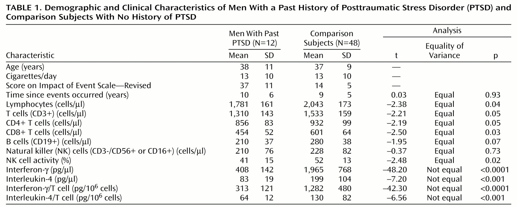
We found 48 comparison subjects for the 12 men with past PTSD (case-comparison ratio=1:4). As shown in
Table 1, there was no significant difference in number of years passed since the exposure to trauma between the men with and without past PTSD. The number of lymphocytes, number of T cells, number of T cell subsets, NK cell activity, and total amounts of IFN-γ and IL-4 as well as amounts produced by T cell were significantly lower in the PTSD group.


