Despite conflicting studies, a recent meta-analysis indicated that estrogen may improve specific cognitive domains, e.g., verbal memory and selected executive cognitions (vigilance, reasoning, motor speed, verbal function)
(1). As well as its potential to elevate mood, the estrogen effects may be partly achieved via the serotonin system. Prefrontal serotonin 2A (5-HT
2A) receptors, shown to be increased in rodents following estrogen administration
(2), are one component relating to cognition/mood and actions of antipsychotics/antidepressants
(3). The receptor modulation by estrogen may account for hormonal regulation of cognition/mood or pathophysiology of menstrually related psychiatric disorders. A recent human positron emission tomography (PET) study
(4), however, failed to demonstrate a statistically significant increase in brain 5-HT
2A receptors after estrogen replacement therapy (17β-estradiol), possibly because of study group size (N=5) and use of a region of interest analysis, which can miss localized change. The current PET study was conducted to verify—by using a voxel-by-voxel image analysis—the hypothesis that estrogen increases human brain 5-HT
2A receptors and that the change mediates the cognitive/mood effects of estrogen.
Method
The ten right-handed subjects (mean age=54.5 years, SD=6.6, range=44–68) were postmenopausal women (plasma levels of follicle-stimulating hormone ≥30 IU/liter and no menstrual cycle for at least a year [mean=6.3 years, SD=7.3]) with no history of psychiatric illness according to the Structured Clinical Interview for DSM-III-R, Non-Patient Edition, given as part of the screening evaluation. Subjects were free of estrogen for at least 2 months and psychoactive drugs for a month. Written informed consent was obtained from all subjects. Subjects received estrogen replacement therapy with a transdermal patch (17β-estradiol, 0.075–0.15 mg; mean dose=0.084 mg [SD=0.012]; mean dose at second PET scan=0.093 mg [SD=0.024]) for a mean of 10.2 weeks (SD=1.8). Plasma estradiol levels were measured by radioimmunoassay. Subjects completed a PET scan following assessments before and after estrogen replacement therapy.
Cognitive assessments, typically performed the day before scans, included tests of verbal memory (paragraph recall and verbal paired associates tests from the Wechsler Memory Scale—Revised
[5]), executive cognition (Trail Making Test A to assess attention/motor speed and Test B to assess sequencing
[6]), and verbal fluency (letters/categories
[7]). Mood was assessed on the PET scan days with the depression/dejection subscale of the Profile of Mood States (POMS)
(8) and Beck Depression Inventory
(9).
Subjects received a bolus injection of [
18F]deuteroaltanserin (mean=211.4 MBq, SD=10.1), followed by a 5-hour continuous infusion (bolus-to-infusion rate ratios: mean=3.21, SD=0.01) before a 45-minute PET scan (Posicam 6.5; 21 slices with 5.125-mm interslice distance)
(10). Magnetic resonance imaging (MRI) scans were acquired with a GE 1.5-T Signa scanner. PET voxel value of 5-HT
2A receptor binding was V
3′ (ml/ml)=(specific binding in cortices minus cerebellum activity)/total plasma parent concentration. At equilibrium, V
3′ is proportional to B
max/K
d (test/retest reproducibility: intrasubject change of 14.1%; reliability: intraclass coefficients of variation of 0.86)
(10) and is not affected by blood flow. Attenuation-corrected PET images were normalized to Talairach space by using MRI (coregistered to PET by the Automated Imaging Registration [Medx4.0]) and “T1.img” within statistical parametric mapping (SPM 99), followed by smoothing (full width at half maximum=20 mm). This statistical parametric mapping method enabled the evaluation of voxel-by-voxel change in receptor binding between two scans and is more suitable to detect localized change than region of interest analysis.
The SPM 99 statistical t map threshold was 2.82 (p=0.01) for comparison and 1.86 (p=0.05) for correlation with estradiol. All p values were two-tailed, and statistical significance was set at p<0.05 (with/without Bonferroni correction) in SPSS 11.0.
Results
Estrogen replacement therapy significantly increased plasma estradiol levels from baseline to endpoint in all subjects (mean=14.7 pg/ml [SD=9.0] versus 176.5 pg/ml [SD=150.0], respectively; p=0.006). There were no significant differences between the two scans in radiotracer plasma parameters (data not shown). 5-HT
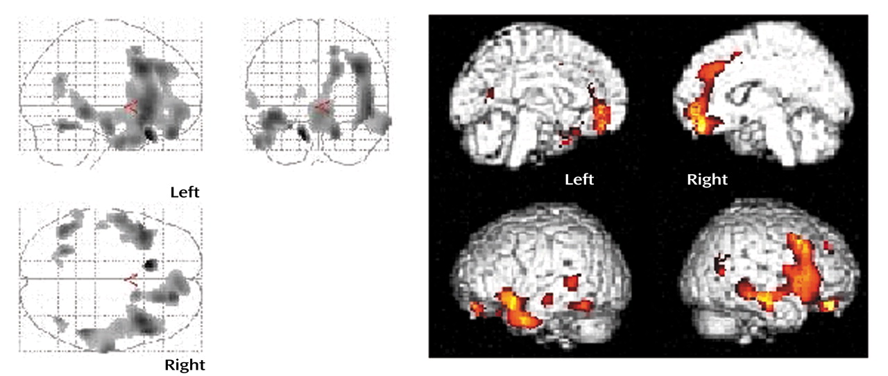
2A receptor binding was increased after estrogen replacement therapy in the regions shown in
Figure 1. The significant increase of 36.5% (V
3′ mean=0.51 ml/ml [SD=0.21] versus mean=0.70 ml/ml [SD=0.26]) (p=0.01) was primarily localized to the right frontal cortex (size=5568 pixels) (corrected p=0.001). The peak t scores were found in the right precentral gyrus (Brodmann’s area 9 [Talairach coordinates: x=40, y=16, z=36]; t=3.79), inferior frontal gyrus (Brodmann’s area 47 [x=50, y=22, z=–2 and x=48, y=22, z=–6]; t=3.74 and 3.71, respectively), medial frontal gyrus (Brodmann’s area 10 [x=4, y=52, z=–6]; t=3.31 and Brodmann’s area 6 [x=16, y=10, z=50]; t=3.31), and anterior cingulate cortex (Brodmann’s area 32 [x=10, y=38, z=16]; t=3.06). Positive and significant correlation with plasma estradiol was found in the right inferior frontal cortex (size 103 pixels) within SPM 99 (peak t=2.63 [x=56, y=6, z=14]; corrected p=0.022).
Significant performance improvement following estrogen replacement therapy was seen for category verbal fluency (baseline: mean=14.8 [SD=3.2], endpoint: mean=18.5 [SD=3.1]; corrected p=0.016) and Trail Making Test A (baseline: mean=34.7 seconds [SD=9.7], endpoint: mean=28.2 seconds [SD=8.3]; corrected p=0.028). No significant improvement was seen in immediate paragraph recall (baseline: mean=19.2 [SD=4.8], endpoint: 17.0 [SD=4.1]), delayed paragraph recall (% retention) (baseline: mean=67.7 [SD=17.9], endpoint: 84.8 [SD=11.4]), or verbal paired associates performance (immediate: mean=12.1 [SD=5.6] versus 15.5 [SD=6.4]; delayed [% retention]: mean=82.5 [SD=22.2] versus 86.3 [SD=17.1]). Baseline mood scales showed euthymic condition (POMS: mean=1.1 [SD=2.1]; Beck Depression Inventory: mean=2.2 [SD=2.7]) and were nonsignificantly decreased after estrogen replacement therapy (p>0.54). These changes in scales were not associated with receptor changes in the above region (p>0.12 in SPSS).
Discussion
The current study found significant, relatively unilateral (right > left) and localized changes in 5-HT
2A receptors after estrogen replacement therapy (dorsal to ventral prefrontal regions including dorsolateral and cingulate cortices). Prefrontal 5-HT
2A receptors have an action on the pyramidal cells via glutamate release and may well modulate cognitive functions as the stimulation of primate prefrontal. 5-HT
2A receptors (caudal area 46 and rostral area 8a) improved spatial working memory
(3). The similarity of our finding to the regions where estrogen evoked human brain activity during a working memory task (Brodmann’s area 46, 47)
(11) strengthens the possibility that 5-HT
2A receptors may be involved in estrogen-induced modulation of these cognitive functions. Subjects’ improvement of executive/prefrontal-related functions (i.e., verbal fluency and Trail Making Test A) may support the possibility. The results are consistent with the meta-analysis
(1) that revealed estrogen effects on motor speed and verbal fluency but not on complex attention tests such as Trails B. Unexpected lack of change in verbal memory might stem from the study group size and noncontrolled design. Future studies would need a control condition and direct measurement of working memory. Other limitations include baseline euthymia, which might decrease the likelihood of correlations with receptor change because of floor effects. Last, use of a ligand-specific template for image normalization may have proven to be more reliable for a study of this type.