Full access
Perspectives
Published Online: 1 September 2010

Psychiatric Manifestations of Paraneoplastic Disorders

Abstract

Paraneoplastic disorders of the CNS result from immune responses to neuronal proteins expressed by tumors found elsewhere in the body. Limbic encephalitis, one of the most common manifestations of paraneoplastic disorders, is characterized by rapid onset of psychiatric and neurological symptoms that often culminate in severe neurological deterioration. Recent work has described paraneoplastic syndromes with prominent, and sometimes isolated, psychiatric symptoms for which patients are fi rst seen by a psychiatrist. Here the authors review the existing literature on psychiatric and behavioral manifestations of paraneoplastic disorders, the cellular mechanisms underlying these syndromes, and current treatment and outcomes. They also discuss the broad behavioral fi ndings that highlight the need for psychiatrists to be aware of initial presentations of paraneoplastic disorders.
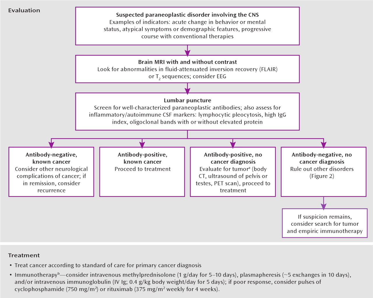
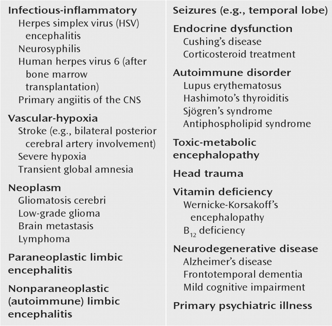
Since the first conceptualization of paraneoplastic syndromes over 50 years ago, our understanding of how neoplasms can influence the nervous system in a manner independent from direct invasion or metastases has advanced significantly (1–(4). Paraneoplastic syndromes may involve the peripheral or central nervous system, resulting in symptoms ranging from sensory neuropathies to profound and diverse neuropsychiatric disturbances, including dysfunction in consciousness, cognition, behavior, mood, and perception (5). Moreover, work during the past two decades indicates that paraneoplastic disorders derive largely from autoimmune phenomena, with production of antineuronal antibodies that recognize various antigens throughout the nervous system (2, 5). Paramount to treatment of paraneoplastic disorders is early recognition and intervention with immunotherapy and tumor treatment (6) (Figure 1). The occurrence of paraneoplastic disorders may be in the setting of a known cancer diagnosis or precede the clinical diagnosis, and they may be observed in isolation or with nonspecific constitutional symptoms, such as weight loss and malaise. While the most complete reviews of these syndromes focus on the neurological manifestations of these disorders (2, 5, 6, 10)–(12), the initial presentation in many instances is psychiatric. Nevertheless, discussion in the psychiatric literature is sparse (13, 14) and has not kept pace with growing knowledge, which increasingly demands that psychiatrists include these syndromes in the differential diagnosis of a wide range of patients (7) (Figure 2). In this article we review the psychiatric and behavioral manifestations of paraneoplastic disorders, the cellular mechanisms underlying these syndromes, and current treatment and outcomes.
FIGURE 1. Approach to Evaluation and Treatment of Suspected Paraneoplastic Disorder Involving the Central Nervous System
aIncreasingly it is appreciated that disorders once thought to be paraneoplastic in nature might in some instances be purely autoimmune and occur in the absence of any identifiable tumor, as is found in approximately 50% of the cases of N-methyl-D-aspartate (NMDA) receptor encephalitis.
bGiven the relative rarity of these disorders, there is no standard of care with regard to immunotherapy. These guidelines are therefore based on clinical experience (7–(9) rather than established care protocols.
FIGURE 2. Differential Diagnosis of Limbic Encephalitis

Evidence for Autoimmunity in Paraneoplastic Disorders

Historically, paraneoplastic syndromes of the CNS were thought to result from neuronal degeneration and secondary inflammation, viral infection, or an immune-mediated mechanism; a preponderance of evidence now points toward the latter as causative (10). In particular, multiple clinical-immunologic relationships have been characterized in which a specific autoantibody occurs exclusively with a given clinical syndrome and (often) cancer type (Table 1) (10, 11); paraneoplastic autoantibodies are virtually never found in comparison subjects (15). The discovery of these antibodies has provided a laboratory method for screening of patients, as well as potential early detection of underlying malignancy (16). Autoantibodies can usually be found in both serum and CSF, although titers are frequently higher in CSF. In further support of immunologic pathogenesis of paraneoplastic disorders, many symptoms respond to immunotherapy, driving the ongoing search for unknown antibodies when no well-described antibodies are found in available screens. The prevalence of paraneoplastic syndromes depends on the type of cancer, and it ranges from below 1% in breast and ovarian cancers to 3%–5% in small cell lung cancer and 20% in thymomas (7), although these numbers may be underestimations of the true prevalence rates.
TABLE 1. Diagnostic, Clinical, and Treatment Characteristics of Paraneoplastic Disorders Involving the Central Nervous System
AntibodyaAssociated Tumor(s)CSF ChangesbMRI Findings of Limbic EncephalitiscPredominant CNS SymptomsOther Behavioral SymptomsPatients' Response to Treatment
HuSmall cell lung cancerCommonCommonShort-term memory deficits, confusionDepressionPoor; 20% survive to 3 years
Ma2Testicular germ cell tumorCommonCommonShort-term memory deficits, diencephalic/brainstem encephalitisREM sleep disorder, obsessive-compulsive disorder (OCD), anxiety50%–70% stabilize or improve
CV2/CRMP5Small cell lung cancer, thymomaCommonCommonSubacute dementia, choreaMemory dysfunction, OCD, disorientationPoor
VGKC∼20% small cell lung cancer, thymomaRareCommonVerbal and visual memory deficits, confusionREM sleep disorder, apathy, irritability80% improve
NMDA receptorOvarian teratomaCommonRarePsychosis (delusional thoughts, hallucinations), seizures, hypoventilation, autonomic instability, dyskinesiasAnxiety, agitation, bizarre behavior70%–80% recover or have mild stable deficits
AMPA receptorBreast or lung tumor or thymomaCommonCommonMemory loss, confusionAgitation, aggressionMany have reduced symptoms, but relapse is frequent
GABA receptorSmall cell lung cancerCommonCommonSeizures, confusion, memory impairmentPsychosis, hallucinationsMost improve if tumor is treated
a
CRMP, collapsin response mediator protein; VGKC, voltage-gated potassium channel; NMDA, N-methyl-D-aspartate; AMPA, α-amino-3-hydroxy-5-methyl-4-isoxazolepropionic acid; GABA, γ-aminobutyric acid.
b
Typical CSF changes in limbic encephalitis include lymphocytic pleocytosis.
c
Typical MRI findings include hyperintensities in medial temporal lobes shown by fluid-attenuated inversion recovery (FLAIR) or T2-weighted MRI.
Antibodies associated with paraneoplastic disorders that often include neuropsychiatric manifestations can be subcategorized on the basis of the cellular locations of the antigens they recognize (Table 1). One group of antibodies is directed against intracellular neuronal antigens. Their pathogenic activity is thought to be mediated by cytotoxic T-cell immunity, rather than by the autoantibodies directly, and is characterized by infiltrates of oligoclonal T-cells in the CNS with deposits of paraneoplastic antibodies in T-cell-dense areas (17, 18). In fact, stereotyped T-cell responses have been demonstrated in response to paraneoplastic antigens, resulting in cell-mediated damage (19). That treatment aimed at the humoral immune response is largely ineffective further suggests that the antibodies themselves are not the primary pathogen, although they might still play a role in generating the abnormal immune response and are crucial for diagnosis (6). Another antibody group recognizes cell-surface antigens and appears to directly cause paraneoplastic syndrome symptoms, likely by disrupting normal synaptic transmission (7, 20). This group can occur with or without an associated cancer, and it includes antibodies recognizing voltage-gated potassium channels and ionotropic glutamate receptors. Syndromes that are directly related to antibody presence appear far more responsive to immunomodulation, although long-term behavioral sequelae and relapse can be significant (20–(22).

Clinical Consideration of Limbic Encephalitis

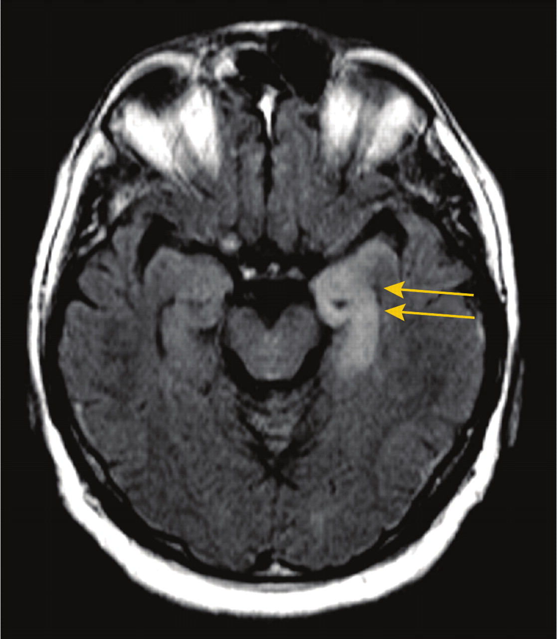
Limbic encephalitis is one of the best-appreciated causes of rapid behavioral dysfunction in association with neurological symptoms, and its clinical manifestations warrant specific discussion. Causes of limbic encephalitis include systemic autoimmune disease, viral disorders, and paraneoplastic disorders (Figure 2). The term “limbic encephalitis” was first used in 1968 to describe patients with memory dysfunction in association with bronchogenic carcinoma (4). Pathology revealed inflammatory and degenerative changes in temporal gray matter in the limbic area of the patients (hippocampus, amygdala, cingulate gyrus). Clinically, the classic syndrome evolves over days to weeks and includes psychiatric changes such as irritability, depression, hallucinations, personality disturbances, and cognitive changes, e.g., short-term memory loss that may progress to dementia. In addition, patients may experience sleep disturbances, confusion, and/or seizures. The CSF often shows a mild lymphocytic pleocytosis (6), 11, 12), and magnetic resonance imaging (MRI) may demonstrate uni- or bilateral medial temporal lobe hyperintensities on fluid-attenuated inversion recovery (FLAIR) and T2-weighted images with progressive hippocampal gyrus volume loss upon repeat imaging (Figure 3) (23).
FIGURE 3. Brain MRI of Patient With Voltage-Gated Potassium Channel Encephalitisa
aAxial MRI fluid-attenuated inversion recovery (FLAIR) sequence demonstrates bilateral medial temporal hyperintensities, primarily involving the left hippocampus (arrows).
Reflecting the variable psychiatric presentation of limbic encephalitis, patients have been described with myriad symptoms, ranging from delusional thought content and paranoid ideation to obsessive-compulsive behavior (24, 25). Although “limbic encephalitis” or “psychiatric manifestations” are often used as blanket generalizations to describe the behavioral features of these patients, detailed case studies yield a rich characterization of the neuropsychiatric symptoms. In the following sections, we discuss specific autoantibody syndromes and their psychiatric manifestations, some but not all of which fall into the category of limbic encephalitis. This overview of paraneoplastic disorders is organized according to antigen location.

Syndromes Associated With Intracellular Antigens

These antigens include anti-Hu antibodies, anti-Ma antibodies, antibodies against collapsin response mediator protein type 5 (CRMP5) and CV2, and rare autoantibodies with intracellular targets.

Anti-Hu Antibodies

The most common known paraneoplastic neurological syndrome is associated with anti-Hu antibodies, which recognize a family of RNA-binding proteins involved in normal neuronal development (26). Anti-Hu encephalitis usually manifests as classic limbic encephalomyelitis occurring with small cell lung cancer (8, 27, 28). Limbic encephalomyelitis entails a wide spectrum of neurological dysfunction, including limbic encephalitis, sensory neuropathy, motor neuron abnormalities, and cerebellar ataxia (8, 16, 27, 28). Patients with anti-Hu antibodies are often in their 50s or 60s with a long history of smoking and complaints of painful sensory neuropathy with recent confusion and amnesia (8, 16, 28, 29). Depression appears as the next most cited symptom, although less prevalent, followed by rare reports of hallucinations (1, 16, 28). Overall, patients with small cell lung cancer and limbic encephalitis fair poorly, with a median survival of about 1 year and a 3-year survival rate of approximately 20% (8, 28). Treatment of the tumor along with immunotherapy for the T-cell response (e.g., cyclophosphamide) can result in symptom stabilization, although not often improvement, and early intervention (within weeks of symptom onset) generally yields better results (8, 30).
Specific case studies present a consistent clinical picture with regard to behavior in paraneoplastic limbic encephalitis with small cell lung cancer. Newman et al. (14) described a 76-year-old man seen in an inpatient psychiatric unit with sudden-onset amnesia and confusion. He noted “feeling low” and developed sleep disturbances and agitation, which responded poorly to treatment with antipsychotics and sedatives. The patient had no history of psychiatric illness or substance abuse aside from a long smoking history. After transfer to another hospital, he became delirious and deteriorated neurologically, unable to stand without assistance. The patient died 4 months after the onset of memory problems, with a diagnosis of small cell lung cancer at autopsy. Byrne et al. (29) also offered a thorough case presentation with profound behavioral disturbances, although a surprisingly positive outcome. The patient was a 62-year-old woman with a 35-year smoking history, and she experienced hypersomnia followed by disorientation, amnesia, and hallucinations, along with myoclonic tremors and episodes of severe weakness. She rapidly deteriorated to variable levels of consciousness, with poorly reactive asymmetric pupils and ataxia. The patient was found to have high levels of anti-Hu antibodies and small cell lung cancer. She made a spontaneous recovery with only mild deficits in short-term memory, and she was ultimately treated with chemotherapy for her cancer.

Anti-Ma Antibodies

A decade ago, an autoantibody syndrome associated with testicular germ cell tumors and paraneoplastic limbic encephalitis was described (31). Antibodies recognize the onconeuronal antigen Ma2, whose cellular localization and structural motifs suggest roles in mRNA processing or biogenesis (32). In contrast to anti-Hu antibodies, isolated anti-Ma2 antibodies are usually seen in young men with CNS-specific disturbances—most often limbic encephalitis along with hypothalamic and/or brainstem encephalitis (2, 6, 9). A review of all published patient reports reveals common traits within this population: severe short-term memory deficits stand out as the most common feature, with a few reports of confusion or disorientation (9, 3134). Symptoms frequently occur with other overt neurological deficits, such as visual dysfunction (diplopia, gaze abnormalities, nystagmus), gait disturbances, and hypokinesis (9, 33). Affective changes (specifically depression and irritability) as well as hallucinations are mentioned in some individual patient descriptions, but these symptoms seem relatively rare (9, 33).
Interestingly, there appears to be a specific connection between anti-Ma2 antibodies and sleep dysregulation of hypothalamic origin (35, 36). As many as one-third of patients have shown excessive daytime sleepiness in addition to other symptoms (9). Generally, patients with narcolepsy are thought to have abnormally low levels of the neuropeptide hormone hypocretin (37, 38). While anti-Ma2 antibodies are not a cause of idiopathic narcolepsy, hypocretin CSF levels have been demonstrated to be low in patients with anti-Ma2 limbic encephalitis (36). One case study (35) describes a 69-year-old man with a 3-month history of progressively severe hypersomnia, memory loss, diplopia, gait unsteadiness, short episodes of fear, and apathy. Further investigation revealed REM sleep behavior disorder, low levels of hypocretin-1, and anti-Ma2 antibodies in CSF and serum.
In a few instances, anti-Ma2 limbic encephalitis has been manifested as a pure psychiatric disturbance, such as a sense of unexplained fear, “nervous breakdowns” and panic attacks, or loss of self-confidence (2, 9). One case describes a 39-year-old previously healthy man who developed obsessive-compulsive behaviors (25). He first had obsessive thoughts of a water leak in his home and began checking equipment over 10 times each day. Within 6 months, he developed additional compulsions and was diagnosed with obsessive-compulsive disorder (OCD). He was ultimately found to have a testicular teratoma and anti-Ma2 antibodies, which the authors suggest as causative, given data indicating involvement of limbic regions in OCD (39).
In comparison to patients with paraneoplastic disorders mediated by other intracellular antibodies, those with isolated anti-Ma2 antibodies may respond well to treatment of the tumor and immunotherapy. While 30%–50% of patients ultimately deteriorate neurologically (the death rate is approximately 15%), one-third experience neurological improvement and 20%–40% stabilize (32, 33). The association of testicular cancer and anti-Ma2 limbic encephalitis in men younger than 50 years is so strong that studies suggest consideration of orchiectomy or testicular irradiation even if a tumor cannot be found (34). In one study, six patients with risk factors for testicular neoplasm and neurological deterioration underwent orchiectomy for questionable ultrasound findings in the absence of an identifiable tumor; all were found to have preinvasive malignant cells in orchiectomy specimens (34).

Anti-CRMP5 and Anti-CV2 Antibodies

Anti-CRMP5 and -CV2 antibodies have been found most commonly in patients with small cell lung cancer but also thymoma and, less frequently, other tumors (40). CRMP localizes to neuronal cell bodies and axons in the mammalian CNS, and it is known to be involved in axon guidance and possibly synapse function (41). Both anti-CRMP5 and anti-CV2 antibodies recognize the CRMP5 antigen, and the presence of these antibodies may accompany anti-Hu antibodies in patients with small cell lung cancer (2). Patients with anti-CV2/CRMP5 have a wide spectrum of neurological symptoms, most notably chorea characterized by involuntary movements often involving the face (42). Optic neuropathy and abnormalities of olfaction and taste are remarkable for their unusually high frequencies in paraneoplastic conditions with these antibodies (40).
Anti-CV2/CRMP5 antibodies are often associated (30%–40% of patients) with various cognitive deficits and limbic encephalitis (40, 42). One of the largest studies of this patient group (40) reported subacute dementia as the most frequent symptom of limbic encephalitis, but it also described rare instances of personality change, depression, confusion, and psychosis. Remarkably, paraneoplastic involvement of striatal and basal ganglia circuitry that results in striatal encephalitis with choreic movements can also affect nonmotor circuitry in the same regions. Cases of patients with manic mood, memory deficits with spatial/temporal disorientation, and obsessive-compulsive behavior have been reported. Nuti et al. (43) described a patient with chorea and progressive neuropsychological deterioration characterized by memory dysfunction and constructional apraxia, with a score of 17 out of 30 on the Mini-Mental State Examination (MMSE), who eventually received a diagnosis of non-Hodgkin's T-cell lymphoma. Muehlschlegel et al. (44) reported a 69-year-old woman who developed irrational and argumentative behavior, followed by obsessive-compulsive-like behaviors. Four weeks later she experienced choreic movements and an unsteady gait. The patient deteriorated neurologically, and CV2 antibodies were reported several days after her death.
Outcomes are less clear for patients with anti-CV2/CRMP5 antibodies than for those with anti-Hu and -Ma2 antibodies. One study described improvement of chorea with tumor treatment, immunotherapy, and antipsychotics, with about 50% of the patients surviving 4–42 months after the onset of chorea (42). Because many patients have psychiatric changes and chorea, differentiating this disorder from Huntington's disease or Wilson's disease (through family history, time line of symptoms, CSF inflammatory markers, and copper metabolism studies) is important given the distinct approach to treatment and potential underlying tumor.

Rare Autoantibodies With Intracellular Targets

Aside from the syndromes described in the preceding sections, other antibodies recognizing intracellular antigens have been identified in patients with paraneoplastic disorders and psychiatric symptoms. Two of these reports involve protein kinases as targets. Sabater and colleagues (45) described a 61-year-old man with a 2-week history of confusion, personality changes, and short-term memory deficits, subsequently diagnosed with small cell lung cancer. Ultimately, an antibody against BR serine/threonine kinase 2 (BRSK2)—a close homologue of the SAD1 kinase in Caenorhabditis elegans, known to be crucial for proper synapse formation—was discovered (46). Similarly, Tuzun et al. (47) reported two male patients in their early 70s with rapidly progressive confusion and memory deficits. One also had delusional thoughts with agitation and aggressive behavior, while the other experienced personality changes and difficulty recognizing faces. Both patients experienced progressive deterioration to dementia that was unaffected by immunotherapy. An autoantibody against adenylate kinase 5, thought to have a role in neuronal energy transfer and RNA/DNA synthesis (48), was identified; no cancer was detected in either patient.
Autoantibodies that are commonly associated with nonbehavioral or cognitive symptoms also have been reported to cause limbic encephalitis in paraneoplastic disorders. Amphiphysin is implicated in paraneoplastic stiff-person syndrome, but it was also described in a woman with small cell lung cancer with memory deficits and an MMSE score of 23 out of 30 (49). She went on to develop agitation, hallucinations, and generalized convulsions but improved significantly following tumor treatment. Antoine and colleagues (50) also described a patient with small cell lung cancer harboring antiamphiphysin antibodies who became abnormally anxious and depressed and then, as her condition deteriorated, developed olfactory and auditory hallucinations and anterograde memory loss. Finally, antibodies recognizing the antigen Ri—commonly associated with brainstem or cerebellar syndromes (51)—were identified in a patient with carcinoid tumor and striking neuropsychological symptoms (52).

Syndromes Associated With Cell-Surface Antigens

This group of antigens includes antibodies against voltage-gated potassium channels, antibodies against the N-methyl-D-aspartate (NMDA) type of glutamate receptors, antibodies against --amino-3-hydroxy-5-methyl-4-isoxazolepropionic acid (AMPA) receptors, and antibodies against s-aminobutyric acid type B (GABAB) receptors.

Antibodies Against Voltage-Gated Potassium Channels

These antibodies are usually associated with nonparaneoplastic limbic encephalitis, but they can occur in the context of an underlying tumor (20% with small cell lung cancer or thymoma) (21, 53). The disorder typically affects middle-aged or older patients, who develop severe verbal and visual memory deficits and confusion or disorientation (21, 22, 54). Seizures are common, but CSF pleocytosis is rare in syndromes with antibodies against voltage-gated potassium channels, which is not the case in classic limbic encephalitis (21, 53).
A survey of case descriptions of patients with voltage-gated potassium channel antibodies illustrates more diffuse psychiatric disturbances than occur in classic limbic encephalitis (described earlier). Behavioral changes are frequent and often include apathy and irritability (21, 22). These symptoms may occur concurrently with signs of autonomic dysfunction, such as excess sweating and salivation (21, 22). Visual hallucinations have also been described (22, 54), although this symptom is rare in comparison to memory dysfunction. In addition, associations between this syndrome and hyponatremia (21, 22), hypothermia (54), and REM sleep behavior disorder (55) have been reported. Despite the clinical variability in presentation, review of the literature highlights memory impairment as a core finding.
Antibodies recognizing voltage-gated potassium channels are well known to underlie the neurological abnormalities in Isaacs and Morvan syndromes, characterized by neuromyotonia, hypersalivation, hyperhidrosis, and insomnia (21). Although some of the clinical symptoms of Isaacs and Morvan syndromes exist in encephalitis caused by antibodies against voltage-gated potassium channels, the predilection for limbic areas is not well understood. Perhaps antibodies in these distinct disorders are subtly different and target peripherally, rather than centrally, expressed potassium channel subtypes (21, 56). It is also not known how antibodies against voltage-gated potassium channels result in memory deficits, although modulation of neuronal excitability likely plays a role (22) and a knockout animal model lacking one of the channels expressed in the hippocampus does display memory impairment (57).
A significant distinction between limbic encephalitis caused by antibodies against voltage-gated potassium channels and limbic encephalitis due to other causes is the rapid response to immunotherapy. About 80% of patients improve neurologically and radiologically following prompt (within 2 months) treatment with high-dose steroids, intravenous immunoglobulins, or plasma exchange (21, 22); neuropsychological improvements have been noted but are less robust. While most patients show a large degree of cognitive and behavioral recovery, the majority still experience short-term memory deficits, although less serious than at symptom onset (21, 22). Given the prognostic outcome with immunotherapy, prompt recognition and diagnosis of this syndrome is crucial, a fact emphasized by work demonstrating relatively poor improvement with more prolonged symptoms (greater than 9 months) prior to treatment (21).

Anti-NMDA Receptor Antibodies

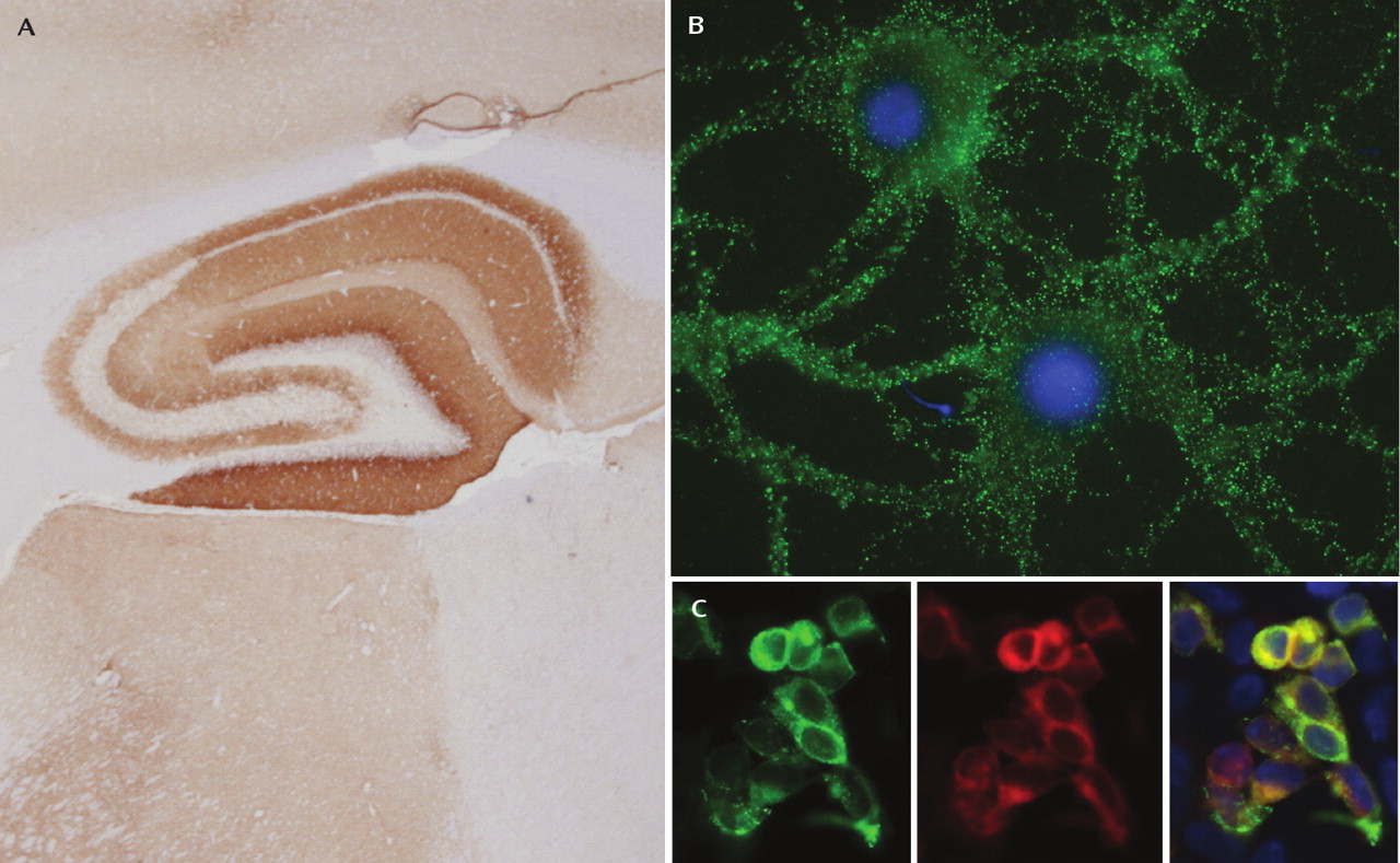
Perhaps the most intriguing disorder with regard to psychiatric symptoms is the autoantibody syndrome involving the NMDA-type glutamate receptor. NMDA receptors are ionotropic glutamate receptors with well-known roles in synaptic transmission and plasticity (58), as well as in neuropsychiatric disease (59). Antibodies in this syndrome recognize the NR1 subunit (Figure 4), which normally combines with various NR2 subunits (A–D) to form heteromers with distinct properties (59). Although the antibody was only identified in the last few years, about 200 patients have now been described with this disorder, and nearly 75% were seen by a psychiatrist or initially admitted to a psychiatric unit (7). Because of the constellation of symptoms, patients were often misdiagnosed with acute psychosis, malingering, or drug abuse (60).
FIGURE 4. Immunohistochemical Evidence for Anti-NMDA Receptor Antibodiesa
aNMDA, N-methyl-D-aspartate. CSF from patients with anti-NMDA receptor encephalitis shows reactivity in three different assays. Part A: CSF incubated with coronal sections of rat brain demonstrates intense staining in the hippocampus, where a high density of NMDA receptors is found. Part B: CSF incubated with live, nonpermeabilized dissociated cultures of rat hippocampal neurons shows punctate cell-surface labeling (green); cell nuclei are stained with DAPI (blue). Part C: HEK293 cells (nonneuronal) transfected with NMDA receptor subunits NR1 and NR2B (forming heteromeric NMDA receptors) and immunostained with patient CSF (green), commercially available NR1 polyclonal antibody (red), and DAPI (blue) demonstrate co-localization of CSF and commercial NMDA receptor antibodies (see reference 7 for methods).
In many ways, the anti-NMDA receptor encephalitis is unique from the other syndromes discussed in this review. Rather than having predominant symptoms of memory changes or disorientation, patients are typically young women (75%) or children (40% younger than 18 years) with prominent psychiatric symptoms, including anxiety and agitation, bizarre behavior, delusional and/or paranoid thoughts, and visual or auditory hallucinations (see clinical vignettes) (7, 61, 62). Recent work has focused specifically on the presentation of anti-NMDA receptor encephalitis in the pediatric population (as young as 23 months old), finding initial symptoms such as temper tantrums, behavioral change, agitation, aggression, and progressive speech deterioration (61). The vast majority of patients in both the adult and pediatric populations experience a viral prodrome-like illness in the weeks prior to psychiatric destabilization. Throughout the first few weeks following admission, patients rapidly deteriorate neurologically with seizures and decreased consciousness, progressing to a catatonic-like state, along with dyskinesias, autonomic instability, and hypoventilation requiring intubation in some patients (more commonly adults) (7, 61). While CSF findings are typical of encephalitis, MRI findings are unreliable, with approximately one-half of patients showing no abnormalities and only about 15% with specific signals in the medial temporal lobes (7).
Initially, the NMDA receptor autoantibody syndrome was thought to be specifically associated with ovarian teratomas (63). Studies now indicate that approximately 50% of patients do not have an identifiable tumor, and this percentage increases to nearly 70% when only those 18 or younger are considered (7, 61). In patients with neoplasms, nearly all develop psychiatric or neurological symptoms prior to tumor diagnosis (7). Although it is somewhat surprising given the severity of symptoms, patients with anti-NMDA receptor encephalitis often recover with treatment. Following tumor resection (if possible) and immunotherapy, nearly half of patients make a full recovery. In two large series, 75% had full or substantial recovery, although those remaining were left with severe deficits or died as a result of neurological complications (7, 61). The presence of a tumor is a positive prognostic factor in this syndrome, as patients with tumor removal within 4 months of neuropsychiatric changes fared better than all other patients (7).
The posthospitalization course in this patient population merits specific discussion. Of patients that ultimately make a full recovery or are left with only mild deficits, most (85%) have significant psychiatric abnormalities at discharge, including poor attention and planning, impulsivity, and behavioral disinhibition; approximately 25% experience sleep dysfunction (7). Recovery is often slow, and it takes many months for patients to return to their baseline behavioral and cognitive status. In addition, 15 out of 100 patients had instances of relapse of encephalitis in one study, and most experienced a persistent amnesia for the entire illness (7). The clinical picture and scientific results in this syndrome suggest that the antibody itself is pathogenic, as antibody titers correlate with disease severity (63) and antibody activity results in a reversible decrease in synaptic NMDA receptors (7). In addition, many drugs that modulate NMDA receptors are known to cause a similar constellation of symptoms, such as psychotic behavior (64), dystonia (65), and autonomic dysfunction (66), while over the past several decades NMDA receptor hypofunction has received increasing scrutiny as a putative mechanism of schizophrenia (67).

Anti-AMPA Receptor Antibodies

A recently discovered paraneoplastic disorder associated with limbic encephalitis is characterized by antibodies against subunits of the AMPA-type glutamate receptor (20). The AMPA receptor mediates the majority of fast glutamatergic neurotransmission in the brain and plays a role in molecular mechanisms of learning and memory (68). In this syndrome, subunits GluR1 and GluR2 are specifically targeted, resulting in the internalization and redistribution of AMPA receptors away from the synapse; the effect is reversible upon removal of antibody-containing CSF from neurons (20). Animal models with similar mistrafficking of AMPA receptors have abnormalities in behavioral paradigms of learning and memory, as well as emotional disturbances (69).
Of the 10 described patients with anti-AMPA receptor limbic encephalitis, nine were women, and the median age was 60 years. Almost all had isolated limbic symptoms: subacute memory loss (less than 8 weeks) and confusion (20). The other most common symptom was agitation and/or aggressive behavior, which was reported in one-half of the patients (another had “behavioral changes” not otherwise specified [70]). Seven of the 10 patients had an associated tumor of the breast or lung or thymoma. All living patients with an identified neoplasm received cancer treatment, and nine patients received immunotherapy. Every patient receiving immunotherapy responded well, with reduced cognitive and behavioral symptoms (20).
One of the most striking features of anti-AMPA receptor limbic encephalitis is the tendency to relapse despite treatment. Seven patients in the preceding series experienced relapsing symptoms, with deficits of short-term memory and persistent behavioral problems, such as aggression, in all but one patient, who had mild persistent depression and apathy (20). All relapse cases occurred in the absence of tumor or tumor recurrence, suggesting that a persistent autoimmune disorder had been triggered. Taken together, these results demonstrate a remarkable similarity between the clinical picture and the behavior one would predict on the basis of AMPA receptor function in animal models.
Anti-GABAB Receptor Antibodies

Paraneoplastic limbic encephalitis can also result from antibodies recognizing receptors involved in inhibitory neurotransmission. Anti-GABAB receptor antibodies have been identified in association with small cell lung cancer, with patients exhibiting seizures, confusion, and severe memory abnormalities (71). Such observations underscore how imbalances in excitation and inhibition can manifest with neurocognitive dysfunction (72).

Disorders With Potential Paraneoplastic Etiologies

In light of how quickly the field of paraneoplastic biology has progressed over the past decade, it is likely that other, as-yet-undefined paraneoplastic disorders exist. Almost certainly, the discovery of new autoantibodies will occur and help define groups of patients with specific symptoms but no clear underlying mechanism. In the following sections we discuss three examples of psychiatric and cognitive syndromes or symptoms hypothesized to have a humoral component independent of the existing cancer itself.

Opsoclonus-Myoclonus-Ataxia and Neuroblastoma in Children

Opsoclonus, myoclonus, and ataxia are characteristic findings in patients with underlying neuroblastoma, occurring almost exclusively in children (73). The symptoms are found in 2%–3% of children with neuroblastoma, although approximately 50% of children experiencing this triad of symptoms are found to have a neuroblastoma (11, 73). While no autoantibody has been identified in association with these symptoms, the opsoclonus-myoclonus syndrome is thought to be mediated by immunologic mechanisms. The survival rate is actually much higher in children with these symptoms and neuroblastoma than in those with the tumor alone, although it is unclear whether this might be due to earlier presentation and diagnosis, rather than the prognostic effect of opsoclonus-myoclonus itself (73).
Opsoclonus-myoclonus symptoms are often accompanied by severe behavioral and developmental abnormalities that persist despite tumor treatment (73). Specifically, in one study most patients exhibited personality changes, such as irritability or inconsolability, coinciding with the onset of opsoclonus-myoclonus (73, 74); sleep disturbances, tantrums, and self-injurious behavior were also prominent. Cognitive function was poor in nearly all children, and abnormalities in vocabulary and language were pervasive. Even after completion of tumor treatment and immunotherapy, most, if not all, patients continued to have expressive language deficits, and many exhibited oppositional and aggressive behavior (73, 74).

Hypothalamic Syndrome in Children With Neural Crest Tumors

Although even more rare than opsoclonus-myoclonus in the setting of neuroblastoma, idiopathic hypothalamic syndrome has been reported by a few groups in children with neural crest cell tumors, and this suggests a paraneoplastic etiology (75, 76). Symptoms include obesity and hyperphagia, hypersomnia and/or reversal of sleep-wake cycles, abnormalities in thermoregulation, and central hypoventilation (76). In addition, two groups have reported a total of seven children with neural crest tumors, with personality and behavioral changes in all but one (75, 76). Mood or personality changes include lability of affect with episodic aggressive behavior, outbursts of euphoria and laughing, and impaired concentration. While treatment aimed at symptoms of typical hypothalamic syndrome is sometimes effective, prognosis is poor because of central hypoventilation (76); the cognitive and psychiatric abnormalities persist regardless of treatment (11, 75).

Pancreatic Cancer and Alterations in Mood

The knowledge that changes of mood in patients with pancreatic cancer are more prevalent than in other gastrointestinal cancers dates back to the 1920s, with observations that depression, anxiety, and an impending sense of doom may often precede or accompany the diagnosis (77, 78). Since that time, numerous studies have demonstrated that depression often occurs before clinical diagnosis and that even in later stages of disease, mood disorders are more pronounced in patients with pancreatic cancer than in those with other abdominal tumors (79–(81). Green and Austin (80) found that approximately 70% of pancreatic cancer patients experience depression, 50% have anxiety, and 30% have both symptoms. It is noteworthy that psychiatric symptoms preceded the initial physical complaint (and the cancer diagnosis) one-third of the time. Panic attacks or manic episodes have also been reported as behavioral predecessors to diagnosis (82, 83). While this body of research is intriguing, more work must be done to address whether the change in mood symptoms is caused by dysregulation of neuroendocrine signaling, as opposed to an autoimmune basis.

Conclusions and Future Directions

Paraneoplastic syndromes are a heterogeneous group of disorders affecting the peripheral or central nervous system with tremendous variability in clinical presentation. Those affecting the CNS often result in impaired neuropsychiatric functions and, to differing degrees, may involve consciousness, cognition, behavior, mood, and perception. The link between specific symptoms and individual autoantibodies accentuates that it is important for clinicians to recognize paraneoplastic disorders (Table 1, Figure 1). This fact is highlighted by the treatable nature of many of these syndromes and the role of early detection in improved outcomes of many cancers. In addition, recent work has demonstrated a generalized increased risk in cancer diagnosis in the first months following first-time admission to a psychiatric hospital (84). This review summarizes the current knowledge of psychiatric symptoms associated with paraneoplastic disorders and is intended to raise awareness that psychiatrists might have a significant role in the diagnostic process (Figure 1). Although psychiatric symptoms are often coupled with cognitive dysfunction, the literature is replete with cases of isolated psychiatric changes in patients later determined to have an underlying neoplasm and limbic encephalitis. Thus, these syndromes must be incorporated into the differential diagnosis of the psychiatrist who encounters a patient with unusual and complex behavioral changes (Figure 2).
Despite a much greater understanding of clinical symptoms and the course of paraneoplastic disorders today than even 10 years ago, the psychiatric characterizations of these syndromes remain relatively cursory. Future work should strive to offer a more rigorous and standardized examination of the behavioral disturbances. The possibility exists that particular combinations and temporal characteristics of psychiatric symptoms might herald oncoming severe neurological disturbances and guide the potential diagnosis. Although this idea has been recognized in the past (13, 14), psychiatry continues to play a small role in the characterization of these disorders.
Finally, although relatively rare, paraneoplastic disorders offer a means to further our understanding of general psychiatric illnesses. A neuroimmunologic basis for psychiatric disease has long been hypothesized (85), and paraneoplastic disorders provide specific examples of how autoantibodies can affect neuronal function. In addition, many of these syndromes open a window onto how disturbance of specific neurotransmitter or intracellular signaling systems within particular neural circuits can affect behavior (7, 20, 45, 47). Researchers might aim to harness identified autoantibodies to explore how NMDA receptor signaling and trafficking, for example, relate to psychosis (7) or how proteins with known roles in neuronal development function in the mature brain (40, 41, 4446). Recent work in the neurosciences demonstrates how targeted disruption of circuit function can dramatically improve understanding of complex behaviors (86, 87). Elucidation of why certain autoantibody syndromes affect only particular areas of the brain might provide yet another tool for studying the neurobiology of psychiatric disorders.

Acknowledgments

The authors thank Dr. Christine Hill-Kayser for discussion and advice.

Footnote

Received Oct. 28, 2009; revision received Dec. 30, 2009; accepted Jan. 28, 2010

References

1.
Bakheit AM, Kennedy PG, Behan PO: Paraneoplastic limbic encephalitis: clinico-pathological correlations. J Neurol Neurosurg Psychiatry 1990; 53:1084–1088
2.
Bataller L, Dalmau J: Paraneoplastic disorders of the memory and cognition, in The Behavioral Neurology of Dementia. Edited by, Miller B, Boeve B. New York, Cambridge University Press, 2009, pp 377–394
3.
Brierley J, Corsellis J, Hierons R, Nevin S: Subacute encephalitis of later adult life: mainly affecting the limbic areas. Brain 1960; 83:357–368
4.
Corsellis JA, Goldberg GJ, Norton AR: "Limbic encephalitis” and its association with carcinoma. Brain 1968; 91:481–496
5.
Darnell RB, Posner JB: Paraneoplastic syndromes involving the nervous system. N Engl J Med 2003; 349:1543–1554
6.
Tuzun E, Dalmau J: Limbic encephalitis and variants: classification, diagnosis and treatment. Neurologist 2007; 13:261–271
7.
Dalmau J, Gleichman AJ, Hughes EG, Rossi JE, Peng X, Lai M, Dessain SK, Rosenfeld MR, Balice-Gordon R, Lynch DR: Anti-NMDA-receptor encephalitis: case series and analysis of the effects of antibodies. Lancet Neurol 2008; 7:1091–1098
8.
Graus F, Keime-Guibert F, Rene R, Benyahia B, Ribalta T, Ascaso C, Escaramis G, Delattre JY: Anti-Hu-associated paraneoplastic encephalomyelitis: analy-sis of 200 patients. Brain 2001; 124(Pt 6):1138–1148
9.
Dalmau J, Graus F, Villarejo A, Posner JB, Blumenthal D, Thiessen B, Saiz A, Meneses P, Rosenfeld MR: Clinical analysis of anti-Ma2-associated encephali-tis. Brain 2004; 127(Pt 8):1831–1844
10.
Dalmau J, Bataller L: Clinical and immunological diversity of limbic encephalitis: a model for paraneoplastic neurologic disorders. Hematol Oncol Clin North Am 2006; 20:1319–1335
11.
Dalmau J, Rosenfeld MR: Paraneoplastic syndromes of the CNS. Lancet Neurol 2008; 7:327–340
12.
Graus F, Delattre JY, Antoine JC, Dalmau J, Giometto B, Grisold W, Honnorat J, Smitt PS, Vedeler C, Verschuuren JJ, Vincent A, Voltz R: Recommended diagnostic criteria for paraneoplastic neurological syndromes. J Neurol Neurosurg Psychiatry 2004; 75:1135–1140
13.
Cornelius JR, Soloff PH, Miewald BK: Behavioral manifestations of paraneoplastic encephalopathy. Biol Psychiatry 1986; 21:686–690
14.
Newman NJ, Bell IR, McKee AC: Paraneoplastic limbic encephalitis: neuropsychiatric presentation. Biol Psychiatry 1990; 27:529–542
15.
Gultekin SH, Rosenfeld MR, Voltz R, Eichen J, Posner JB, Dalmau J: Paraneoplastic limbic encephalitis: neurological symptoms, immunological findings and tumour association in 50 patients. Brain 2000; 123(Pt 7):1481–1494
16.
Alamowitch S, Graus F, Uchuya M, Rene R, Bescansa E, Delattre JY: Limbic encephalitis and small cell lung cancer: clinical and immunological features. Brain 1997; 120(Pt 6):923–928
17.
Bernal F, Graus F, Pifarre A, Saiz A, Benyahia B, Ribalta T: Immunohistochemical analysis of anti-Hu-associated paraneoplastic encephalomyelitis. Acta Neu-ropathol 2002; 103:509–515
18.
Voltz R, Dalmau J, Posner JB, Rosenfeld MR: T-cell receptor analysis in anti-Hu associated paraneoplastic encephalomyelitis. Neurology 1998; 51:1146–1150
19.
Benyahia B, Liblau R, Merle-Beral H, Tourani JM, Dalmau J, Delattre JY: Cell-mediated autoimmunity in paraneoplastic neurological syndromes with anti-Hu antibodies. Ann Neurol 1999; 45:162–167
20.
Lai M, Hughes EG, Peng X, Zhou L, Gleichman AJ, Shu H, Mata S, Kremens D, Vitaliani R, Geschwind MD, Bataller L, Kalb RG, Davis R, Graus F, Lynch DR, Balice-Gordon R, Dalmau J: AMPA receptor antibodies in limbic encephalitis alter synaptic receptor location. Ann Neurol 2009; 65:424–434
21.
Thieben MJ, Lennon VA, Boeve BF, Aksamit AJ, Keegan M, Vernino S: Potentially reversible autoimmune limbic encephalitis with neuronal potassium channel antibody. Neurol 2004; 62:1177–1182
22.
Vincent A, Buckley C, Schott JM, Baker I, Dewar BK, Detert N, Clover L, Parkinson A, Bien CG, Omer S, Lang B, Rossor MN, Palace J: Potassium channel antibody-associated encephalopathy: a potentially immunotherapy-responsive form of limbic encephalitis. Brain 2004; 127(Pt 3):701–712
23.
Lawn ND, Westmoreland BF, Kiely MJ, Lennon VA, Vernino S: Clinical, magnetic resonance imaging, and electroencephalographic findings in paraneoplastic limbic encephalitis. Mayo Clin Proc 2003; 78:1363–1368
24.
Koide R, Shimizu T, Koike K, Dalmau J: EFA6A-like antibodies in paraneoplastic encephalitis associated with immature ovarian teratoma: a case report. J Neurooncol 2007; 81:71–74
25.
Scheid R, Voltz R, Guthke T, Bauer J, Sammler D, von Cramon DY: Neuropsychiatric findings in anti-Ma2-positive paraneoplastic limbic encephalitis. Neurol-ogy 2003; 61:1159–1161
26.
Szabo A, Dalmau J, Manley G, Rosenfeld M, Wong E, Henson J, Posner JB, Furneaux HM: HuD, a paraneoplastic encephalomyelitis antigen, contains RNA-binding domains and is homologous to Elav and Sex-lethal. Cell 1991; 67:325–333
27.
Dalmau J, Graus F, Rosenblum MK, Posner JB: Anti-Hu-associated paraneoplastic encephalomyelitis/sensory neuronopathy: a clinical study of 71 patients. Medicine (Baltimore) 1992; 71:59–72
28.
Sillevis Smitt P, Grefkens J, de Leeuw B, van den Bent M, van Putten W, Hooijkaas H, Vecht C: Survival and outcome in 73 anti-Hu positive patients with paraneoplastic encephalomyelitis/sensory neuronopathy. J Neurol 2002; 249:745–753
29.
Byrne T, Mason WP, Posner JB, Dalmau J: Spontaneous neurological improvement in anti-Hu associated encephalomyelitis. J Neurol Neurosurg Psychiatry 1997; 62:276–278
30.
Vernino S, O'Neill BP, Marks RS, O'Fallon JR, Kimmel DW: Immunomodulatory treatment trial for paraneoplastic neurological disorders. Neuro Oncol 2004; 6:55–62
31.
Voltz R, Gultekin SH, Rosenfeld MR, Gerstner E, Eichen J, Posner JB, Dalmau J: A serologic marker of paraneoplastic limbic and brain-stem encephalitis in patients with testicular cancer. N Engl J Med 1999; 340:1788–1795
32.
Rosenfeld MR, Eichen JG, Wade DF, Posner JB, Dalmau J: Molecular and clinical diversity in paraneoplastic immunity to Ma proteins. Ann Neurol 2001; 50:339–348
33.
Hoffmann LA, Jarius S, Pellkofer HL, Schueller M, Krumbholz M, Koenig F, Johannis W, la Fougere C, Newman T, Vincent A, Voltz R: Anti-Ma and anti-Ta as-sociated paraneoplastic neurological syndromes: 22 newly diagnosed patients and review of previous cases. J Neurol Neurosurg Psychiatry 2008; 79:767–773
34.
Mathew RM, Vandenberghe R, Garcia-Merino A, Yamamoto T, Landolfi JC, Rosenfeld MR, Rossi JE, Thiessen B, Dropcho EJ, Dalmau J: Orchiectomy for suspected microscopic tumor in patients with anti-Ma2-associated encephalitis. Neurology 2007; 68:900–905
35.
Compta Y, Iranzo A, Santamaria J, Casamitjana R, Graus F: REM sleep behavior disorder and narcoleptic features in anti-Ma2-associated encephalitis. Sleep 2007; 30:767–769
36.
Overeem S, Dalmau J, Bataller L, Nishino S, Mignot E, Verschuuren J, Lammers GJ: Hypocretin-1 CSF levels in anti-Ma2 associated encephalitis. Neurology 2004; 62:138–140
37.
Peyron C, Faraco J, Rogers W, Ripley B, Overeem S, Charnay Y, Nevsimalova S, Aldrich M, Reynolds D, Albin R, Li R, Hungs M, Pedrazzoli M, Padigaru M, Kucherlapati M, Fan J, Maki R, Lammers GJ, Bouras C, Kucherlapati R, Nishino S, Mignot E: A mutation in a case of early onset narcolepsy and a general-ized absence of hypocretin peptides in human narcoleptic brains. Nat Med 2000; 6:991–997
38.
Thannickal TC, Moore RY, Nienhuis R, Ramanathan L, Gulyani S, Aldrich M, Cornford M, Siegel JM: Reduced number of hypocretin neurons in human narco-lepsy. Neuron 2000; 27:469–474
39.
Breiter HC, Rauch SL, Kwong KK, Baker JR, Weisskoff RM, Kennedy DN, Kendrick AD, Davis TL, Jiang A, Cohen MS, Stern CE, Belliveau JW, Baer L, O'Sullivan RL, Savage CR, Jenike MA, Rosen BR: Functional magnetic resonance imaging of symptom provocation in obsessive-compulsive disorder. Arch Gen Psychiatry 1996; 53:595–606
40.
Yu Z, Kryzer TJ, Griesmann GE, Kim K, Benarroch EE, Lennon VA: CRMP-5 neuronal autoantibody: marker of lung cancer and thymoma-related autoimmu-nity. Ann Neurol 2001; 49:146–154
41.
Goshima Y, Nakamura F, Strittmatter P, Strittmatter SM: Collapsin-induced growth cone collapse mediated by an intracellular protein related to UNC-33. Na-ture 1995; 376:509–514
42.
Vernino S, Tuite P, Adler CH, Meschia JF, Boeve BF, Boasberg P, Parisi JE, Lennon VA: Paraneoplastic chorea associated with CRMP-5 neuronal antibody and lung carcinoma. Ann Neurol 2002; 51:625–630
43.
Nuti A, Ceravolo R, Salvetti S, Gambaccini G, Bonuccelli U, Capochiani E: Paraneoplastic choreic syndrome during non-Hodgkin's lymphoma. Mov Disord 2000; 15:350–352
44.
Muehlschlegel S, Okun MS, Foote KD, Coco D, Yachnis AT, Fernandez HH: Paraneoplastic chorea with leukoencephalopathy presenting with obsessive-compulsive and behavioral disorder. Mov Disord 2005; 20:1523–1527
45.
Sabater L, Gomez-Choco M, Saiz A, Graus F: BR serine/threonine kinase 2: a new autoantigen in paraneoplastic limbic encephalitis. J Neuroimmunol 2005; 170(1–2):186–190
46.
Crump JG, Zhen M, Jin Y, Bargmann CI: The SAD-1 kinase regulates presynaptic vesicle clustering and axon termination. Neuron 2001; 29:115–129
47.
Tuzun E, Rossi JE, Karner SF, Centurion AF, Dalmau J: Adenylate kinase 5 autoimmunity in treatment refractory limbic encephalitis. J Neuroimmunol 2007; 186(1–2):177–180
48.
Ren H, Wang L, Bennett M, Liang Y, Zheng X, Lu F, Li L, Nan J, Luo M, Eriksson S, Zhang C, Su XD: The crystal structure of human adenylate kinase 6: an adenylate kinase localized to the cell nucleus. Proc Natl Acad Sci USA 2005; 102:303–308
49.
Dorresteijn LD, Kappelle AC, Renier WO, Gijtenbeek JM: Anti-amphiphysin associated limbic encephalitis: a paraneoplastic presentation of small-cell lung car-cinoma. J Neurol 2002; 249:1307–1308
50.
Antoine JC, Absi L, Honnorat J, Boulesteix JM, de Brouker T, Vial C, Butler M, De Camilli P, Michel D: Antiamphiphysin antibodies are associated with various paraneoplastic neurological syndromes and tumors. Arch Neurol 1999; 56:172–177
51.
Pittock SJ, Lucchinetti CF, Lennon VA: Anti-neuronal nuclear autoantibody type 2: paraneoplastic accompaniments. Ann Neurol 2003; 53:580–587
52.
Harloff A, Hummel S, Kleinschmidt M, Rauer S: Anti-Ri antibodies and limbic encephalitis in a patient with carcinoid tumour of the lung. J Neurol 2005; 252:1404–1405
53.
Jarius S, Hoffmann L, Clover L, Vincent A, Voltz R: CSF findings in patients with voltage gated potassium channel antibody associated limbic encephalitis. J Neurol Sci 2008; 268(1–2):74–77
54.
Jacob S, Irani SR, Rajabally YA, Grubneac A, Walters RJ, Yazaki M, Clover L, Vincent A: Hypothermia in VGKC antibody-associated limbic encephalitis. J Neurol Neurosurg Psychiatry 2008; 79:202–204
55.
Iranzo A, Graus F, Clover L, Morera J, Bruna J, Vilar C, Martinez-Rodriguez JE, Vincent A, Santamaria J: Rapid eye movement sleep behavior disorder and potassium channel antibody-associated limbic encephalitis. Ann Neurol 2006; 59:178–181
56.
Buckley C, Oger J, Clover L, Tuzun E, Carpenter K, Jackson M, Vincent A: Potassium channel antibodies in two patients with reversible limbic encephalitis. Ann Neurol 2001; 50:73–78
57.
Gratacos E, Ghelardini C, Gherardini LM, Galeotti N, Murphy KJ, Bartolini A, Regan CM: Kv1.1 channel antisense attenuates learning and modulation of den-tate polysialylated NCAM. Neuroreport 1998; 9:2727–2731
58.
Malenka RC, Bear MF: LTP and LTD: an embarrassment of riches. Neuron 2004; 44:5–21
59.
Lau CG, Zukin RS: NMDA receptor trafficking in synaptic plasticity and neuropsychiatric disorders. Nat Rev Neurosci 2007; 8:413–426
60.
Vitaliani R, Mason W, Ances B, Zwerdling T, Jiang Z, Dalmau J: Paraneoplastic encephalitis, psychiatric symptoms, and hypoventilation in ovarian teratoma. Ann Neurol 2005; 58:594–604
61.
Florance NR, Davis RL, Lam C, Szperka C, Zhou L, Ahmad S, Campen CJ, Moss H, Peter N, Gleichman AJ, Glaser CA, Lynch DR, Rosenfeld MR, Dalmau J: Anti-N-methyl-D-aspartate receptor (NMDAR) encephalitis in children and adolescents. Ann Neurol 2009; 66:11–18
62.
Gable MS, Gavali S, Radner A, Tilley DH, Lee B, Dyner L, Collins A, Dengel A, Dalmau J, Glaser CA: Anti-NMDA receptor encephalitis: report of ten cases and comparison with viral encephalitis. Eur J Clin Microbiol Infect Dis 2009; 28:1421–1429
63.
Dalmau J, Tuzun E, Wu HY, Masjuan J, Rossi JE, Voloschin A, Baehring JM, Shimazaki H, Koide R, King D, Mason W, Sansing LH, Dichter MA, Rosenfeld MR, Lynch DR: Paraneoplastic anti-N-methyl-D-aspartate receptor encephalitis associated with ovarian teratoma. Ann Neurol 2007; 61:25–36
64.
Manahan-Vaughan D, von Haebler D, Winter C, Juckel G, Heinemann U: A single application of MK801 causes symptoms of acute psychosis, deficits in spa-tial memory, and impairment of synaptic plasticity in rats. Hippocampus 2008; 18:125–134
65.
Hallett PJ, Dunah AW, Ravenscroft P, Zhou S, Bezard E, Crossman AR, Brotchie JM, Standaert DG: Alterations of striatal NMDA receptor subunits associated with the development of dyskinesia in the MPTP-lesioned primate model of Parkinson's disease. Neuropharmacology 2005; 48:503–516
66.
McCarron MM, Schulze BW, Thompson GA, Conder MC, Goetz WA: Acute phencyclidine intoxication: clinical patterns, complications, and treatment. Ann Emerg Med 1981; 10:290–297
67.
Olney JW, Newcomer JW, Farber NB: NMDA receptor hypofunction model of schizophrenia. J Psychiatr Res 1999; 33:523–533
68.
Bredt DS, Nicoll RA: AMPA receptor trafficking at excitatory synapses. Neuron 2003; 40:361–379
69.
Kessels HW, Malinow R: Synaptic AMPA receptor plasticity and behavior. Neuron 2009; 61:340–350
70.
Graus F, Saiz A, Lai M, Bruna J, Lopez F, Sabater L, Blanco Y, Rey MJ, Ribalta T, Dalmau J: Neuronal surface antigen antibodies in limbic encephalitis: clini-cal-immunologic associations. Neurology 2008; 71:930–936
71.
Lancaster E, Lai M, Peng X, Hughes E, Constantinescu R, Raizer J, Friedman D, Skeen MB, Grisold W, Kimura A, Ohta K, Iizuka T, Guzman M, Graus F, Moss SJ, Balice-Gordon R, Dalmau J: Antibodies to the GABA(B) receptor in limbic encephalitis with seizures: case series and characterisation of the anti-gen. Lancet Neurology 2010; 9:67–76
72.
Hussman JP: Suppressed GABAergic inhibition as a common factor in suspected etiologies of autism. J Autism Dev Disord 2001; 31:247–248
73.
Mitchell WG, Davalos-Gonzalez Y, Brumm VL, Aller SK, Burger E, Turkel SB, Borchert MS, Hollar S, Padilla S: Opsoclonus-ataxia caused by childhood neuroblastoma: developmental and neurologic sequelae. Pediatrics 2002; 109:86–98
74.
Turkel SB, Brumm VL, Mitchell WG, Tavare CJ: Mood and behavioral dysfunction with opsoclonus-myoclonus ataxia. J Neuropsychiatry Clin Neurosci 2006; 18:239–241
75.
Nunn K, Ouvrier R, Sprague T, Arbuckle S, Docker M: Idiopathic hypothalamic dysfunction: a paraneoplastic syndrome? J Child Neurol 1997; 12:276–281
76.
Sirvent N, Berard E, Chastagner P, Feillet F, Wagner K, Sommelet D: Hypothalamic dysfunction associated with neuroblastoma: evidence for a new paraneo-plastic syndrome? Med Pediatr Oncol 2003; 40:326–328
77.
Scholtz T, Pfeiffer F: Roentgenologic diagnosis of carcinoma of the tail of the pancreas. JAMA 1923; 81:275–277
78.
Yaskin J: Nervous symptoms at earliest manifestations of cancer of the pancreas. JAMA 1931; 96:1664–1668
79.
Fras I, Litin EM, Pearson JS: Comparison of psychiatric symptoms in carcinoma of the pancreas with those in some other intra-abdominal neoplasms. Am J Psychiatry 1967; 123:1553–1562
80.
Green AI, Austin CP: Psychopathology of pancreatic cancer: a psychobiologic probe. Psychosomatics 1993; 34:208–221
81.
Holland JC, Korzun AH, Tross S, Silberfarb P, Perry M, Comis R, Oster M: Comparative psychological disturbance in patients with pancreatic and gastric can-cer. Am J Psychiatry 1986; 143:982–986
82.
Murru A, Castro P, Sanchez R, Benabarre A, Vieta E: Late-onset manic episode in a 91-year-old man without previous psychiatric history as a unique clinical manifestation of a pancreatic neoplasm. J Clin Psychiatry 2009; 70:296–297
83.
Passik SD, Breitbart WS: Depression in patients with pancreatic carcinoma: diagnostic and treatment issues. Cancer 1996; 78(3 suppl):615–626
84.
Benros ME, Laursen TM, Dalton SO, Mortensen PB: Psychiatric disorder as a first manifestation of cancer: a 10-year population-based study. Int J Cancer 2009; 124:2917–2922
85.
Goldsmith CA, Rogers DP: The case for autoimmunity in the etiology of schizophrenia. Pharmacotherapy 2008; 28:730–741
86.
Luo L, Callaway EM, Svoboda K: Genetic dissection of neural circuits. Neuron 2008; 57:634–660
87.
Zhang F, Aravanis AM, Adamantidis A, de Lecea L, Deisseroth K: Circuit-breakers: optical technologies for probing neural signals and systems. Nat Rev Neu-rosci 2007; 8:577–581

Information & Authors

Information

Published In

Go to American Journal of Psychiatry
American Journal of Psychiatry
Pages: 1039 - 1050
PubMed: 20439389

History

Received: 28 October 2009
Revision received: 30 December 2009
Accepted: 28 January 2010
Published online: 1 September 2010
Published in print: September 2010

Authors

Details

Matthew S. Kayser, M.D., Ph.D.
From the Departments of Psychiatry and Neurology, University of Pennsylvania, Philadelphia.
Christian G. Kohler, M.D.
From the Departments of Psychiatry and Neurology, University of Pennsylvania, Philadelphia.
Josep Dalmau, M.D., Ph.D.
From the Departments of Psychiatry and Neurology, University of Pennsylvania, Philadelphia.

Notes

Address correspondence and reprint requests to Dr. Kayser, Department of Psychiatry, University of Pennsylvania, 3535 Market St., 2nd Floor, Philadelphia, PA 19104; [email protected] (e-mail).

Funding Information

Dr. Dalmau receives patent royalties from Memorial Sloan-Kettering Cancer Center, is on the editorial board of UpToDate, and has received grant support for research from Euroimmun and NIH. The other authors report no financial relationships with commercial interests.Supported by National Cancer Institute grants RO1 CA-89054-06A2 and RO1 CA-107192 to Dr. Dalmau.

Metrics & Citations

Metrics

Citations

Export Citations

If you have the appropriate software installed, you can download article citation data to the citation manager of your choice. Simply select your manager software from the list below and click Download.

For more information or tips please see 'Downloading to a citation manager' in the Help menu.

Format
Citation style
Style
Copy to clipboard

View Options

View options

PDF/EPUB

View PDF/EPUB

Login options

Already a subscriber? Access your subscription through your login credentials or your institution for full access to this article.

Personal login Institutional Login Open Athens login
Purchase Options

Purchase this article to access the full text.

PPV Articles - American Journal of Psychiatry

PPV Articles - American Journal of Psychiatry

Not a subscriber?

Subscribe Now / Learn More

PsychiatryOnline subscription options offer access to the DSM-5-TR® library, books, journals, CME, and patient resources. This all-in-one virtual library provides psychiatrists and mental health professionals with key resources for diagnosis, treatment, research, and professional development.

Need more help? PsychiatryOnline Customer Service may be reached by emailing [email protected] or by calling 800-368-5777 (in the U.S.) or 703-907-7322 (outside the U.S.).

Media

Figures

Other

Tables

Share

Share

Share article link

Share