Full access
New Research
Published Online: 1 November 2011

An fMRI Study of Self-Regulatory Control and Conflict Resolution in Adolescents With Bulimia Nervosa

Abstract

A self-regulatory control fMRI paradigm was employed to study adolescent girls with a short duration of bulimia nervosa compared with healthy comparison subjects. Adolescents with and without bulimia nervosa performed similarly on the behavioral study task. Those with bulimia nervosa showed less activation in the right frontostriatal regions and greater deactivation in left inferior and superior frontal gyri and right posterior cingulate cortex. The degree of neural deactivation among adolescents with bulimia nervosa was positively correlated with frequency of binge eating and vomiting and degree of preoccupation with body weight and shape.

Abstract

Objective:

The authors examined functional activity in the frontostriatal systems that mediate self-regulatory capacities and conflict resolution in adolescents with bulimia nervosa.

Method:

Functional magnetic resonance imaging was used to compare blood-oxygen-level-dependent response in 18 female adolescents with bulimia nervosa and 18 healthy female age-matched subjects during performance on a Simon spatial incompatibility task. Bayesian analyses were used to compare the two groups on patterns of brain activation during correct responses to conflict stimuli and to explore the effects of antecedent stimulus context on group differences in self-regulation and conflict resolution.

Results:

Adolescents with and without bulimia nervosa performed similarly on the task. During correct responses in conflict trials, frontostriatal circuits—including the right inferolateral and dorsolateral prefrontal cortices and putamen—failed to activate to the same degree in adolescents with bulimia nervosa as in healthy comparison subjects. Instead, deactivation was seen in the left inferior frontal gyrus as well as a neural system encompassing the posterior cingulate cortex and superior frontal gyrus. Group differences in cortical and striatal regions were driven by the differential responses to stimuli preceded by conflict and nonconflict stimuli, respectively.

Conclusions:

When engaging the self-regulatory control processes necessary to resolve conflict, adolescents with bulimia nervosa displayed abnormal patterns of activation in frontostriatal and default-mode systems. Their abnormal processing of the antecedent stimulus context conditioned their brain response to conflict differently from that of healthy comparison subjects, specifically in frontal regions. It is suspected that functional disturbances in frontal portions of frontostriatal systems may release feeding behaviors from regulatory control, thereby perpetuating the conflicting desires to consume fattening foods and avoid weight gain that characterize bulimia nervosa.
Bulimia nervosa typically begins in adolescence and primarily affects women. It is characterized by recurrent episodes of binge eating (consumption of an excessive amount of food) followed by self-induced vomiting or another compensatory behavior to avoid weight gain.
An extreme sense of loss of control accompanies these binge eating episodes (1, 2). Mood disturbances and behavioral dyscontrol that includes impulsive, aggressive, and compulsory behaviors are also common in persons with bulimia nervosa, suggesting the presence of pervasive difficulties in behavioral self-regulation (2).
Our previous functional neuroimaging findings in adult female patients with bulimia nervosa suggest that frontostriatal systems that are not engaged may contribute to impaired self-regulation in this population (3). Women with bulimia nervosa responded more impulsively, making more errors than healthy comparison subjects on the Simon spatial incompatibility task, which requires participants to ignore a task-irrelevant feature of a stimulus (e.g., the side of the screen on which an arrow appears) when it conflicts with a more task-relevant feature (e.g., the direction that the arrow is pointing). Therefore, self-regulatory control is required to respond correctly on conflict-laden incongruent trials. During correct responses on these trials, frontostriatal circuits—including the inferolateral prefrontal cortex, inferior frontal gyrus, anterior cingulate cortex, putamen, and caudate—failed to activate to the same extent in women with bulimia nervosa as in healthy comparison subjects. In addition, these regions were engaged the least among individuals with the most severe bulimic symptoms, who performed most poorly on the task. These findings suggest that the inability of fronto-striatal systems to engage appropriately likely contributed to the origins of their binge eating and other impulsive behaviors in women with bulimia nervosa. However, we could not know when dysfunction in frontostriatal systems arose or how it characterized the early disease process, disentangled from the effects of chronic illness.
In the present article, we report on an event-related functional magnetic resonance imaging (fMRI) study in which we used a modified version of the Simon task to investigate the neural substrates of self-regulatory control in female adolescents with and without bulimia nervosa. This shorter task consisted of three rather than 10 scan runs, making it more suitable for younger participants. In addition, congruent and incongruent stimuli appeared equally as often, thereby eliminating potential oddball effects associated with the infrequent presentation of incongruent stimuli and reducing the priming effects associated with long repeated trials of congruent stimuli. Because this modified task is easier than the standard version used in our prior study (3), we suspected that the bulimia nervosa and healthy comparison groups would perform with similar reaction times and with few response errors on both incongruent and congruent trials. However, we hypothesized that similar to our findings from adults with the disorder, frontostriatal regions in adolescents with bulimia nervosa would not activate to the same extent as in healthy adolescents during correct responses to incongruent stimuli, reflecting an inability of these circuits to engage in the service of self-regulation and conflict resolution. Behavioral studies of the Simon task suggest that healthy individuals experience more cognitive conflict (i.e., they respond more slowly) in response to incongruent stimuli that are preceded by congruent stimuli than in response to incongruent stimuli that are preceded by incongruent stimuli (4, 5). Conflict in a preceding incongruent trial tends to enhance self-regulatory control in a current incongruent trial, facilitating processing and making responses faster and more accurate. Thus, the preceding stimulus context likely contributes to greater engagement of frontostriatal regions needed to respond correctly in postcongruent conflict trials (6, 7). We therefore suspected that the preceding stimulus context would contribute to group differences in brain activation associated with correct responses to incongruent stimuli relative to congruent stimuli.

Method

Participants

Female adolescents with bulimia nervosa were recruited through flyers posted in the local community and advertisements posted on Internet sites (e.g., craigslist.com and eating disorder-specific websites) as well as through the New York State Psychiatric Institute Eating Disorders Clinic, where some were receiving outpatient treatment. Individuals receiving medication treatment were not excluded. Healthy comparison female adolescents were also recruited through flyers and Internet advertisements. Participants with bulimia nervosa and healthy comparison subjects were group-matched by age and body mass index. The Institutional Review Board of the New York State Psychiatric Institute approved this study, and all participants gave informed consent prior to study entry.
The details regarding exclusion criteria, clinical and behavioral assessments, MRI pulse sequence, image processing, behavioral analysis, and exploratory image analyses are described in the data supplement accompanying the online version of this article.

fMRI Paradigm

Stimuli were presented through nonmagnetic goggles (Resonance Technologies, Inc., Los Angeles) using E-Prime software (Psychology Software Tools, Inc., Sharpsburg, Pa.). A series of white arrows pointing either left or right was displayed against a black background either to the left or to the right of a white gaze fixation crosshair positioned at midline. Stimuli subtended 1 vertical degree and 3.92 horizontal degrees of the visual field. Stimuli were congruent (pointing in the same direction as their position on the screen), incongruent (pointing in a direction opposite their position on the screen), or blank (a crosshair positioned at midline).
Participants were instructed to respond quickly to the direction of the arrow by pressing a button on a response box using the index finger of their right hand for a left-pointing arrow and the middle finger of that hand for a right-pointing arrow. The button press recorded responses and reaction times for each trial containing congruent or incongruent stimuli. Stimulus duration was 1,300 msec, with a jittered interstimulus interval (mean=5,352 msec [SD=842]; range=4,009–6,857 msec). Each scan run contained 55 stimuli (duration: 5 minutes, 7 seconds), with 22 congruent stimuli (11 left-pointing arrows presented to the left of midline and 11 right-pointing arrows presented to the right of midline), 22 incongruent stimuli (11 left-pointing arrows presented to the right of midline and 11 right-pointing arrows presented to the left of midline), and 11 blank stimuli (longer periods of fixation). These stimuli were arranged and presented in a pseudorandom order. Each experiment contained three scan runs, totaling 66 congruent and 66 incongruent stimuli.

Image Analysis

First-level parametric analyses were performed individually for each participant using a modified version of the general linear model function in SPM8 (Wellcome Department of Imaging Neuroscience, London [http://www.fil.ion.ucl.ac.uk/spm/]). Preprocessed blood-oxygen-level-dependent (BOLD) time series data at each voxel, concatenated from all three scan runs of the Simon task (420 volumes), were modeled using a general linear model, with the following five predictors corresponding to each of the different trial types (a total of 15 independent variables): 1) congruent correct trials, 2) incongruent correct trials, 3) blank trials, 4) fixation trials, and 5) all incorrect trials (either congruent or incongruent). All of these events were then convolved with the canonical hemodynamic response function (8). For each participant, least-squares regression was used to estimate parameters for each of the 15 independent variables. The parameter estimates for the three scan runs were summed to produce an incongruent correct versus congruent correct contrast for each participant that was used to assess brain activity during the engagement of self-regulatory control. Additional planned contrasts (incongruent correct versus blank, incongruent correct versus fixation, congruent correct versus blank, and congruent correct versus fixation) were also included to establish the direction of BOLD signal associated with congruent and incongruent events relative to baseline events (blank or fixation trials). Because of the limited number of incorrect trials, contrasts involving these trials were not assessed.
Group differences in brain activations were tested using a second-level Bayesian analysis (911). We report voxels that were identified as having a posterior probability of 97.5%, using a p-value threshold <0.025 and a cluster filter of at least 25 adjacent pixels, a conjoint requirement that, based on an approximation formula (12), yields a conservative effective p value that is <0.000005. The combined application of a statistical threshold and cluster filter reduces substantially the false positive identification of activated pixels at any given threshold (13).

Hypothesis Testing

We tested whether female adolescents with and without bulimia nervosa differed in brain activity during correct responses for incongruent trials relative to correct responses for congruent trials. A Bayesian analysis was conducted to identify significant group differences in frontostriatal regions. Further details of our exploratory hypotheses are provided in the data supplement.

Results

Participants

Eighteen female adolescents with bulimia nervosa and 18 age-matched healthy female comparison subjects participated in the study. All participants were right-handed. The bulimia nervosa group consisted of five outpatients and 13 individuals who were symptomatic but not receiving treatment. The mean duration of illness was less than 3 years. Three individuals with bulimia nervosa met criteria for major depressive disorder and were receiving treatment with selective serotonin reuptake inhibitors, and one patient met criteria for attention deficit hyperactivity disorder (ADHD) and was receiving treatment with methylphenidate. Additional demographic and clinical characteristics of both groups are presented in Table 1.
TABLE 1. Demographic and Clinical Characteristics of Female Adolescents With Bulimia Nervosa and Age-Matched Female Healthy Comparison Subjects
 Bulimia Nervosa Adolescentsa (N=18)Healthy Comparison Adolescents (N=18)Analysis
CharacteristicMeanSDMeanSDtdfp
Age (years)18.42.117.32.4–1.36340.18
Height (inches)64.782.764.292.4–0.56340.57
Weight (lbs)130.4516.7128.7219.1–0.28340.77
Body mass index (kg/m2)22.032.021.981.9–0.67340.23
Duration of illness (years)2.831.7 
Wechsler Abbreviated Scale of Intelligence IQ score 
    Full-4112.3310.7115.7714.4–0.81340.42
    Verbal112.014.0120.017.5–1.51340.14
    Performance109.949.2108.511.40.41340.68
Eating Disorders Examination ratings 
    Objective bulimic episodes (past 28 days)39.8354.6 
    Vomiting episodes (past 28 days)62.2974.7 
    Preoccupation with body shape and weight2.42.5 
HAM-D scoreb5.755.70.881.22.47190.02
Children's Depression Rating Scale score3715.717.51.13.57110.00
BDI score15.4712.191.52.44.76330.00
Attention deficit hyperactivity disorder ratings 
    Total current16.1310.74.783.14.32340.00
    Inattention9.734.62.01.86.48340.00
    Hyperactivity7.56.52.52.13.08340.00
a
In the bulimia nervosa group, three participants had a history of anorexia nervosa (16%), four were receiving medication treatment (22%), and four presented with subclinical bulimia nervosa (22%); among those presenting with subclinical symptoms, three experienced less than three objective binge eating episodes and one experienced less than three vomiting episodes per week over the past 28 days prior to participation in the study.
b
HAM-D scores were available for 12 adolescents with bulimia nervosa and nine healthy comparison subjects, and Children's Depression Rating Scale scores were available for five adolescents with bulimia nervosa and five healthy comparison subjects.

Behavioral Performance

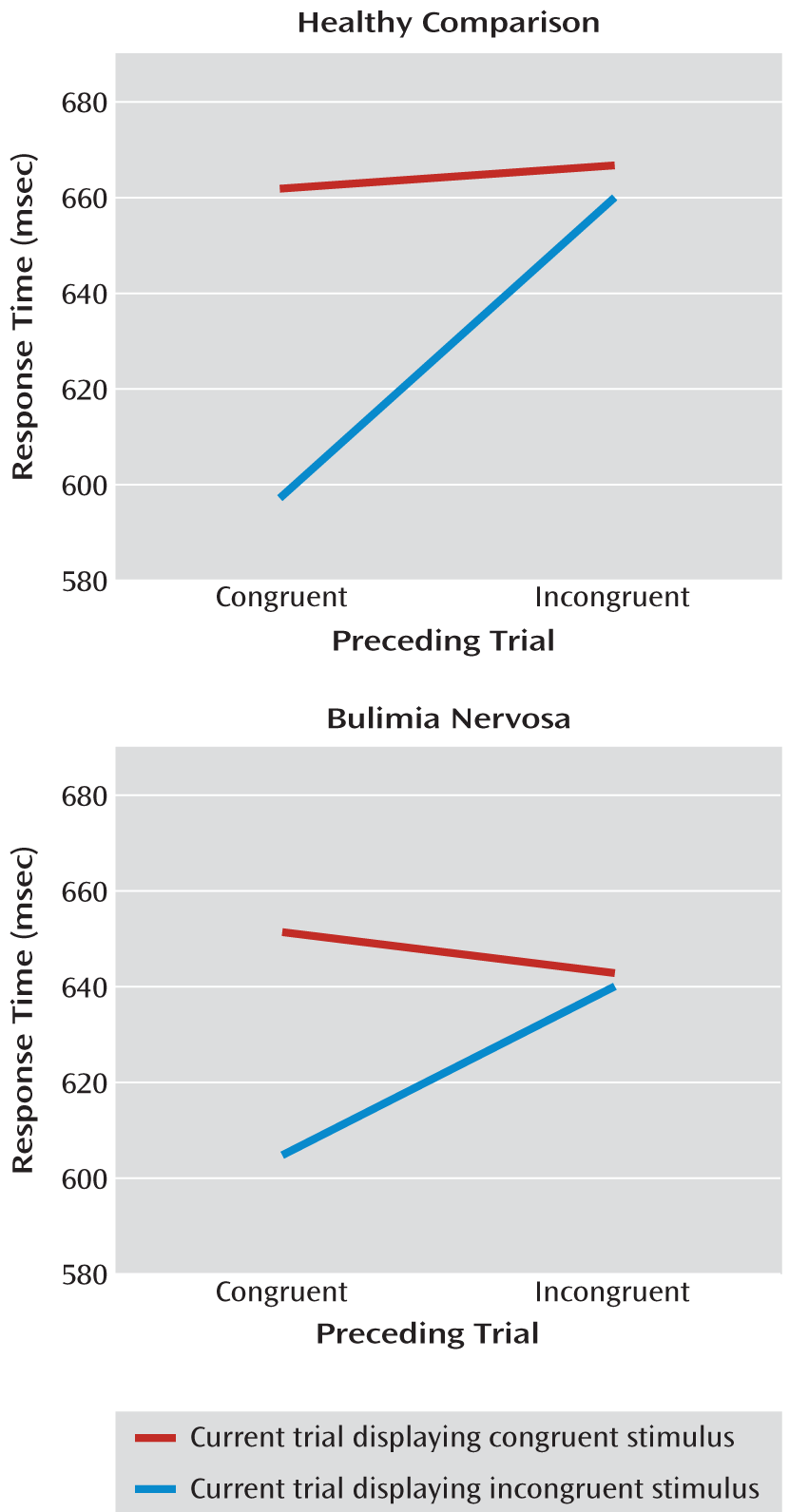
No significant main effect of group or interaction of group-by-stimulus type was detected in either model, indicating that there were no group differences specific to stimulus type; namely, response latency and accuracy scores were similar for congruent and incongruent trials in both groups. All participants demonstrated interference on the task (mean response latency for incongruent trials > mean response latency for congruent trials), and as predicted, both groups made few errors in congruent and incongruent trials (Table 2). In both groups, interference was greater following congruent trials than incongruent trials (stimulus type-by-repetition interactions: bulimia nervosa, p<0.001; healthy comparison, p=0.002 [Figure 1]), and this interaction effect did not differ significantly across groups. Finally, interference scores did not correlate significantly with symptom severity in the bulimia nervosa group.
TABLE 2. Group Differences on Performance Measures in Female Adolescents With Bulimia Nervosa and Age-Matched Female Healthy Comparison Subjects
 Bulimia Nervosa Adolescents (N=18)Healthy Comparison Adolescents (N=18)Analysis
MeasureMeanSDMeanSDtdfp
Mean reaction time incongruent (msec)633.8985.16656.0793.60.74340.46
Mean reaction time congruent (msec)624.1384.9641.5093.10.58340.56
Mean reaction time interference (msec)9.7532.914.56330.43340.66
Error incongruent (%)0.630.60.631.20.0341.00
Error congruent (%)0.00.10.110.40.82340.41
Error effect0.630.60.541.1–0.28340.77
FIGURE 1. Response Times Among Female Adolescents With Bulimia Nervosa Relative to Age-Matched Female Healthy Comparison Subjects on the Simon Spatial Incompatibility Taska
a Response times are depicted as a function of preceding and current trial stimulus type (congruent, incongruent).

A Priori Hypothesis

Neural activity during correct responses.

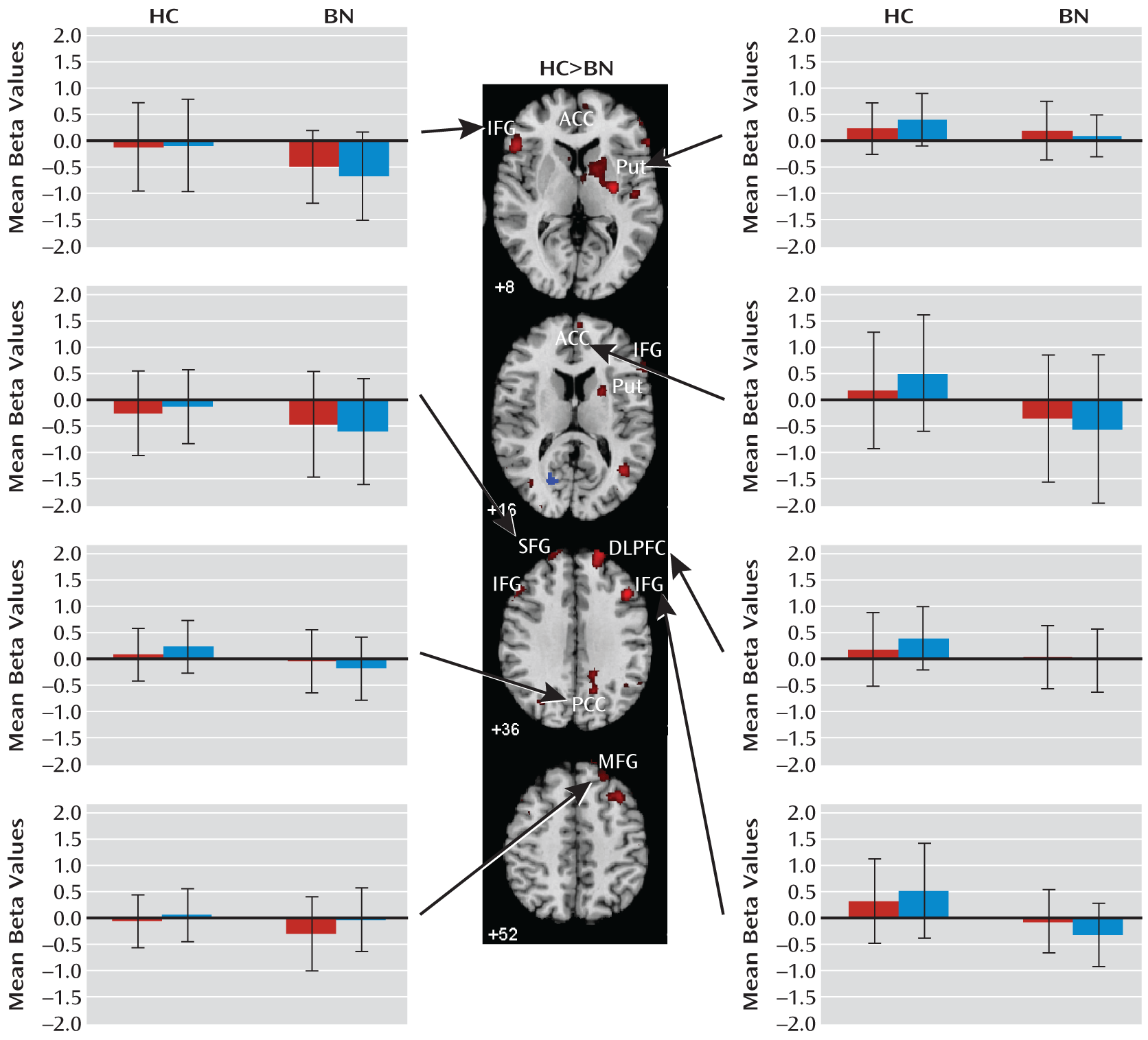
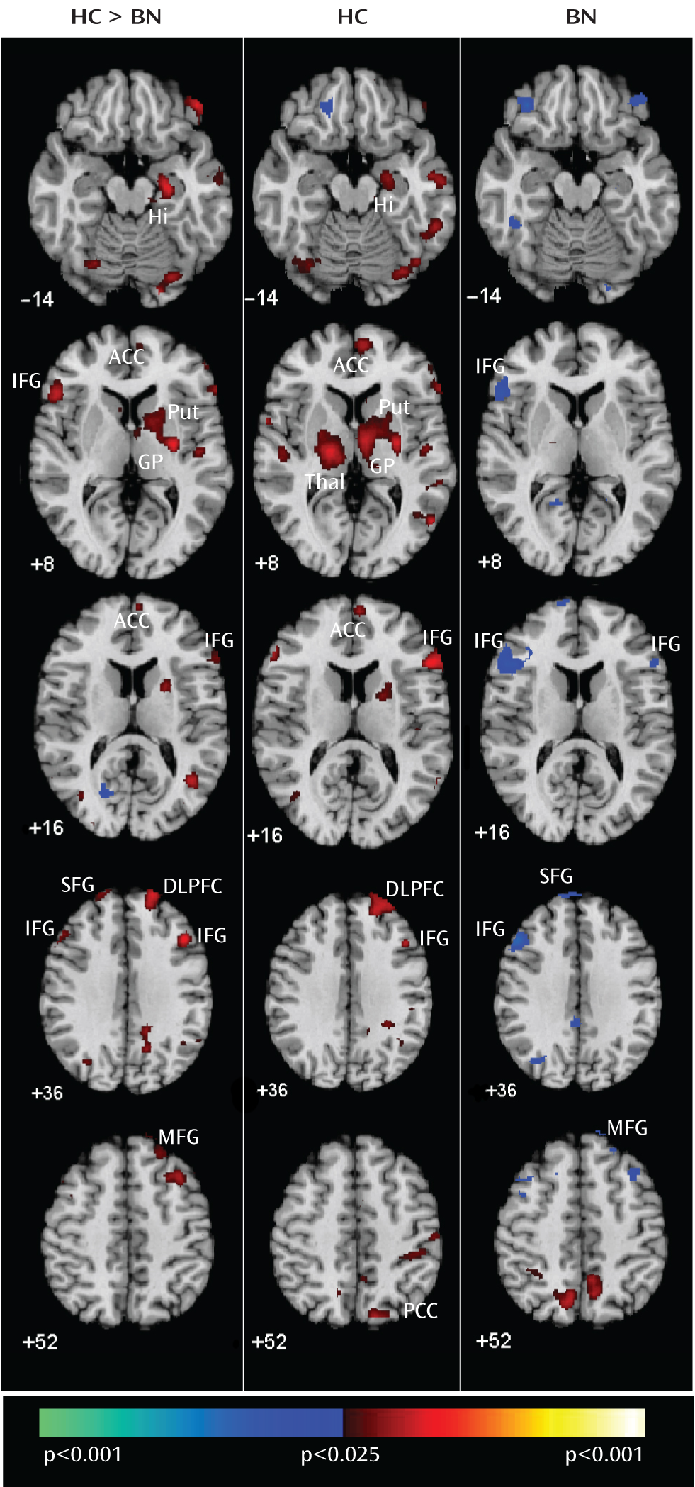
Significant group-by-stimulus interactions that derived from greater activations in healthy comparison subjects were detected in right hemispheric frontostriatal regions, including the anterior cingulate cortex (Brodmann's area 32), dorsolateral prefrontal cortex (Brodmann's area 9), inferior frontal gyrus (Brodmann's area 46), and putamen as well as the right hippocampus (Table 3, Figure 2). Significant group-by-stimulus interactions that derived from greater deactivation in participants with bulimia nervosa were detected in the left inferior frontal (Brodmann's area 46), left superior frontal (Brodmann's area 8), and right medial frontal (Brodmann's area 6/8) gyri as well as in the posterior cingulate cortex (Brodmann's area 31). Plots of the mean beta estimates for congruent and incongruent stimuli (Figure 3) revealed deactivations relative to baseline events in each participant in the bulimia nervosa group. For participants with bulimia nervosa, greater deactivation in the left inferior and superior frontal gyri as well as the right posterior cingulate cortex was seen in response to incongruent stimuli relative to congruent stimuli, and greater deactivation in the right medial frontal gyrus was seen in response to congruent stimuli relative to incongruent stimuli.
TABLE 3. Activations That Differed Significantly Across Female Adolescents With Bulimia Nervosa and Age-Matched Female Healthy Comparison Subjects During Responses to Congruent and Incongruent Stimuli
Activated RegionSideBrodmann's AreaaNumber of VoxelsPeak Location (x, y, z)t
Greater activations in healthy comparison subjects 
PutamenRight 86930, –16, 83.12
HippocampusRight2857525, –23, –143.16
Inferior frontal gyrusRight4561154, 20, 163.20
Anterior cingulate cortexRight241687, 53, 82.64
Dorsolateral prefrontal cortexRight9/461,65020, 54, 383.22
Greater deactivations in bulimia nervosa patients 
Inferior frontal gyrusLeft45483–52, 22, 83.24
Medial frontal gyrusRight6/855523, 35, 523.41
Superior frontal gyrusLeft9137–19, 50, 392.41
Posterior cingulate cortexRight3114114, –52, 362.82
a
Data were not available for frontostriatal analysis in the putamen.
FIGURE 2. Group Average Brain Activations During Correct Trials Among Female Adolescents With Bulimia Nervosa Relative to Age-Matched Female Healthy Comparison Subjectsa
a The images depict axial slices positioned inferiorly to superiorly (top to bottom). Group-by-stimulus (congruent versus incongruent) interactions (left) were detected in frontostriatal regions (red). Main effects of stimulus condition (congruent versus incongruent) are shown for healthy (center) and bulimia nervosa (right) adolescents. Increases in signal during correct incongruent relative to correct congruent trials are shown in red, and decreases are shown in blue. Abbreviations: ACC=anterior cingulate cortex; BN=bulimia nervosa; DLPFC=dorsolateral prefrontal cortex; GP=globus pallidus; HC=healthy comparison; Hi=hippocampus; IFG=inferior frontal gyrus; MFG=medial frontal gyrus; PCC=posterior cingulate cortex; Put=putamen; SFG=superior frontal gyrus; Thal=thalamus.
FIGURE 3. Region-of-Interest Analysis in Female Adolescents With Bulimia Nervosa Relative to Age-Matched Female Healthy Comparison Subjectsa
a The map of group-by-stimulus interactions is displayed again (as in Figure 2), along with plots of the mean beta estimates for congruent and incongruent stimuli for each region, plotted separately for the bulimia nervosa and healthy comparison groups. Error bars indicate standard error of the mean. The right side of the image shows the right lateralized frontostriatal regions that were more active during correct responses to incongruent versus congruent stimuli in healthy comparison subjects than in adolescents with bulimia nervosa. The left side of the image shows the regions in which deactivation associated with responses to incongruent stimuli (left inferior frontal gyrus [IFG], left superior frontal gyrus [SFG], and right posterior cingulate cortex [PCC]) and congruent stimuli (right SFG) was greater in adolescents with bulimia nervosa relative to healthy comparison subjects. Abbreviations: ACC=anterior cingulate cortex; BN=bulimia nervosa; DLPFC=dorsolateral prefrontal cortex; HC=healthy comparison; MFG=medial frontal gyrus; Put=putamen.

Exploratory Analyses

Context effects.

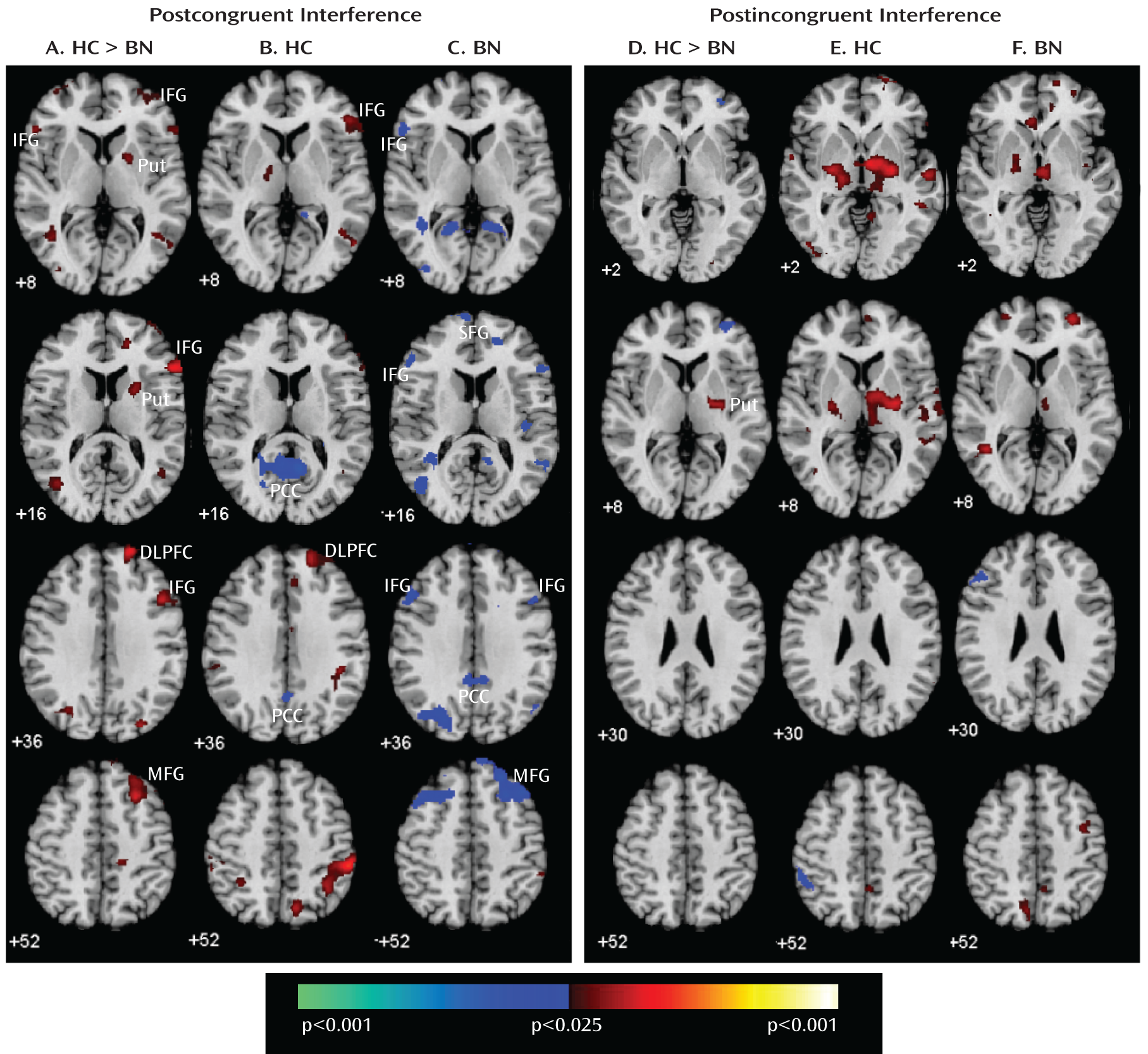
The group differences in activation (greater frontostriatal activation in healthy comparison subjects and greater deactivation of inferior and superior prefrontal cortices bilaterally in participants with bulimia nervosa) derived from stimuli that were preceded by congruent stimuli (postcongruent contrast [Figure 4]), when interference effects were greatest in both groups (Figure 1). Because deactivation was seen in the posterior cingulate cortex in both groups in this contrast, no interaction with group was detected in this region. A group-by-stimulus interaction from stimuli that were preceded by incongruent stimuli (postincongruent contrast [Figure 4]) was detected in the right posterior putamen and derived from greater activation in healthy comparison subjects. In addition, group differences in prefrontal regions were driven by postcongruent interference effects in the bulimia nervosa group (significant group-by-preceding-by-current stimulus interactions in the right inferolateral and dorsolateral prefrontal cortices and in the bilateral superior frontal gyri [see Figure 1 in the data supplement]). Finally, a group-by-stimulus interaction from congruent stimuli preceded by incongruent stimuli compared with congruent stimuli preceded by congruent stimuli was detected in the superior frontal gyrus and derived from greater deactivation in participants with bulimia nervosa (see Figure 2 in the data supplement).
FIGURE 4. Context Effects in Female Adolescents With Bulimia Nervosa Relative to Age-Matched Female Healthy Comparison Subjectsa
a Posterior probability maps depict the group average brain activity associated with the processing and resolution of cognitive interference. Postcongruent interference=activation contrast between incongruent and congruent stimuli in trial following a congruent trial; Postincongruent interference=activation contrast between incongruent and congruent stimuli in trial following an incongruent trial. Column A shows group-by-stimulus (congruent versus incongruent) interactions and main effects of stimulus following congruent trials in healthy comparison subjects (column B) and bulimia nervosa participants (column C). Column D shows group-by-stimulus interactions and main effects of stimulus following incongruent trials in healthy comparison subjects (column E) and bulimia nervosa participants (column F). Increases in signal during incongruent relative to congruent trials are shown in red; decreases are shown in blue (columns A–F). Abbreviations: BN=bulimia nervosa; DLPFC=dorsolateral prefrontal cortex; HC=healthy comparison; IFG=inferior frontal gyrus; MFG=medial frontal gyrus; PCC=posterior cingulate cortex; Put=putamen; SFG=superior frontal gyrus.

Correlations with symptom severity.

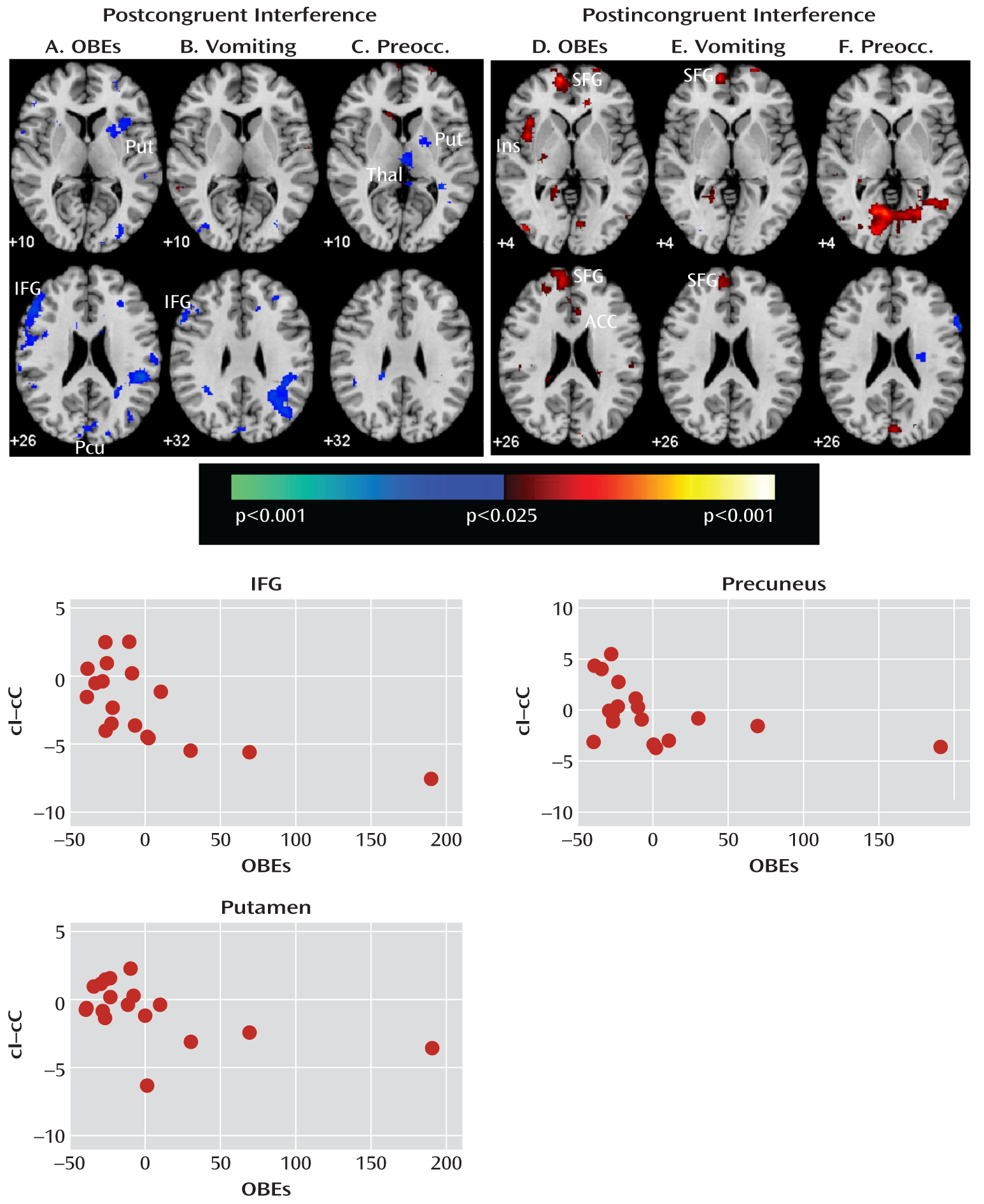
Nonparametric analyses revealed that the number of objective bulimic and vomiting episodes in the bulimia nervosa group correlated inversely with postcongruent brain activity in the left inferior frontal gyrus and bilateral precuneus (Figure 5). As shown in Figure 5, scatterplots of these correlations indicated that in response to postcongruent interference, the most symptomatic patients deactivated the inferior frontal gyrus and precuneus. The number of bulimic episodes and ratings of preoccupation with body shape and weight correlated inversely with postcongruent conflict activity in the right putamen, indicating that this subcortical region was engaged the least among the most preoccupied adolescents. Postincongruent conflict activity correlated positively with bulimic and vomiting episodes in the left superior frontal gyrus and with bulimic episodes in the left insula and right anterior cingulate cortex. Those with more severe symptoms showed the most activation in these regions, consistent with the postincongruent activation of cortical portions of frontostriatal circuits in the bulimia group (Figure 4).
FIGURE 5. Main Effects of Symptom Severity in Female Adolescents With Bulimia Nervosaa
a Inverse correlations of the magnitude of activation during correct responding to postcongruent interference (incongruent preceded by congruent-congruent preceded by congruent contrast) with A) the number of objective bulimic episodes (OBEs), B) the number of vomiting episodes, and C) ratings of preoccupation with body shape and weight (from the Eating Disorders Examination) are shown. Positive correlations (red) of the magnitude of activation during correct responding to postincongruent interference (incongruent preceded by incongruent-congruent preceded by incongruent contrast) with D) the number of OBEs, E) the number of vomiting episodes, and F) ratings of preoccupation with body shape and weight are also shown. Scatterplots depict the associations of OBEs with postcongruent interference activity. Abbreviations: IFG=inferior frontal gyrus; Ins=insula; Preocc=preoccupation ratings; Pcu=precuneus; Put=putamen; SFG=superior frontal gyrus; Thal=thalamus.

Medication and comorbidity effects.

A comparison of group average activation maps for only those bulimia nervosa participants who were not receiving medication treatment with a map for healthy comparison subjects suggested that medication did not contribute to our findings. Likewise, a history of anorexia nervosa, depressive symptoms, or ADHD symptoms was not associated with group differences in brain activations (see Figure 3 and Figure 4 in the data supplement).

Discussion

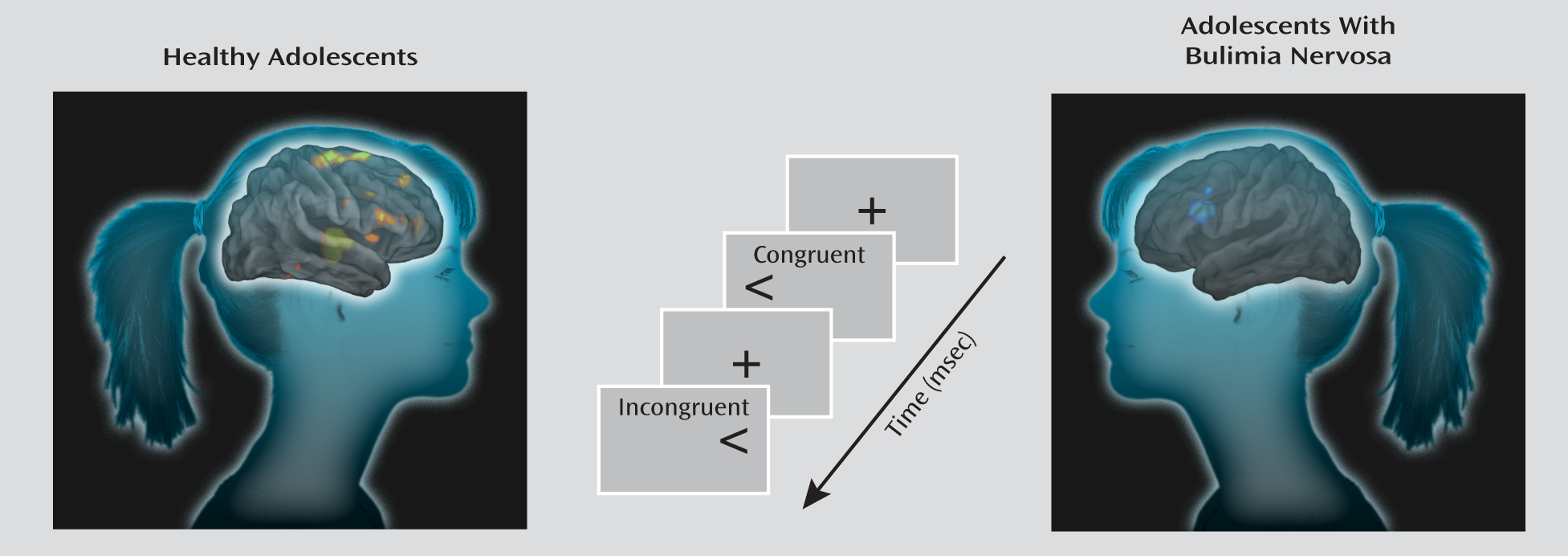
Adolescents with and without bulimia nervosa performed similarly on a rapid version of the Simon task. Both groups responded more slowly to incongruent stimuli relative to congruent stimuli and made few errors on the task (Figure 6). Nevertheless, participants with bulimia nervosa did not demonstrate the same magnitude of neural activity as healthy comparison subjects in the fronto-striatal systems known to subserve self-regulatory control, including the right inferior frontal gyrus, dorsolateral prefrontal cortex, and putamen. These findings are consistent with our prior fMRI findings in adult female patients (3). We previously speculated that the inability of these systems to engage appropriately in women with bulimia nervosa may exacerbate and interfere with their ability to resolve their conflicting desires to consume fattening foods and avoid weight gain. This inability may also contribute to their difficulty regulating binge-type eating and other impulsive behaviors. Our findings in adolescents with bulimia nervosa suggest that the inability to engage frontostriatal systems to resolve conflict and regulate behaviors may arise early in the course of the illness.
FIGURE 6. Processing of Congruent and Incongruent Stimuli on the Simon Spatial Incompatibility Task Among Female Adolescents With Bulimia Nervosa Relative to Age-Matched Female Healthy Comparison Subjects
When responding correctly to incongruent stimuli compared with congruent stimuli, deactivation was seen in the left inferior frontal gyrus as well as a neural system encompassing the posterior cingulate cortex and superior frontal gyrus in adolescents with bulimia nervosa. The posterior cingulate cortex is a major node of the default-mode network of brain areas that activate when individuals are engaged in internally driven thoughts and decrease in activity when attention is directed to a task (14). The superior frontal gyrus is also part of this network (15), and recent findings suggest that the posterior cingulate cortex and superior frontal gyrus are functionally connected to the medial and inferior frontal gyri (16). Deactivation of all of these brain areas in response to the presentation of both congruent and incongruent stimuli was observed in adolescents with bulimia nervosa, perhaps suggesting that they may have been attending to internally driven thoughts, such as thoughts about eating or body image that preoccupy individuals with eating disorders.
Our analyses suggest that functional abnormalities in frontostriatal and default-mode systems in female adolescents with bulimia nervosa derived from altered processing of cognitive interference, which depended on the presence or absence of antecedent conflict, and were particularly driven by abnormal processing of cognitive interference that followed congruent stimuli, when interference effects were greatest in both the bulimia nervosa and healthy comparison groups (postcongruent interference: incongruent preceded by congruent-congruent preceded by congruent contrast [Table 4, Figure 1]). In this contrast, deactivation in the inferior and superior prefrontal cortices was seen in female adolescents with bulimia nervosa, whereas activation in the right frontostriatal regions—including the inferior and dorsal prefrontal cortices and putamen—was seen in healthy female adolescents (Figure 4). Deactivation in the posterior cingulate cortex in response to postcongruent interference was found in both groups, but deactivation in the posterior cingulate cortex and medial and superior frontal gyri in response to both congruent and incongruent stimuli was seen only in the bulimia nervosa group (Figure 3). In addition, greater deactivation of these cortical regions was associated with more severe symptoms, suggesting that cortical deactivation in the bulimia nervosa group may have represented the intrusion of self-referential and illness-related thoughts when processing interference in the absence of prior conflict. In the bulimia nervosa group, deactivation of the superior frontal gyrus and posterior cingulate cortex in response to congruent stimuli that were preceded by incongruent stimuli relative to congruent stimuli (congruent preceded by incongruent-congruent preceded by congruent contrast [see Figure 2 in the data supplement]) suggests that deactivation of these default-mode regions was specific to the anteceding conflict-laden context rather than current conflict. This finding further emphasizes the contextual origins of the group differences in this network.
TABLE 4. Activations That Differed Significantly Across Female Adolescents With Bulimia Nervosa and Age-Matched Female Healthy Comparison Subjects During Correct Responses to Incongruent Stimuli by Preceding Trial Stimulus Type
Activated RegionSideBrodmann's AreaaNumber of VoxelsPeak Location (x, y, z)t
Postcongruent interferenceb 
Greater activations in healthy comparison subjects 
    Inferior frontal gyrusRight4529654, 20, 163.18
    Dorsolateral prefrontal cortexRight9/4640320, 52, 383.30
Greater deactivations in bulimia nervosa patients 
    Inferior frontal gyrusLeft45109–54, 21, 162.52
    Medial frontal gyrusRight6/855526, 18, 673.69
Postincongruent interferencec 
Greater activations in healthy comparison subjects 
Posterior putamenRight 17026, –16, 82.75
a
Data were not available for frontostriatal analysis in the putamen.
b
Activation contrast between incongruent and congruent stimuli in trial following a congruent trial.
c
Activation contrast between incongruent and congruent stimuli in trial following an incongruent trial.
The number of objective bulimic and vomiting episodes correlated inversely with activity associated with postcongruent interference in the left inferior frontal gyrus, a region that was deactivated in response to incongruent stimuli in the bulimia nervosa group. The number of bulimic episodes and ratings of preoccupation with body shape and weight also correlated inversely with postcongruent interference in the right putamen. Thus, in the bulimia nervosa group, deactivation in the inferior frontal gyrus was seen the most and activation in the subcortical portions of frontostriatal circuits was seen the least among the most symptomatic individuals when responding to postcongruent interference, consistent with our prior findings that frontostriatal activation correlated with symptom severity in adult female patients with bulimia nervosa (3). The task used in our previous study consisted of more congruent trials than incongruent trials, and thus the analysis of interference effects (incongruent versus congruent comparison) in that study was most similar to the postcongruent interference contrast in the present study.
Group differences in subcortical activations were driven by the differential processing of cognitive interference following incongruent stimuli (incongruent preceded by incongruent-congruent preceded by incongruent contrast [Table 4]), which is the contrast that produced the least interference in both groups. Activation of frontostriatal regions in this condition was observed in both groups (i.e., postincongruent interference), but activation of the subcortical portions of these circuits was seen in the healthy comparison group more than the bulimia nervosa group, suggesting that the healthy subjects relied more on an automatic response tendency based within the striatum (17) when responding to repeated conflict over successive trials and that this adaptive habitual responding may be deficient in adolescents with bulimia nervosa. These differential patterns of brain activation when resolving cognitive interference following congruent or incongruent stimuli, respectively, contributed to the overall group differences in our primary analyses of cortical and subcortical brain activation in processing cognitive interference that we detected for analyses of all trials, regardless of the presence or absence of antecedent conflict (Figure 2).
Our findings suggest that frontostriatal functioning is impaired in bulimia nervosa early in the course of illness. When engaging the self-regulatory control processes necessary to resolve cognitive interference and respond correctly in incongruent trials, adolescents with bulimia nervosa did not display normal patterns of activation in frontostriatal circuits or deactivation in default-mode systems. Abnormal processing of the antecedent stimulus context conditioned their brain responses to conflict differently, particularly in frontal regions when preceding stimuli were congruent and conflict-free. Given that the bulimia nervosa and healthy comparison groups performed similarly on the task and that our analyses were limited to activations associated with correct responses, these differences in brain activation did not derive from performance differences and instead represented functional disturbances in neural systems that subserve self-regulation and the resolution of interference. The Simon task used in this study differed from the longer version of the task used in our prior study of adults with bulimia nervosa (3). Congruent and incongruent stimuli appeared equally as often using the shorter version, thereby making it easier to inhibit the prepotent response tendency, and as hypothesized, the groups performed similarly on the task. In addition, context effects could be assessed with this task design, contributing to our additional finding that abnormal contextual processing likely affects the neural processing of conflict in adolescents with bulimia nervosa. These findings of impaired frontostriatal functioning may inform clinical practice, suggesting that strategies for improving self-regulatory control and conflict resolution through cognitive remediation therapy may be a promising treatment for bulimia nervosa in adolescents.
A limitation of this study was the absence of a comparison group consisting of impulsive individuals with normal weight and eating behaviors, which would permit assessment of the specificity of frontostriatal abnormalities in adolescents with bulimia nervosa. Other limitations were the modest sample size and presence of comorbid illnesses in participants with bulimia nervosa, some of whom were receiving medication treatment. In addition, participants had varying symptom severities, and individuals with full- and partial-syndrome bulimia were included. However, given that subthreshold eating disorders are most prevalent among adolescents (18), our sample represented well the general population of adolescents with bulimia nervosa. Moreover, our inclusion criteria were consistent with the lower and more developmentally sensitive thresholds that are more appropriate for adolescents and that have been proposed for DSM-5 (19). Nevertheless, future studies should include larger samples of adolescents with bulimia nervosa and assess changes in the functioning of frontostriatal and default-mode systems in the same participants over time to understand whether disturbances in these systems may contribute to the development and progression of the illness.
Footnote

Received Jan. 18, 2011; revision received March 3, 2011; accepted March 17, 2011.

References

1.
Walsh BT, Klein DA: Eating disorders. Int Rev Psychiatry 2003; 15:205–216
2.
Kaye W, Strober M, Jimerson DC: The neurobiology of eating disorders, in The Neurobiology of Mental Illness. Edited by, Charney D, Nestler EJ. New York, Oxford University Press, 2004, pp 1112–1128
3.
Marsh R, Steinglass JE, Gerber AJ, O'Leary KG, Walsh BT, Peterson BS: Deficient activity in the neural systems that mediate self-regulatory control in bulimia nervosa. Arch Gen Psychiatry 2009; 66:1–13
4.
Wühr P, Ansorge U: Exploring trial-by-trial modulations of the Simon effect. Q J Exp Psychol A 2005; 58:705–731
5.
Sturmer B, Leuthold H, Soetens E, Schroter H, Sommer W: Control over location-based response activation in the Simon task: behavioral and electrophysiological evidence. J Exp Psychol Hum Percept Perform 2002; 28:1345–1363
6.
Botvinick M, Nystrom LE, Fissell K, Carter CS, Cohen JD: Conflict monitoring versus selection-for-action in anterior cingulate cortex. Nature 1999; 402:179–181
7.
Kerns JG, Cohen JD, MacDonald AW, Cohen JD, Cho RY, Stenger VA, Carter CS: Anterior cingulate conflict monitoring and adjustments in control. Science 2004; 303:1023–1026
8.
Henson RN, Price CJ, Rugg MD, Turner R, Friston KJ: Detecting latency differences in event-related BOLD responses: application to words versus nonwords and initial versus repeated face presentations. Neuroimage 2002; 15:83–97
9.
Neumann J, Lohmann G: Bayesian second-level analysis of functional magnetic resonance images. Neuroimage 2003; 20:1346–1355
10.
Friston KJ, Penny W: Posterior probability maps and SPMs. Neuroimage 2003; 19:1240–1249
11.
Thirion B, Pinel P, Meriaux S, Roche A, Dehaene S, Poline JB: Analysis of a large fMRI cohort: statistical and methodological issues for group analyses. Neuroimage 2007; 35:105–120
12.
Friston KJ, Worsley KJ, Frackowiak RSJ, Mazziotta JC, Evans AC: Assessing the significance of focal activations using their spatial extent. Hum Brain Mapp 1994; 1:214–220
13.
Forman SD, Cohen JD, Fitzgerald M, Eddy WF, Mintun MA, Noll DC: Improved assessment of significant activation in functional magnetic resonance imaging (fMRI): use of a cluster-size threshold. Magn Reson Med 1995; 33:636–647
14.
Greicius MD, Krasnow B, Reiss AL, Menon V: Functional connectivity in the resting brain: a network analysis of the default mode hypothesis. Proc Natl Acad Sci USA 2003; 100:253–258
15.
Buckner RL, Andrews-Hanna JR, Schacter DL: The brain's default network: anatomy, function, and relevance to disease. Ann NY Acad Sci 2008; 1124:1–38
16.
Grigg O, Grady CL: The default network and processing of personally relevant information: converging evidence from task-related modulations and functional connectivity. Neuropsychologia 2010; 48:3815–3823
17.
Packard MG, Knowlton BJ: Learning and memory functions of the basal ganglia. Annu Rev Neurosci 2002; 25:563–593
18.
Stice E, Marti CN, Shaw H, Jaconis M: An 8-year longitudinal study of the natural history of threshold, subthreshold, and partial eating disorders from a community sample of adolescents. J Abnorm Psychol 2009; 118:587–597
19.
Bravender T, Bryant-Waugh R, Herzog D, Katzman D, Kriepe RD, Lask B, Le Grange D, Lock J, Loeb KL, Marcus MD, Madden S, Nicholls D, O'Toole J, Pinhas L, Rome E, Sokol-Burger M, Wallin U, Zucker N Workgroup for Classification of Eating Disorders in Children and Adolescents: Classification of eating disturbance in children and adolescents: proposed changes for the DSM-V. Eur Eat Disord Rev 2010; 18:79–89

Information & Authors

Information

Published In

Go to American Journal of Psychiatry
American Journal of Psychiatry
Pages: 1210 - 1220
PubMed: 21676991

History

Received: 18 January 2011
Revision received: 3 March 2011
Accepted: 17 March 2011
Published online: 1 November 2011
Published in print: November 2011

Authors

Details

Rachel Marsh, Ph.D.
From the Division of Child and Adolescent Psychiatry, Department of Psychiatry, New York State Psychiatric Institute and the College of Physicians and Surgeons, Columbia University, New York; and the Eating Disorders Research Unit, Department of Psychiatry, New York State Psychiatric Institute and the College of Physicians and Surgeons, Columbia University, New York.
Guillermo Horga, M.D.
From the Division of Child and Adolescent Psychiatry, Department of Psychiatry, New York State Psychiatric Institute and the College of Physicians and Surgeons, Columbia University, New York; and the Eating Disorders Research Unit, Department of Psychiatry, New York State Psychiatric Institute and the College of Physicians and Surgeons, Columbia University, New York.
Zhishun Wang, Ph.D.
From the Division of Child and Adolescent Psychiatry, Department of Psychiatry, New York State Psychiatric Institute and the College of Physicians and Surgeons, Columbia University, New York; and the Eating Disorders Research Unit, Department of Psychiatry, New York State Psychiatric Institute and the College of Physicians and Surgeons, Columbia University, New York.
Pengwei Wang, Ph.D.
From the Division of Child and Adolescent Psychiatry, Department of Psychiatry, New York State Psychiatric Institute and the College of Physicians and Surgeons, Columbia University, New York; and the Eating Disorders Research Unit, Department of Psychiatry, New York State Psychiatric Institute and the College of Physicians and Surgeons, Columbia University, New York.
Kristin W. Klahr, M.A.
From the Division of Child and Adolescent Psychiatry, Department of Psychiatry, New York State Psychiatric Institute and the College of Physicians and Surgeons, Columbia University, New York; and the Eating Disorders Research Unit, Department of Psychiatry, New York State Psychiatric Institute and the College of Physicians and Surgeons, Columbia University, New York.
Laura A. Berner, A.B.
From the Division of Child and Adolescent Psychiatry, Department of Psychiatry, New York State Psychiatric Institute and the College of Physicians and Surgeons, Columbia University, New York; and the Eating Disorders Research Unit, Department of Psychiatry, New York State Psychiatric Institute and the College of Physicians and Surgeons, Columbia University, New York.
B. Timothy Walsh, M.D.
From the Division of Child and Adolescent Psychiatry, Department of Psychiatry, New York State Psychiatric Institute and the College of Physicians and Surgeons, Columbia University, New York; and the Eating Disorders Research Unit, Department of Psychiatry, New York State Psychiatric Institute and the College of Physicians and Surgeons, Columbia University, New York.
Bradley S. Peterson, M.D.
From the Division of Child and Adolescent Psychiatry, Department of Psychiatry, New York State Psychiatric Institute and the College of Physicians and Surgeons, Columbia University, New York; and the Eating Disorders Research Unit, Department of Psychiatry, New York State Psychiatric Institute and the College of Physicians and Surgeons, Columbia University, New York.

Notes

Address correspondence to Dr. Marsh ([email protected]).

Funding Information

Supported in part by the National Alliance for Research on Schizophrenia and Depression, the Sackler Institute for Developmental Psychobiology, and NIMH grant K01-MH-077652 (Dr. Marsh); the Alicia Koplowitz Foundation of Spain (Dr. Horga); and NIMH grant K02-MH-074677 (Dr. Peterson).Dr. Walsh has received research support from AstraZeneca. All other authors report no financial relationships with commercial interests.

Metrics & Citations

Metrics

Citations

Export Citations

If you have the appropriate software installed, you can download article citation data to the citation manager of your choice. Simply select your manager software from the list below and click Download.

For more information or tips please see 'Downloading to a citation manager' in the Help menu.

Format
Citation style
Style
Copy to clipboard

View Options

View options

PDF/EPUB

View PDF/EPUB

Get Access

Login options

Already a subscriber? Access your subscription through your login credentials or your institution for full access to this article.

Personal login Institutional Login Open Athens login
Purchase Options

Purchase this article to access the full text.

PPV Articles - American Journal of Psychiatry

PPV Articles - American Journal of Psychiatry

Not a subscriber?

Subscribe Now / Learn More

PsychiatryOnline subscription options offer access to the DSM-5-TR® library, books, journals, CME, and patient resources. This all-in-one virtual library provides psychiatrists and mental health professionals with key resources for diagnosis, treatment, research, and professional development.

Need more help? PsychiatryOnline Customer Service may be reached by emailing [email protected] or by calling 800-368-5777 (in the U.S.) or 703-907-7322 (outside the U.S.).

Media

Figures

Other

Tables

Share

Share

Share article link

Share