Abstract

Objective:

Individuals with autism as young as 2 years have been observed to have larger brains than healthy comparison subjects. Studies using head circumference suggest that brain enlargement is a postnatal event that occurs around the latter part of the first year. To the authors' knowledge, no previous brain imaging studies have systematically examined the period prior to age 2. In this study they used magnetic resonance imaging (MRI) to measure brain volume in 6-month-olds at high familial risk for autism.

Method:

The Infant Brain Imaging Study (IBIS) is a longitudinal imaging study of infants at high risk for autism. This cross-sectional analysis compared brain volumes at 6 months of age in high-risk infants (N=98) and infants without family members with autism (N=36). MRI scans were also examined for radiologic abnormalities

Results:

No group differences were observed for intracranial, cerebrum, cerebellum, or lateral ventricle volume or for head circumference.

Conclusions:

The authors did not observe significant group differences for head circumference, brain volume, or abnormalities in radiologic findings from a group of 6-month-old infants at high risk for autism. The authors are unable to conclude that these abnormalities are not present in infants who later go on to receive a diagnosis of autism; rather, abnormalities were not detected in a large group at high familial risk. Future longitudinal studies of the IBIS study group will examine whether brain volume differs in infants who go on to develop autism.
Converging evidence from postmortem, magnetic resonance imaging (MRI), and head circumference studies has documented that individuals with autism spectrum disorders (ASDs) have larger brains and heads than comparison subjects (110). Brain MRI studies have revealed generalized enlargement in cerebral cortical gray and white matter (911) and selected subcortical structures (12) that is present as early as 2 years of age. A longitudinal MRI study of 2-year-old children with ASDs followed up at 4–5 years of age demonstrated enlargement at age 2 with no increase in growth rate during the subsequent 2-year interval (10), and this pattern is consistent with the conclusion that high brain volume is the result of overgrowth occurring before 2 years of age. A recent study of a group of 2–50-year-olds with ASDs (13) also showed that early brain enlargement was associated with autism and that it was characterized by an early postnatal period of overgrowth followed by a period of slower or arrested growth.
Longitudinal studies of head circumference suggest that the onset of brain overgrowth is in the latter part of the first year of life (9, 14), and high rates of macrocephaly have been observed in parents of probands with ASDs (7). Data from behavioral studies of infant siblings of autistic individuals (infant siblings who are therefore at high genetic risk for developing ASDs) suggest that most of the defining features of this condition (e.g., social deficits) are not present at 6 months of age but are first evident by 12 months of age (15). These two independent lines of research (brain studies, developmental behavioral studies) appear to converge on the latter part of the first year of life as a period of both changes in brain structure and the onset of behavioral symptoms of autism.
Structural imaging studies of early brain development in typically developing children highlight that the first year of life is a time of dynamic growth, where total brain volume increases 100% (16). As autism is not typically diagnosed until about age 2 years or later, studies of abnormal brain development during infancy in autism must wait until the diagnosis is established. Given the high recurrence risk of autism in subsequently born siblings (17), a strategy that has recently been employed is the examination of the infant siblings of autistic children (i.e., infants at risk for autism before the diagnosis of autism). Virtually nothing is known about early brain development in infants at risk for autism from neuroimaging studies. It could be valuable to know whether there are observable differences in brain volume between these high-risk infants and infants from the normal population who have no known genetic risk for autism and therefore most often display typical development, i.e., “low-risk” subjects.
In the present study we assessed volumetric brain MRI measures in a large group of 6-month-old infants at high genetic risk for autism and in infants at low risk for autism. To our knowledge, this study is the first to measure brain volume in 6-month-olds at high familial risk for autism, approximately 20% of whom may receive a later diagnosis of an ASD (17), to test whether brain volume differences may be associated with high risk for ASDs.

Method

Study Group

This investigation included data acquired from an Autism Center of Excellence network study funded by the National Institutes of Health. Informally called the Infant Brain Imaging Study (IBIS), the network includes four clinical data collection sites (University of North Carolina at Chapel Hill, University of Washington, Children's Hospital of Philadelphia, Washington University in St Louis), a data coordinating center (Montreal Neurological Institute at McGill University), and two image processing sites (University of Utah, University of North Carolina). The network study collects neuroimaging and behavioral data on infants who are at genetic risk for ASDs by virtue of having an older sibling with autism, referred to as “high-risk infant siblings.” The study enrolls 1) 6-month-old high-risk infant siblings who are seen for follow-up assessments at 12 and 24 months of age, 2) high-risk infant siblings who enter the study at 12 months of age and are followed up at 24 months, and 3) comparison subjects who are typically developing infants considered to be at low risk for autism (i.e., an older sibling is developing typically), who are seen at 6, 12 and 24 months. Diagnostic outcome data for ASD status is determined at 24 months of age (and again at 36 months of age if the timeline of the project allows). For the purposes of this article, we have included cross-sectional data on the 6-month-olds who were scanned from the start of the study through the fall of 2010. Two groups were included: infants at high risk for autism and typically developing children considered to be at low risk for autism. Subjects were combined from all four clinical data collections sites.
The subjects include all children with imaging data collected and processed through the end of September 2010. There were 318 children who attempted the MRI scans, and 88% of the high-risk infants and 83% of the low-risk subjects were successfully scanned, for a total of 276 scans. Owing to difficulties in scheduling visits and missed visits (e.g., due to illness, changes in travel plans), some children were seen in a wider age window, up to 8 months of age. Infants develop rapidly during the first year, and so for the purpose of this analysis, a narrow, optimal age window was defined as from 1 week before age 6 months to 3 weeks after age 6 months. There were 134 infants who were in this optimal age window (98 high-risk, 36 low-risk), and all results are based on this group. Of note, the wider age group at the first time point will be included in later longitudinal analyses of these data.
Subjects were characterized as having high risk if they had an older sibling with a diagnosis of an ASD that was documented in a clinical diagnostic report and confirmed by the Autism Diagnostic Interview-Revised (18) administered at enrollment. Subjects were enrolled in the low-risk group if they had an older sibling without evidence of ASDs and no family history of a first- or second-degree relative with an ASD. Exclusion criteria for both groups included the following: 1) diagnosis or physical signs strongly suggestive of a genetic condition or syndrome reported to be associated with ASDs (e.g., fragile X syndrome), 2) significant medical or neurological condition affecting growth, development, or cognition (e.g., CNS infection, seizure disorder, congenital heart disease), 3) sensory impairment, such as vision or hearing loss, 4) low birth weight (<2,000 g) or prematurity (<36 weeks gestation), 5) possible perinatal brain injury from exposure to in utero exogenous compounds reported to likely affect brain adversely in at least some individuals (e.g., alcohol, selected prescription medications), 6) non-English-speaking family, 7) contraindication for MRI (e.g., metal implants), 8) adoption, and 9) family history of intellectual disability, psychosis, schizophrenia, or bipolar disorder in a first-degree relative.

Assessment Protocols

Behavioral assessment.

The infants were assessed directly at ages 6, 12, and 24 months, and a telephone interview was conducted with the parents at 18 months to assess their infant's development. The direct assessments included brain MRI scans in addition to a battery of behavioral and developmental tests. The assessment battery for the visit at 6 months included the Mullen Scales of Early Learning (19), the Vineland Adaptive Behavior Scales-II (20), the Autism Observation Scale for Infants (21), various questionnaires examining behavior, temperament, and family characteristics, and a medical record review. See Table 1 for a summary of subject characteristics.
TABLE 1. Characteristics of 6-Month-Old Infants at Low or High Familial Risk for Autism
CharacteristicLow Risk (N=36)High Risk (N=98)Difference
 N%N%pa
Male215861620.69
Maternal education    0.01
  Not answered51444 
  Less than college degree10283637 
  College degree8224142 
  Graduate degree13361717 
Family annual income    0.76
  Not answered7191010 
  <$75,00015424445 
  $75,000–$150,0009253031 
  >$150,0005141414 
 MeanSDMeanSDpb
Mother's age at infant's birth (years)32.95.733.74.70.37
Gestational age at birth (weeks)39.51.239.01.30.47
Age at MRI scan (months)6.30.26.20.30.37
Mullen Scales of Early Leaning composite score from MRI visit100.010.095.212.50.13
a
Fisher's exact test.
b
Analysis of variance controlling for site differences.

MRI acquisition.

All the brain MRI scans were completed at each of the clinical sites on a 3-T Siemens Tim Trio scanner with a 12-channel head coil. All scans were completed while the infants were naturally sleeping. A specific structured preparation was completed by the families at home before the scanning; it included conditioning to the scanner sounds on a compact disc played to the infants while sleeping. The imaging protocol was designed to maximize tissue contrast for volumetric analysis across three time points (ages 6, 12, 24 months). The protocol included 1) a localizer scan, 2) a three-dimensional T1 magnetization-prepared rapid acquisition gradient-echo scan: TR=2,400 ms, TE=3.16 ms, 160 sagittal slices, field of view (FOV)=256 mm, voxel size=1 mm3, 3) a three-dimensional T2 fast spin echo scan: TR=3,200 ms, TE=499 ms, 160 sagittal slices, FOV=256 mm, voxel size=1 mm3, and 4) a 25-direction diffusion tensor imaging scan: TR=12,800 ms, TE=102 ms, slice thickness=2 mm isotropic, variable b value=maximum of 1,000 s/mm2, FOV=190 mm.
A number of quality control procedures were employed to assess scanner stability and reliability across sites, time, and procedures. A LEGO phantom (22) was scanned monthly at each location and analyzed to assess image quality and quantitatively address site-specific regional distortions. Two adult subjects (“human phantoms”) were scanned once per year per scanner (twice in year 1). The data for these phantoms were evaluated for scanner stability across sites and time (23). The results indicated excellent stability across sites, with covariates of variation for intracranial volume below 1% and with intraclass correlations for intracranial volume at 0.98 for intersite and 0.99 for intrasite reliability. Finally, all scans were blindly reviewed for image quality by a single rater (D. Louis Collins, McGill University) and again rated by a single reviewer (Rachel Gimpel Smith, University of North Carolina) in a preprocessing stage before image analysis.

Radiologic review.

All scans were reviewed locally by a pediatric neuroradiologist for radiologic findings that, if present, could be communicated to the participants. In addition, a board-certified pediatric neuroradiologist (R.C.M., Washington University) blindly reviewed all MRI scans across the IBIS network and rated the incidental findings. A third neuroradiologist (D.W.W.S., University of Washington) provided a second blind review for the Washington University site and contributed to a final consensus rating if there were discrepancies between the local site reviews and the network review. The final consensus review was used to evaluate whether there were group differences in the number and/or type of incidental findings.

Image processing.

The following brain volumes were obtained: intracranial, total brain (gray matter plus white matter), total CSF, cerebrum, cerebellum, and lateral ventricles. There is minimal tissue contrast in the 6-month-old brain because of ongoing myelination and maturation. As a result, reliable separation of white matter and gray matter tissue is highly limited. Thus, we chose to initially focus on the total brain volume measure without separating white and gray matter. We also parcellated the brain into large regions (e.g., cerebrum, cerebellum, hemispheres) but omitted a fine-grained lobar or cortical segmentation, which is less reliable at this young age.
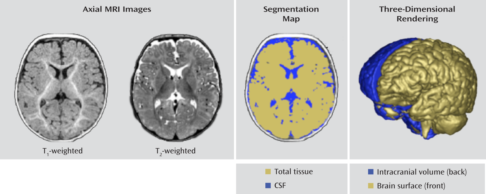
The brain volume segmentations were obtained following a rigid transformation to a normative brain space and preprocessing for bias correction and intensity normalization. These steps are part of the expectation-maximization (EM) tissue segmentation method (24) using the AutoSeg tool (25). Tissue probability maps for white matter, gray matter, and CSF were obtained. Intracranial volume was defined to be the sum of white matter, gray matter, and CSF, and an example of the segmentation labeling is shown in Figure 1.
FIGURE 1. Brain Segmentation at 6 Months of Age
Measures of head circumference were obtained by using a semiautomated image processing tool according to an established protocol (9). Head circumference was measured by a single rater using the HeadCirc tool developed by collaborators at the University of North Carolina. HeadCirc computes head circumference by extracting the appropriate contour from head segmentation of MRI images (using Fourier harmonics to parameterize the contour of the head on MRI). Intra- and interrater reliability for the head circumference measures in the data set from the 6-month-old infants was exceptional, with an intraclass correlation (ICC) of 0.99 for each one. The ITK-SNAP tool (26) was used by a single rater to obtain measurements of the lateral ventricles. ICC values for intra- and interrater reliability for the lateral ventricles were 0.99 (for combined volume and for right and left hemispheres). The AutoSeg, HeadCirc, and ITK-SNAP tools are available at http://www.ia.unc.edu/dev/download and http://www.nitrc.org.
Scans from 12 infants were excluded because they did not pass the initial scan review and/or preprocessing steps. The problems included misalignment (N=2), off-center or distorted image (N=2), poor registration (N=3), and poor image quality or artifact (N=5). There were no group differences in the rate or type of scan failures.

Statistical Analysis

Power consideration.

The design of the study was based on findings from our previous comparison of 2-4-year-olds with ASDs and typically developing comparison subjects (10). The percentage difference in the volume of relevant structures (e.g., cerebral cortex, cerebral cortical white matter, and temporal lobe white matter) was 4% to 9%, with effect sizes (Cohen's d) from 0.59 to 0.95, in cross-sectional groups of 45–51 children with ASDs and 15–26 comparison subjects. With our current groups of 36 low-risk and 98 high-risk infants, we expected to have 85%–99% power to achieve effect sizes similar to those in the 2–4-year-old children with ASDs.

Analysis.

Our research goal was to examine whether there were significant group differences in head circumference, brain volumes, and radiologic findings in a group of infants at high genetic risk for autism. Group differences in sex were tested with Fisher's exact test, and separate analysis of variance (ANOVA) models were used to assess group differences in the Mullen early learning composite score, gestational age at birth, age at MRI scan, and maternal age at birth while controlling for site differences. Cross-sectional group differences in brain volumes were analyzed by using a general linear model in SAS (SAS Institute, Cary, N.C.). Covariates included in the model were age at scan, sex (male/female), site, and the Mullen early learning composite standard score. The dependent variables of interest included head circumference, intracranial volume, and volumes of the total tissue (gray plus white matter), CSF, cerebrum (left and right total tissue plus CSF), cerebellum (left and right total tissue plus CSF), and left and right lateral ventricles. To test the influence of sex differences on group effect, we included the additional interaction term (group by sex) in the model.

Results

As shown in Table 1, there were no significant group differences for age, sex, Mullen early learning composite standard score, gestational age, maternal age at childbirth, or socioeconomic status as measured by parental report of family income. There was a significant difference between groups in level of maternal education, with the high-risk group containing more college-educated mothers. However, the low-risk group had a higher rate of mothers with graduate degrees.

Radiologic Reviews

The scans were rated as normal, abnormal, or noted with incidental findings. One child was excluded from the study because of a radiologic abnormality (Chiari I malformation, 5 mm). In the high-risk group, 44% of the scans were read as normal and 56% had incidental findings. In the low-risk group, 39% were read as normal and 61% had incidental findings. There were no differences between groups in these proportions (χ2=0.27, df=1, p=0.60).
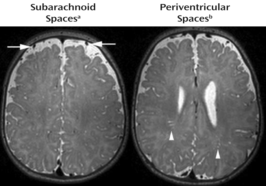
The types of incidental findings observed included enlargement of perivascular spaces (low risk: N=4, 11%; high risk: N=5, 5%), enlargement of subarachnoid spaces (low risk: N=17, 47%; high risk: N=49, 50%), Chiari I malformation <5 mm (low risk: N=1, 3%), pineal cyst (low risk: N=1, 3%), cavum septum pellucidum (low risk: N=1, 3%), plagiocephaly (low risk: N=1, 3%), cavum velum interpositum (high risk: N=2, 2%), and pars intermedia cyst of the pituitary (high risk: N=1, 1%). There were six infants in the high-risk group (6%) and three in the low-risk group (8%) who had more than one atypicality observed (they are included in the preceding list). Ratings (range=0–4) were given to the subarachnoid spaces and perivascular spaces of each infant, to indicate the degree or severity of enlargement, so that the groups could be compared. Examples of mild enlargement are shown in Figure 2. No significant group difference was found in the proportion of infants in each severity category for enlargement of either the subarachnoid spaces (χ2=3.1, df=3, p=0.37) or the perivascular spaces (χ2=2.5, df=2, p=0.29).
FIGURE 2. Grading of Subarachnoid Spaces and Periventricular Spaces
a This image demonstrates mild enlargement of the subarachnoid spaces. Minimal enlargement (rating=1) is defined as one vessel width of subarachnoid space and brain that does not touch the inner table of the calvaria on multiple consecutive sections. Mild enlargement (rating=2) has CSF space over the convexities more than one vessel thick. Moderate enlargement (rating=3) of the subarachnoid spaces extends to significantly enlarge the CSF space between the hemispheres along the falx cerebri. Marked enlargement (rating=4) would be profound pathological enlargement of the CSF spaces over and between the hemispheres.
b This image demonstrates mild enlargement of the perivascular spaces. Minimal enlargement (rating=1) is defined as fewer than five small, linear T2 hyperintensities or one or two larger foci. Mild enlargement (rating=2) is 5–10 small, linear or 3–5 larger, rounded hyperintensities. Moderate enlargement (rating=3) is more than 10 linear or 5–10 larger, rounded hyperintensities. Marked enlargement (rating=4) involves pathological number, size, and location (e.g., corpus callosum) of the hyperintensities associated with low white matter volume.

Head Circumference

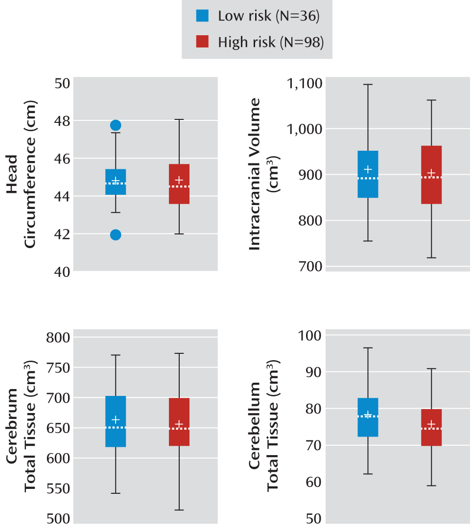
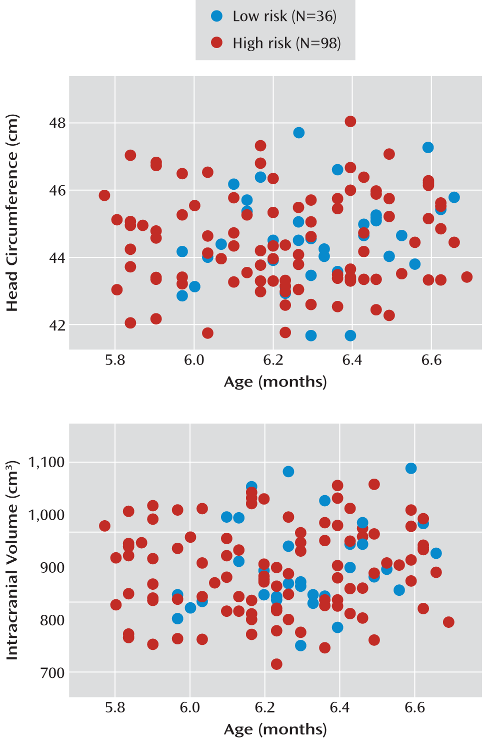
After adjustment for age and site, the high- and low-risk groups did not differ in head circumference (F=0.37, df=1, 125, p=0.55). The mean head circumference for the high-risk group was 44.5 cm (SD=1.4), and for the low-risk group it was 44.6 cm (SD=1.3). There was no group-by-sex interaction for head circumference (F=0.84, df=1, 125, p=0.36). The overall group values are shown in Figure 3, and individual values appear in Figure 4.
FIGURE 3. Box Plots of Head Circumference and Total Brain Volumes for 6-Month-Old Infants at Low and High Familial Risk for Autisma
a The horizontal line in each box represents the median for the group; the cross indicates the mean.
FIGURE 4. Scatterplots of Head Circumference and Intracranial Volume for 6-Month-Old Infants at Low and High Familial Risk for Autism

Total Brain Volume, Total Tissue, CSF

After adjustment for age, sex, site, and cognitive development, there were no significant differences between the high- and low-risk groups in total brain volume (intracranial volume), total tissue, or total CSF (Table 2). Box plots displaying group values for head circumference, intracranial volume, and total tissue for the cerebrum and cerebellum are displayed in Figure 3. Individual values for head circumference and intracranial volume are shown in Figure 4.
TABLE 2. Adjusted Brain Volumes of 6-Month-Old Infants at Low or High Familial Risk for Autism
 Adjusteda Volume (cm3)
 Low Risk (N=36)High Risk (N=98)Group Difference
Measurement RegionLeast Squares MeanSELeast Squares MeanSEF (df=1, 125)p
Intracranial volume897.212.1889.57.90.280.60
  Total tissue (gray plus white matter)737.99.2730.96.00.390.53
  Total CSF159.35.1158.63.30.020.90
Cerebrum 
  Total (tissue plus CSF)798.611.4792.67.40.190.66
  Left (gray plus white matter)326.14.3323.12.80.360.55
  Right (gray plus white matter)327.44.2325.42.80.160.69
  Total CSF145.05.1144.13.30.020.89
Cerebellum 
  Total (tissue plus CSF)89.01.287.30.81.260.26
  Left (gray plus white matter)38.50.637.60.41.650.20
  Right (gray plus white matter)38.20.637.30.41.800.18
  Total CSF12.30.412.40.30.050.82
Lateral ventricles 
  Left5.60.45.70.20.040.84
  Right5.10.34.90.20.220.64
a
Adjusted for age, sex, Mullen Scales of Early Leaning composite score, and site.

Cerebral Cortex and Cerebellum

Total tissue volume, total CSF volume, and volumes within each hemisphere of the cerebrum and cerebellum were examined. The groups did not differ significantly in any of the measures, and there were no significant group-by-sex effects. Mean group volumes for these measures are displayed in Table 2.

Lateral Ventricles

The volumes of the lateral ventricles in each hemisphere for both groups are also presented in Table 2. There was no group difference for either the left or right ventricular volume and no significant interaction of group and sex (left: p=0.80, right: p=0.46).

Discussion

First-degree relatives of autistic individuals are reported to have high rates of macrocephaly (6). In addition, it is anticipated that approximately 20% of high-risk infants in this group will meet criteria for an ASD by 36 months of age (17). To our knowledge, this is the first report of brain volume in 6-month-old infants at high familial risk for autism from an ongoing longitudinal neuroimaging study. We found no differences between these infants and low-risk peers in overall brain tissue volumes, head circumference, or ventricular volumes.
This investigation does not address the question of whether early brain volume differences can be observed in high-risk children who are later diagnosed with an ASD, which will be examined in the longitudinal data set from this study. Rather, our intent was to study whether differences in brain volume could be detected in children at high genetic risk for autism, an approach that has been used previously to examine familial rates of macrocephaly in ASDs (7). There is, therefore, a chance that we will be able to detect significant differences at 6 months once we are able to examine trajectories of growth in the various subgroups at 24 months of age.
The absence of brain volume differences in 6-month-olds at high familial risk for autism in this large cohort is consistent with our hypothesis that brain enlargement in autism and possibly in those with high genetic liability is a later-occurring postnatal process. Disruptions of cortical maturation related to impaired experience-dependent synaptic development have been observed in mouse models of both Angelman syndrome (27) and fragile X syndrome (28), neurogenetic developmental disorders with behavioral features that overlap with those of ASDs. There has also been a report of mutations associated with the defective expression of activity-driven genes in individuals with ASDs (29), although it is unclear whether this mechanism accounts for more than a very small proportion of genetic variance underlying ASDs. Other hypotheses about brain overgrowth, based on the observation that it is associated with increases in cortical surface area (10), suggest that brain overgrowth in the latter part of the first year may be the result of an increase in neuronal precursors, perhaps related to aberrant molecular regulatory processes (30). The findings from the current study suggest that early postnatal events may underlie later-occurring brain overgrowth and raise the possibility that there is a window of opportunity in which early postnatal intervention, during a period of tremendous brain plasticity, may have an important impact on later emergence of autistic behavior.
Studies examining early behavioral markers of autism have shown that infants who later go on to receive a diagnosis of an ASD do not exhibit overt autistic behaviors at 6 months of age (15, 17). However, by 12 months of age, a time when we anticipate brain differences may begin to emerge, behavioral deficits can be detected in the infants who are later diagnosed with autism (15, 17, 31). Our group is currently examining the behavioral characteristics of this study group. In the current study, the high-risk group likely includes children who will later receive a diagnosis of autism. With the later addition of diagnostic outcome data for this group, it will be possible to examine whether brain volumes for children in the high-risk group who eventually manifest ASDs differ from volumes for high-risk children who do not go on to develop ASDs, to specifically test our hypothesis of brain overgrowth in the latter part of the first year of life or early in the second year of life.
We also did not detect any clinically significant radiologic findings specific to the high-risk group. The types and rates of incidental findings observed were similar in the two groups, as were the degrees of enlargement for the subarachnoid spaces and the periventricular spaces. Most investigations of clinical neuroradiologic abnormalities in individuals with autism have not detected a significant type or pattern of findings associated with the disorder. Scattered reports of individuals with neuronal migration abnormalities (32), subcortical abnormalities in the corpus callosum (33), and high rates of dilated Virchow-Robin spaces (34) exist. However, interpretation of this research has been complicated by inconsistent findings across studies and by numerous methodological shortcomings, such as the etiologic heterogeneity known to be inherent in autism, small study groups, and variations in subject age. One large-scale retrospective study (35) showed higher rates of clinically reported abnormalities in children with autism than in patients without developmental or neurological disorders. Forty-eight percent of the children with ASDs were rated as having abnormalities. The primary abnormalities included white matter signal intensity abnormalities, dilated Virchow-Robin spaces, and temporal lobe abnormalities (e.g., subcortical hyperintensities in the temporal poles). White matter abnormalities (e.g., punctate or posterior T2 hyperintensities) were present in younger children (e.g., 5-year-olds versus 7-year-olds), while the temporal lobe abnormalities showed no developmental trend. It should be noted that the current study differs from the investigation by Boddaert et al. (35); whereas they studied children 2 to 16 years of age with ASDs, we examined infants at high risk for autism. The developmental nature of the incidental findings we observed and their association with ASDs therefore remain questions worthy of future investigation.
A limitation of this study, due in part to our focus on 6-month-olds, is that because of the difficulty associated with segmenting gray and white matter at this early age in development, we were unable to define regionally specific brain volumes. White matter structure and myelination may best be captured with other methods, such as diffusion tensor imaging, and we recently reported significant differences in white matter development between high-risk infants who did and did not later receive a diagnosis of an ASD (36). More dynamic methods, such as resting-state functional MRI, may also detect early functional changes before the occurrence of more grossly observable changes in volume. These methods may be more sensitive to early brain differences and may be able to detect abnormalities in structural or functional development that are not possible to identify by means of standard tissue classification methods. Last, our current study was cross-sectional, and at this point the study group is characterized in terms of high-risk and low-risk status. Only a portion of our current high-risk group may later be diagnosed with an ASD. We will include diagnostic outcome data and longitudinal imaging data from later time points (e.g., 12, 24, and/or 36 months of age) to examine the groups for more specific information on the behavioral and biological mechanisms underlying the development of ASDs.

Acknowledgments

The Infant Brain Imaging Study (IBIS) Network is an NIH-funded Autism Center of Excellence and consists of a consortium of seven universities in the United States and Canada. Clinical sites are located at the University of North Carolina (J. Piven, IBIS Network primary investigator; H.C. Hazlett, C. Chappell); the University of Washington (S.R. Dager, A.M. Estes, D. Shaw); Washington University (K.N. Botteron, R.C. McKinstry, J. Constantino, J. Pruett); Children's Hospital of Philadelphia (R.T. Schultz, S.J. Paterson); and the University of Alberta (L. Zwaigenbaum). The data coordinating center is at the Montreal Neurological Institute (A.C. Evans, D.L. Collins, G.B. Pike, V. Fonov, P. Kostopoulos, S. Das). The image processing core is at the University of Utah (G. Gerig) and the University of North Carolina (M. Styner). The statistical analysis core is at the University of North Carolina (H. Gu). The genetics analysis core is at the University of North Carolina (P. Sullivan, F. Wright).
The authors thank Sun Hyung Kim, Rachel Gimpel Smith, Michael Graves, and Ryan Scotton for assistance in processing these data; Penelope Kostopoulus and Samir Das for assistance with database management; and the families who participated in this study.
Footnote

Received Sept. 23, 2011; revisions received Jan. 23 and Feb. 28, 2012; accepted Feb. 29, 2012.

References

1.
Piven J, Nehme E, Simon J, Barta P, Pearlson G, Folstein SE: Magnetic resonance imaging in autism: measurement of the cerebellum, pons, and fourth ventricle. Biol Psychiatry 1992; 31:491–504
2.
Piven J, Arndt S, Bailey J, Andreasen N: Regional brain enlargement in autism: a magnetic resonance imaging study. J Am Acad Child Adolesc Psychiatry 1996; 35:530–536
3.
Courchesne E, Karns CM, Davis HR, Ziccardi R, Carper RA, Tigue ZD, Chisum HJ, Moses P, Pierce K, Lord C, Lincoln AJ, Pizzo S, Schreibman L, Haas RH, Akshoomoff NA, Courchesne RY: Unusual brain growth patterns in early life in patients with autistic disorder: an MRI study. Neurology 2001; 57:245–254
4.
Sparks BF, Friedman SD, Shaw DW, Aylward EH, Echelard D, Artru AA, Maravilla KR, Giedd JN, Munson J, Dawson G, Dager SR: Brain structural abnormalities in young children with autism spectrum disorder. Neurology 2002; 59:184–192
5.
Bailey A, Le Couteur A, Gottesman I, Bolton P, Simonoff E, Yuzda E, Rutter M: Autism as a strongly genetic disorder: evidence from a British twin study. Psychol Med 1995; 25:63–77
6.
Lainhart JE, Piven J, Wzorek M, Landa R, Santegelo SL, Coon H, Folstein SE: Macrocephaly in children and adults with autism. J Am Acad Child Adolesc Psychiatry 1997; 36:282–289
7.
Lainhart JE, Bigler ED, Bocian M, Coon H, Dinh E, Dawson G, Deutsch CK, Dunn M, Estes A, Tager-Flusberg H, Folstein S, Hepburn S, Hyman S, McMahon W, Minshew N, Munson J, Osann K, Ozonoff S, Rodier P, Rogers S, Sigman M, Spence MA, Stodgell CJ, Volkmar F: Head circumference and height in autism: a study by the Collaborative Program of Excellence in Autism. Am J Med Genet A 2006; 140:2257–2274
8.
Bailey A, Luthert P, Bolton P, LeCouteur A, Rutter M: Autism and megalencephaly. Lancet 1993; 34:1225–1226
9.
Hazlett HC, Poe M, Gerig G, Smith RG, Provenzale J, Ross A, Gilmore J, Piven J: Magnetic resonance imaging and head circumference study of brain size in autism. Arch Gen Psychiatry 2005; 62:1366–1376
10.
Hazlett HC, Poe MD, Gerig G, Styner M, Chappell C, Smith RG, Vachet C, Piven J: Early brain overgrowth in autism associated with an increase in cortical surface area before age 2 years. Arch Gen Psychiatry 2011; 68:467–476
11.
Schumann CM, Bloss CS, Barnes CC, Wideman GM, Carper RA, Akschoomoff N, Pierce K, Hagler D, Schork N, Lord C, Courchesne E: Longitudinal magnetic resonance imaging study of cortical development through early childhood in autism. J Neurosci 2010; 30:4419–4427
12.
Mosconi MW, Hazlett HC, Poe MD, Gerig G, Smith RG, Piven J: Longitudinal study of amygdala volume and joint attention in 2- to 4- year old children with autism. Arch Gen Psychiatry 2009; 66:509–516
13.
Courchesne E, Campbell K, Solso S: Brain growth across the life span in autism: age-specific changes in anatomical pathology. Brain Res 2011; 22:138–145
14.
Constantino JN, Majmudar P, Bottini A, Arvin M, Virkud Y, Simons P, Spitznagel E: Infant head growth in male siblings of children with and without autism spectrum disorders. J Neurodev Disord 2010; 2:39–46
15.
Zwaigenbaum L, Bryson S, Rogers T, Roberts W, Brain J, Szatmari P: Behavioral manifestations of autism in the first year of life. Int J Dev Neurosci 2005; 23:143–152
16.
Knickmeyer RC, Gouttard S, Kang C, Evans D, Wilber K, Smith JK, Hamer RM, Lin W, Gerig G, Gilmore JH: A structural MRI study of human brain development from birth to 2 years. J Neurosci 2008; 28:12176–12182
17.
Ozonoff S, Young GS, Carter A, Messinger D, Yirmiya N, Zwaigenbaum L, Bryson S, Carver LJ, Constantino J, Dobkins K, Hutman T, Iverson JM, Landa R, Rogers SJ, Sigman M, Stone WL: Recurrence risk for autism spectrum disorders: a Baby Siblings Research Consortium study. Pediatrics 2011; 128:e488–e495
18.
Lord C, Rutter M, Le Couteur A: Autism Diagnostic Interview-Revised: a revised version of a diagnostic interview for caregivers of individuals with possible pervasive developmental disorders. J Autism Dev Disord 1994; 24:659–685
19.
Mullen EM: Mullen Scales of Early Learning: AGS Edition. Circle Pines, Minn, AGS Publishing, 1995
20.
Sparrow SS, Balla D, Cicchetti DV: Vineland Adaptive Behavior Scales, 2nd ed. Shoreview, Minn, American Guidance Service, 2005
21.
Bryson SE, Zwaigenbaum L, McDermott C, Rombough V, Brian J: The Autism Observation Scale for Infants: scale development and reliability data. J Autism Dev Disord 2008; 38:731–738
22.
Fonov V, Janke A, Caramanos Z, Arnold D, Narayanan S, Pike GB, Collins DL: Improved precision in the measurement of longitudinal global and regional volumetric changes via a novel MRI gradient distortion characterization and correction technique, in Medical Imaging and Augmented Reality: Lecture Notes in Computer Science, vol 6326. Edited by, Liao H, Edwards PJ, Pan X, Fan Y, Yang G-Z. New York, Springer, 2010, pp 324–333
23.
Gouttard S, Styner M, Prastawa M, Gerig G: Assessment of reliability of multi-site neuroimaging via traveling phantom study. Med Image Comput Assist Interv 2008; 11(pt 2):263–270
24.
Van Leemput K, Maes F, Vandermeulen D, Suetens P: Automated model-based tissue classification of MR images of the brain. IEEE Trans Med Imaging 1999; 18:897–908
25.
Gouttard S, Styner M, Joshi S, Smith RG, Hazlett HC, Gerig G: Subcortical structure segmentation using probabilistic atlas priors, in SPIE Medical Imaging 2007: Image Processing: Proceedings, vol 6512. Edited by, Pluim JPW, Reinhardt JM. Bellingham, Wash, SPIE, 2007, p 88
26.
Yushkevich PA, Piven J, Hazlett HC, Smith RG, Ho S, Gee JC, Gerig G: User-guided 3D active contour segmentation of anatomical structures: significantly improved efficacy and reliability. Neuroimage 2006; 31:1116–1128
27.
Yashiro K, Riday TT, Condon KH, Roberts AC, Bernardo DR, Prakash R, Weinberg RJ, Ehlers MD, Philpot BD: Ube3a is required for experience-dependent maturation of the neocortex. Nat Neurosci 2009; 12:777–783
28.
Dolen G, Osterweil E, Rao BS, Smith GB, Auerbach BD, Chattarji S, Bear MF: Correction of fragile X syndrome in mice. Neuron 2007; 56:955–962
29.
Morrow EM, Yoo SY, Flavell SW, Kim TK, Lin Y, Hill RD, Mukaddes NM, Balkhy S, Glascon G, Hashmi A, Al-Saad S, Ware J, Joseph RM, Greenblatt R, Gleason D, Ertelt JA, Apse KA, Bodell A, Partlow JN, Barry B, Yao H, Markianos K, Ferland RJ, Greenberg ME, Walsh CA: Identifying autism loci and genes by tracing recent shared ancestry. Science 2008; 321:218–223
30.
Chenn A, Walsh CA: Regulation of cerebral cortical size by control of cell cycle exit in neural precursors. Science 2002; 297:365–369
31.
Wetherby AM, Brosnan-Maddox S, Peace V, Newton L: Validation of the Infant-Toddler Checklist as a broadband screener for autism spectrum disorders from 9 to 24 months of age. Autism 2008; 12:487–511
32.
Zeegers M, Van Der Grond J, Durston S, Nievelstein RJ, Witkamp T, Van Daalen E, Buitelaar J, Engeland HV: Radiological findings in autistic and developmentally delayed children. Brain Dev 2006; 28:495–499
33.
Steiner CE, Guerreiro MM, Marques-de-Faria AP: Acrocallosal syndrome and autism. J Autism Dev Disord 2004; 34:723–726
34.
Taber KH, Shaw JB, Loveland KA, Pearson DA, Lane DM, Hayman LA: Accentuated Virchow-Robin spaces in the centrum semiovale in children with autistic disorder. J Comput Assist Tomogr 2004; 28:263–268
35.
Boddaert N, Zilbovicius M, Philipe A, Robel L, Bourgeois M, Barthelemy C, Seidenwurm D, Meresse I, Laurieer L, Desguerre I, Bahi-Buisson N, Brunelle F, Munnich A, Samson Y, Mouren M-C, Chabane N: MRI findings in 77 children with non-syndromic autistic disorder. PLoS One 2009; 4(2):e4415
36.
Wolff JJ, Gu H, Gerig G, Elison JT, Styner M, Gouttard S, Botteron KN, Dager SR, Dawson G, Estes AM, Evans A, Hazlett HC, Kostopoulos P, McKinstry RC, Paterson SJ, Schultz RT, Zwaigenbaum L, Piven J: Differences in white matter fiber tract development present from 6 to 24 months in infants with autism. Am J Psychiatry 2012; 169:589–600

Information & Authors

Information

Published In

Go to American Journal of Psychiatry
American Journal of Psychiatry
Pages: 601 - 608
PubMed: 22684595

History

Received: 23 September 2011
Revision received: 23 January 2012
Revision received: 28 February 2012
Accepted: 29 February 2012
Published online: 1 June 2012
Published in print: June 2012

Authors

Details

Heather Cody Hazlett, Ph.D.
From the Carolina Institute for Developmental Disabilities and the Department of Psychiatry, University of North Carolina, Chapel Hill; the Department of Psychiatry and the Department of Radiology, Mallinckrodt Institute of Radiology, Washington University School of Medicine, St. Louis; the Department of Radiology and the Department of Speech and Hearing Sciences, University of Washington, Seattle; the Scientific Computing and Imaging Institute, University of Utah, Salt Lake City; the Center for Autism Research, Children's Hospital of Philadelphia, University of Pennsylvania; and the Montreal Neurological Institute, McGill University, Montreal.
Hongbin Gu, Ph.D.
From the Carolina Institute for Developmental Disabilities and the Department of Psychiatry, University of North Carolina, Chapel Hill; the Department of Psychiatry and the Department of Radiology, Mallinckrodt Institute of Radiology, Washington University School of Medicine, St. Louis; the Department of Radiology and the Department of Speech and Hearing Sciences, University of Washington, Seattle; the Scientific Computing and Imaging Institute, University of Utah, Salt Lake City; the Center for Autism Research, Children's Hospital of Philadelphia, University of Pennsylvania; and the Montreal Neurological Institute, McGill University, Montreal.
Robert C. McKinstry, M.D., Ph.D.
From the Carolina Institute for Developmental Disabilities and the Department of Psychiatry, University of North Carolina, Chapel Hill; the Department of Psychiatry and the Department of Radiology, Mallinckrodt Institute of Radiology, Washington University School of Medicine, St. Louis; the Department of Radiology and the Department of Speech and Hearing Sciences, University of Washington, Seattle; the Scientific Computing and Imaging Institute, University of Utah, Salt Lake City; the Center for Autism Research, Children's Hospital of Philadelphia, University of Pennsylvania; and the Montreal Neurological Institute, McGill University, Montreal.
Dennis W.W. Shaw, M.D.
From the Carolina Institute for Developmental Disabilities and the Department of Psychiatry, University of North Carolina, Chapel Hill; the Department of Psychiatry and the Department of Radiology, Mallinckrodt Institute of Radiology, Washington University School of Medicine, St. Louis; the Department of Radiology and the Department of Speech and Hearing Sciences, University of Washington, Seattle; the Scientific Computing and Imaging Institute, University of Utah, Salt Lake City; the Center for Autism Research, Children's Hospital of Philadelphia, University of Pennsylvania; and the Montreal Neurological Institute, McGill University, Montreal.
Kelly N. Botteron, M.D.
From the Carolina Institute for Developmental Disabilities and the Department of Psychiatry, University of North Carolina, Chapel Hill; the Department of Psychiatry and the Department of Radiology, Mallinckrodt Institute of Radiology, Washington University School of Medicine, St. Louis; the Department of Radiology and the Department of Speech and Hearing Sciences, University of Washington, Seattle; the Scientific Computing and Imaging Institute, University of Utah, Salt Lake City; the Center for Autism Research, Children's Hospital of Philadelphia, University of Pennsylvania; and the Montreal Neurological Institute, McGill University, Montreal.
Stephen R. Dager, M.D.
From the Carolina Institute for Developmental Disabilities and the Department of Psychiatry, University of North Carolina, Chapel Hill; the Department of Psychiatry and the Department of Radiology, Mallinckrodt Institute of Radiology, Washington University School of Medicine, St. Louis; the Department of Radiology and the Department of Speech and Hearing Sciences, University of Washington, Seattle; the Scientific Computing and Imaging Institute, University of Utah, Salt Lake City; the Center for Autism Research, Children's Hospital of Philadelphia, University of Pennsylvania; and the Montreal Neurological Institute, McGill University, Montreal.
Martin Styner, Ph.D.
From the Carolina Institute for Developmental Disabilities and the Department of Psychiatry, University of North Carolina, Chapel Hill; the Department of Psychiatry and the Department of Radiology, Mallinckrodt Institute of Radiology, Washington University School of Medicine, St. Louis; the Department of Radiology and the Department of Speech and Hearing Sciences, University of Washington, Seattle; the Scientific Computing and Imaging Institute, University of Utah, Salt Lake City; the Center for Autism Research, Children's Hospital of Philadelphia, University of Pennsylvania; and the Montreal Neurological Institute, McGill University, Montreal.
Clement Vachet, M.S.
From the Carolina Institute for Developmental Disabilities and the Department of Psychiatry, University of North Carolina, Chapel Hill; the Department of Psychiatry and the Department of Radiology, Mallinckrodt Institute of Radiology, Washington University School of Medicine, St. Louis; the Department of Radiology and the Department of Speech and Hearing Sciences, University of Washington, Seattle; the Scientific Computing and Imaging Institute, University of Utah, Salt Lake City; the Center for Autism Research, Children's Hospital of Philadelphia, University of Pennsylvania; and the Montreal Neurological Institute, McGill University, Montreal.
Guido Gerig, Ph.D.
From the Carolina Institute for Developmental Disabilities and the Department of Psychiatry, University of North Carolina, Chapel Hill; the Department of Psychiatry and the Department of Radiology, Mallinckrodt Institute of Radiology, Washington University School of Medicine, St. Louis; the Department of Radiology and the Department of Speech and Hearing Sciences, University of Washington, Seattle; the Scientific Computing and Imaging Institute, University of Utah, Salt Lake City; the Center for Autism Research, Children's Hospital of Philadelphia, University of Pennsylvania; and the Montreal Neurological Institute, McGill University, Montreal.
Sarah J. Paterson, Ph.D.
From the Carolina Institute for Developmental Disabilities and the Department of Psychiatry, University of North Carolina, Chapel Hill; the Department of Psychiatry and the Department of Radiology, Mallinckrodt Institute of Radiology, Washington University School of Medicine, St. Louis; the Department of Radiology and the Department of Speech and Hearing Sciences, University of Washington, Seattle; the Scientific Computing and Imaging Institute, University of Utah, Salt Lake City; the Center for Autism Research, Children's Hospital of Philadelphia, University of Pennsylvania; and the Montreal Neurological Institute, McGill University, Montreal.
Robert T. Schultz, Ph.D.
From the Carolina Institute for Developmental Disabilities and the Department of Psychiatry, University of North Carolina, Chapel Hill; the Department of Psychiatry and the Department of Radiology, Mallinckrodt Institute of Radiology, Washington University School of Medicine, St. Louis; the Department of Radiology and the Department of Speech and Hearing Sciences, University of Washington, Seattle; the Scientific Computing and Imaging Institute, University of Utah, Salt Lake City; the Center for Autism Research, Children's Hospital of Philadelphia, University of Pennsylvania; and the Montreal Neurological Institute, McGill University, Montreal.
Annette M. Estes, Ph.D.
From the Carolina Institute for Developmental Disabilities and the Department of Psychiatry, University of North Carolina, Chapel Hill; the Department of Psychiatry and the Department of Radiology, Mallinckrodt Institute of Radiology, Washington University School of Medicine, St. Louis; the Department of Radiology and the Department of Speech and Hearing Sciences, University of Washington, Seattle; the Scientific Computing and Imaging Institute, University of Utah, Salt Lake City; the Center for Autism Research, Children's Hospital of Philadelphia, University of Pennsylvania; and the Montreal Neurological Institute, McGill University, Montreal.
Alan C. Evans, Ph.D.
From the Carolina Institute for Developmental Disabilities and the Department of Psychiatry, University of North Carolina, Chapel Hill; the Department of Psychiatry and the Department of Radiology, Mallinckrodt Institute of Radiology, Washington University School of Medicine, St. Louis; the Department of Radiology and the Department of Speech and Hearing Sciences, University of Washington, Seattle; the Scientific Computing and Imaging Institute, University of Utah, Salt Lake City; the Center for Autism Research, Children's Hospital of Philadelphia, University of Pennsylvania; and the Montreal Neurological Institute, McGill University, Montreal.
Joseph Piven, M.D.
From the Carolina Institute for Developmental Disabilities and the Department of Psychiatry, University of North Carolina, Chapel Hill; the Department of Psychiatry and the Department of Radiology, Mallinckrodt Institute of Radiology, Washington University School of Medicine, St. Louis; the Department of Radiology and the Department of Speech and Hearing Sciences, University of Washington, Seattle; the Scientific Computing and Imaging Institute, University of Utah, Salt Lake City; the Center for Autism Research, Children's Hospital of Philadelphia, University of Pennsylvania; and the Montreal Neurological Institute, McGill University, Montreal.
the IBIS Network
From the Carolina Institute for Developmental Disabilities and the Department of Psychiatry, University of North Carolina, Chapel Hill; the Department of Psychiatry and the Department of Radiology, Mallinckrodt Institute of Radiology, Washington University School of Medicine, St. Louis; the Department of Radiology and the Department of Speech and Hearing Sciences, University of Washington, Seattle; the Scientific Computing and Imaging Institute, University of Utah, Salt Lake City; the Center for Autism Research, Children's Hospital of Philadelphia, University of Pennsylvania; and the Montreal Neurological Institute, McGill University, Montreal.

Notes

Address correspondence to Dr. Hazlett ([email protected]).

Funding Information

Dr. Evans is cofounder of and holds equity in Biospective, Inc., a company that performs image analysis for pharmaceutical companies; he has also received consulting fees from Johnson & Johnson and Pfizer within the last 36 months. Dr. Schultz reports receiving CME funding from Shire Pharmaceuticals, advisory panel funding from Seaside Therapeutics, and advisory panel funding from Roche. All other authors report no financial relationships with commercial interests.Supported by Autism Center of Excellence grant HD-055741 from NIMH and the National Institute of Child Health and Development to Dr. Piven, by Autism Speaks, and by the Simons Foundation. Further support was provided by the National Alliance for Medical Image Computing, funded through grant U54 EB-005149 from the National Institute of Biomedical Imaging and Bioengineering.

Metrics & Citations

Metrics

Citations

Export Citations

If you have the appropriate software installed, you can download article citation data to the citation manager of your choice. Simply select your manager software from the list below and click Download.

For more information or tips please see 'Downloading to a citation manager' in the Help menu.

Format
Citation style
Style
Copy to clipboard

View Options

View options

PDF/EPUB

View PDF/EPUB

Login options

Already a subscriber? Access your subscription through your login credentials or your institution for full access to this article.

Personal login Institutional Login Open Athens login
Purchase Options

Purchase this article to access the full text.

PPV Articles - American Journal of Psychiatry

PPV Articles - American Journal of Psychiatry

Not a subscriber?

Subscribe Now / Learn More

PsychiatryOnline subscription options offer access to the DSM-5-TR® library, books, journals, CME, and patient resources. This all-in-one virtual library provides psychiatrists and mental health professionals with key resources for diagnosis, treatment, research, and professional development.

Need more help? PsychiatryOnline Customer Service may be reached by emailing [email protected] or by calling 800-368-5777 (in the U.S.) or 703-907-7322 (outside the U.S.).

Media

Figures

Other

Tables

Share

Share

Share article link

Share