Posttraumatic stress disorder (PTSD) may result from various kinds of trauma, such as assault, rape, childhood maltreatment, terrorist attack, and combat-related stress. Individuals with PTSD suffer from distressing symptoms after direct experience or witnessing of traumatizing events (
1).
Evidence from MRI studies suggests that PTSD is associated with abnormalities in brain structure. Meta-analyses of region-of-interest studies have primarily highlighted reductions in the volume of the hippocampus. These studies, however, examined small numbers of brain structures, and the study with the most brain regions analyzed (nine) was conducted more than 10 years ago (
2). Voxel-based morphometry (VBM), a complementary approach to analyzing structural MRI data, surveys the entire brain to examine regional changes in brain volume. A small number of VBM meta-analyses of PTSD have been conducted, and they all used coordinate values, which are effective in summarizing consistently reported peaks, but, critically, they ignore nonsignificant data. Newer methods have been developed that allow the inclusion of three-dimensional statistical maps, obtained from the study authors, which include nonsignificant data and increase accuracy when added to coordinate data. This approach has not yet been applied in PTSD, however. Furthermore, region-of-interest and VBM meta-analyses analyze the MRI data in different ways, and these methods have not yet been formally compared in a single study.
PTSD is highly comorbid with depression, and epidemiological studies have reported that half or more individuals with PTSD have experienced a major depressive episode (
3,
4). If brain abnormalities are identified in PTSD, they could conceivably be associated with the comorbid major depressive disorder rather than the diagnosis of PTSD. Therefore, it is informative to compare brain changes in PTSD with those in major depressive disorder (hereafter referred to as depression) to try to determine whether the abnormalities may be related to a comorbid diagnosis of depression or are unique to PTSD.
In the present study, we first constructed a comprehensive online database of 113 MRI studies comparing patients with PTSD to control subjects. We conducted a region-of-interest meta-analysis examining the volumes of 56 brain regions. Within this meta-analysis, we also conducted three subanalyses that included control groups with and without trauma to tease apart the effects of experiencing a traumatic event and the diagnosis of PTSD. Comparing patients with PTSD to control subjects who have experienced trauma may reveal abnormalities associated with the diagnosis of PTSD, and comparing control subjects who have experienced trauma to control subjects with no trauma may highlight brain changes associated with trauma per se. To compare the PTSD region-of-interest meta-analysis to data from subjects with major depressive disorder, we used data from our previously published meta-analysis in depression (
5) and made statistical comparisons between the disorders to determine which abnormalities are specific to PTSD. Finally, in a VBM meta-analysis, we examined regional gray matter volume from published coordinates; additional t-maps were provided by study authors to increase accuracy and to compare with the main region-of-interest meta-analysis.
Method
The study was divided into four parts: the construction of an online database of 113 studies that investigated structural brain abnormalities in PTSD, the main region-of-interest meta-analysis, a stratified meta-analysis comparing PTSD with depression, and a meta-analysis of VBM studies using seed-based d mapping. We followed the PRISMA checklist in reporting this meta-analysis (
6).
Database of Imaging Studies in PTSD
Published studies that measured brain structure using MRI in patients with PTSD and a control group were included in the database. A MEDLINE search of studies published from 1992 through June 2016 was performed, combining Medical Subject Heading (MeSH) terms and free text searches. A total of 800 publications were identified, of which 113 met inclusion criteria and were included in the database. Further details, including search terms and a study inclusion flow chart (Figure S1), are provided in the online supplement.
PTSD Region-of-Interest Meta-Analysis
From all 89 region-of-interest studies in the database, more than 100 different brain regions were reported. Of these, we selected 56 regions that were reported by three or more studies to ensure that each meta-analysis was sufficiently powered. Because the inclusion of pediatric data may increase heterogeneity, these have been excluded from the meta-analysis; these data were included in the sensitivity analysis, however. Publications were excluded if they included sample overlap with other publications. The final number of studies included in the meta-analysis was 66.
Control groups.
Of the 66 studies that were included in the region-of-interest meta-analysis, 28 selected nontraumatized control participants, 22 selected traumatized control participants, and 16 included both of these control groups. We used a strategy to balance the advantages of pooling the maximum number of studies with the need to examine potential differences in selecting a control group. In the main meta-analysis, we combined all 66 studies using the available case-control comparison; where both types of control groups were available, the nontraumatized control group was selected, as it was the most commonly available comparison group. However, in order to examine the effects of different control groups, three additional pairwise sub-meta-analyses were conducted: PTSD compared with traumatized control subjects, PTSD compared with nontraumatized control subjects, and traumatized compared with nontraumatized control subjects.
Combining study estimates.
For continuous measures, we used Hedges’ g, which is the Cohen’s effect size with a correction for bias from small samples (
7). Outcome measures were combined using a random-effects inverse-weighted variance model (
8). The Cochran Q statistic was calculated to examine the heterogeneity between studies (
9). The I
2 statistic was also calculated, which is equal to the percentage of total variation between studies due to heterogeneity (
10). The effect of small-study bias (which may include publication bias) was investigated for regions where at least five studies were included to ensure that the test was sufficiently powered. Small-study bias was assessed using Egger’s regression test. Brain regions with evidence of small-study bias were adjusted using a trim-and-fill method (
11). For brain regions with a significant pooled effect size, we examined how robust the result was by excluding one individual effect size at a time, a leave-one-out approach.
Effect of clinical variables on hippocampal volume.
The number of brain regions and clinical variables included in the database allows a potentially high number of correlations to be examined, which may lead to type I errors. Thus, the analysis was limited to the effect of clinical variables on total hippocampal volume. We selected this region because of the robust evidence of volumetric reduction in PTSD and because many studies have measured this structure, ensuring adequate statistical power. A random-effects meta-regression was implemented (METAREG command in Stata, version 9.2 [
12]) to examine age at illness onset, time since trauma, Clinician-Administered PTSD Scale (CAPS) score, percentage of patients using antidepressants, percentage of patients who are drug free, and patient age.
Sensitivity analysis.
To test how robust the results were to variations in the meta-analytic method, the effects of the following were examined: 1) setting the correlation coefficient between the left and right regional volumes as 0.1, 0.5, and 1 (see the online supplement for further details); 2) excluding studies that reported volumes divided by intracranial volume; 3) excluding cortical thickness measures; and 4) including 11 pediatric studies that were excluded in the main meta-analysis to reduce heterogeneity.
Stratified Region-of-Interest Meta-Analysis Comparing PTSD With Depression
Two meta-analytic approaches may be taken to examine differences between PTSD and depression: 1) meta-analysis of studies directly comparing the same brain structure in patients with PTSD and patients with depression and 2) indirect analysis comparing the pooled effect size from studies comparing patients with PTSD versus all control subjects with that from studies comparing patients with depression versus control subjects. We adopted the second approach, which has the advantage of including more studies and brain structures, because there are very few direct comparisons in the literature. First, from our previously published meta-analysis in major depressive disorder (
5), we excluded pediatric samples and a study that included two patients with comorbid PTSD. To compare the results, we combined the effect sizes from PTSD patients versus all control subjects and depression patients versus control subjects and performed a stratified meta-analysis using a z test to compare across the two disorders. To reduce the number of comparisons, we focused on brain regions that were significantly different from those of control subjects in either the PTSD or depression meta-analysis. The PTSD versus all controls comparison was chosen to include a larger number of studies and increase power; however, because the depression meta-analysis did not include traumatized control subjects, we also performed an additional analysis comparing patients with PTSD versus nontraumatized control subjects to depression patients versus control subjects.
VBM Meta-Analysis Using Seed-Based d Mapping
Thirteen VBM studies from the database were included in the meta-analysis (the inclusion criteria and details of the analysis are provided in the
online supplement). Coordinates signifying gray matter volume changes were extracted from each study, and t-maps from authors were analyzed using seed-based d mapping (
13) (SDM, version 5.14;
http://www.sdmproject.com). A jackknife sensitivity analysis was performed to assess the robustness of the results, which was achieved by excluding one study in each of the analyses.
Comparison between region-of-interest and VBM meta-analysis.
Seed-based d mapping used in the VBM meta-analysis allows the selection of brain regions from a standard atlas for meta-analysis. The regions are then used to extract data from the voxel-wise meta-analysis, producing pooled estimates of effect size, using Hedges’ g, for the selected brain region. We used this functionality to make a comparison with the region-of-interest meta-analysis. The pooled effect size regions were compared if they were flagged as significant in either the region-of-interest or VBM meta-analysis.
Results
Database of Imaging Studies in PTSD
The database comprised 113 studies (see Table S1 in the
online supplement) that included a total of 2,689 patients with PTSD, 2,250 nontraumatized control subjects, and 1,646 traumatized control subjects.
Table 1 summarizes the variables extracted from the studies. The bases for defining PTSD in the studies were DSM-IV (101 studies), DSM-III-R (four studies), the CAPS (five studies), ICD-10 (two studies), and the World Health Organization Composite International Diagnostic Interview (one study). In the studies’ MRI acquisition, 69% used a 1.5-T scanner and 29% used a higher magnetic field strength. The mean MRI slice thickness was 1.5 mm (SD=0.9).
PTSD Region-of-Interest Meta-Analysis
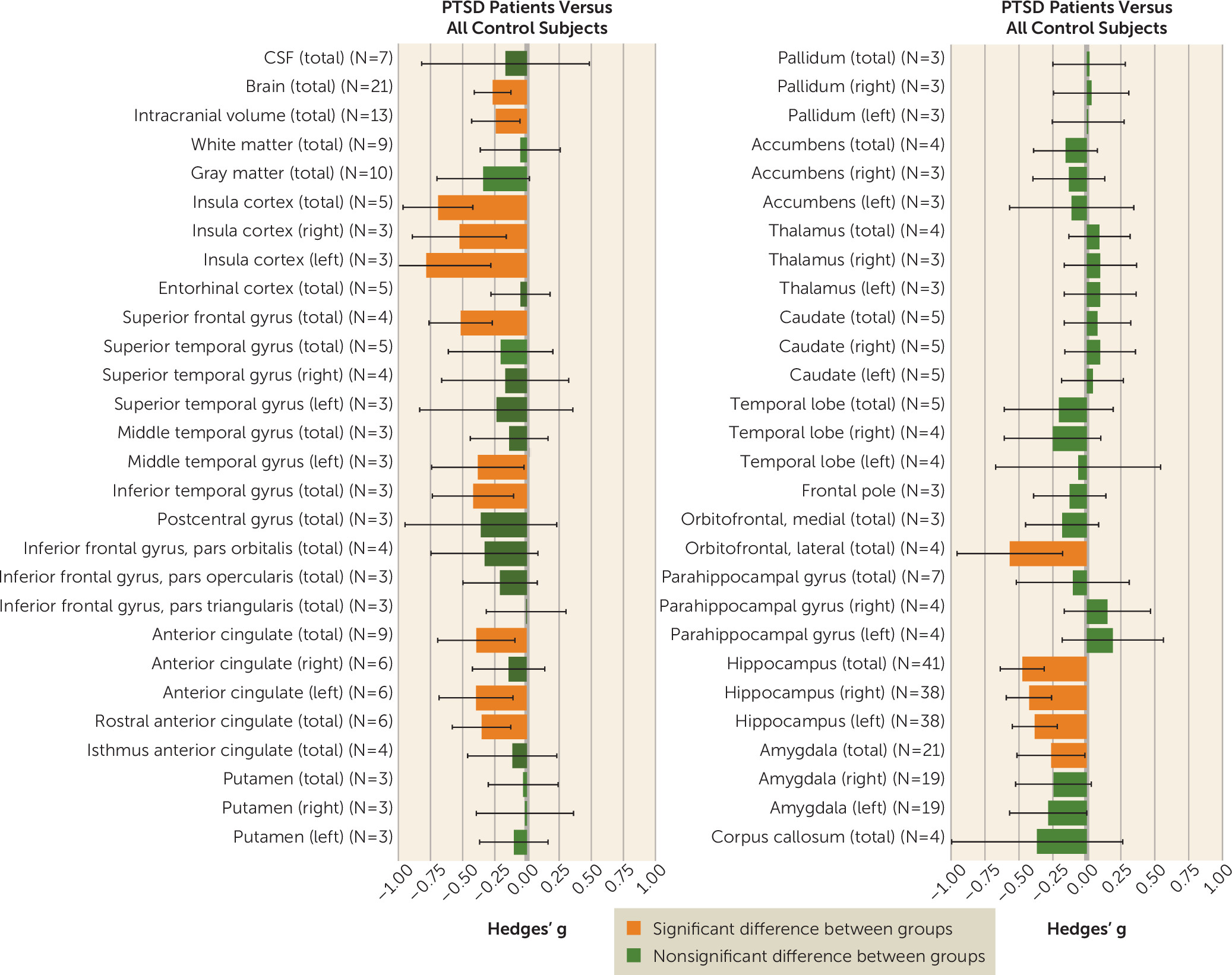
Results from the main meta-analysis comparing patients with PTSD with all control subjects are summarized in
Table 2 and
Figure 1. Compared with control subjects, PTSD patients had reduced brain volume, intracranial volume, and volumes of the insula (left, right, and total), superior frontal gyrus, left middle temporal gyrus, inferior temporal gyrus, anterior cingulate (left and total), rostral anterior cingulate cortex, lateral orbitofrontal cortex, and total amygdala. The left, right, and total hippocampal volumes were reduced in PTSD; however, these results were associated with significant small-sample bias. Subsequent trim-and-fill analysis for the hippocampal meta-analyses resulted in no additional imputed studies, although after the exclusion of two outlier studies (
14,
15) associated with large negative effect sizes (g<−2.6) for the left, right, and total hippocampus, small-sample bias was no longer significant.
Effect of clinical variables on hippocampal volume.
Because outliers may have a disproportionate effect on meta-regression analysis, the two outliers (
14,
15) were removed before we investigated the effect of clinical variables on total hippocampal volume (effect sizes of outliers, −3.0 and −3.2; effect size range of the remaining 39 studies, −1.4 to 0.4; pooled effect size after two studies were excluded, −0.44, p<0.001). There was no significant effect of the following clinical variables on the difference in total hippocampal volume between patients and control subjects: CAPS score (25 studies, p=0.16), age at illness onset (nine studies, p=0.45), time since trauma (13 studies, p=0.45), percentage of patients using antidepressants (27 studies, p=0.099), percentage of patients drug free (24 studies, p=0.11), and patient age (38 studies, p=0.52).
Pairwise sub-meta-analyses.
PTSD versus nontraumatized control group:
Patients with PTSD compared with nontraumatized control subjects had smaller total brain volume and volumes of gray matter, total, left, and right insula, total parahippocampal gyrus, and total, left, and right hippocampus (see Table S2 and Figure S2 in the online supplement).
PTSD versus traumatized control group:
Compared with the traumatized control group, patients with PTSD had smaller total brain volume, intracranial volume, superior frontal gyrus volume, total insula volume, anterior cingulate volume, lateral orbitofrontal cortex volume, and total and right hippocampal volume. Contrary to the general direction of results, right and left parahippocampal gyri were significantly larger in the PTSD group compared with the traumatized control group (see Table S3 and Figure S3 in the online supplement).
Traumatized control group versus nontraumatized control group:
Compared with the nontraumatized control subjects, the traumatized control group showed significant reductions in total, left, and right hippocampal volumes (see Table S4 and Figure S4 in the online supplement).
Sensitivity analysis.
When the correlation coefficient between left and right regions was changed from 0.8 to 0, 0.5, or 1.0, there was no change in any of the results. When three studies that divided brain volumes by intracranial volume were excluded, there was no change in any of the results. When the cortical thickness measures were excluded, the total parahippocampal gyrus volume decrease observed in the PTSD versus nontraumatized controls comparison was no longer significant and the reduction of the lateral orbitofrontal cortex volume in the PTSD versus traumatized controls comparison was no longer significant. When 11 pediatric studies were included in the meta-analysis, there were a number of new results, which are detailed in the online supplement.
Comparison of PTSD Region-of-Interest Meta-Analysis With Depression
Ten brain structures in both the present PTSD versus all controls region-of-interest meta-analysis and the previously published depression versus controls region-of-interest meta-analysis (
5) showed significant differences compared with control subjects (
Table 3). Two of these regions significantly differed between PTSD and depression. Compared with depressed patients and control subjects, PTSD patients had significantly reduced total brain volume. Compared with PTSD patients and control subjects, depressed patients had significantly reduced volume of the thalamus. Both PTSD and depression patients had significantly smaller hippocampal volume compared with control subjects, with no difference between the patient groups in this brain region. In an analysis using the smaller PTSD versus nontraumatized controls contrast, PTSD patients had reduced total brain volume compared with depression patients, although this difference fell short of significance (p=0.07) (see Table S8 in the
online supplement).
VBM Meta-Analysis Using Seed-Based d-Mapping
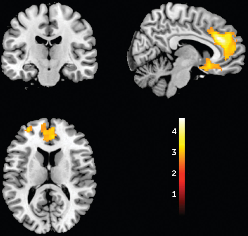
The VBM main meta-analysis (
Figure 2; see also Table S9 in the
online supplement) revealed that PTSD patients compared with all control subjects exhibited significant gray matter volume reductions in a large cluster in the medial prefrontal cortex encompassing the left and right anterior cingulate and extending to the subgenual prefrontal cortex. Reductions in volume were also observed in the left superior frontal gyrus and other smaller clusters, including the amygdala bilaterally, extending to the head of the hippocampi. The jackknife sensitivity analysis indicated that the large clusters were robust (see Figure S8 in the
online supplement). The meta-analysis was repeated for PTSD versus nontraumatized control subjects (see Table S10 and Figures S9 and S10 in the
online supplement), PTSD versus traumatized control subjects (see Table S11 and Figures S11 and S12 in the
online supplement), and PTSD versus all control subjects, including pediatric data (see Table S12 and Figures S13 and S14 in the
online supplement).
Comparison Between Region-of-Interest and VBM Meta-Analyses of PTSD
To compare the region-of-interest and VBM meta-analyses, regions were included if they were flagged as significant in either the region-of-interest (
Table 2) or the VBM meta-analysis (
Figure 2) and the brain region was available using the SDM extract tool, which gives pooled effect sizes for a region. A total of eight bilateral regions were compared (see Table S13 in the
online supplement). The region-of-interest meta-analysis reported volume reductions in the PTSD group in the insula bilaterally, the left anterior cingulate, and the hippocampus bilaterally but not the right anterior cingulate or amygdala. Conversely, the VBM meta-analysis reported reductions in the anterior cingulate and amygdala bilaterally but not the insula or hippocampus.
The database, forest plots for each meta-analysis, and three-dimensional image files from the SDM analysis are available at
http://www.ptsdmri.uk (see Figure S15 in the
online supplement).
Discussion
This study is, to our knowledge, the most comprehensive meta-analysis of MRI studies in PTSD to date, in terms of both number of brain regions and number of included studies. New techniques used in this study include use of t-maps in the VBM analysis, a direct comparison between VBM and region-of-interest techniques, a statistical comparison between PTSD and depression, and a freely available online database and meta-analysis. In the main region-of-interest meta-analysis, PTSD patients compared with all control subjects had volumetric reductions of the brain, intracranial volume, insula, superior frontal gyrus, temporal gyri, anterior cingulate, rostral anterior cingulate, hippocampus, and amygdala. PTSD patients compared with nontraumatized control subjects showed a similar pattern of reduced volumes, including total brain, insula, and hippocampus. PTSD patients compared with traumatized control subjects showed a similar pattern of volumetric reductions, including total brain, intracranial volume, insula, anterior cingulate, and hippocampus. This subanalysis showed a volumetric increase of the parahippocampal gyri bilaterally in PTSD, a finding related to higher early trauma scores (
16). Traumatized control subjects compared with nontraumatized control subjects showed only smaller volumes of the hippocampus. When compared with a meta-analysis of patients with depression, the distinguishing feature of PTSD was a reduction in total brain volume. We found a different pattern of regions highlighted in the region-of-interest analysis compared with the VBM meta-analysis.
Hippocampus
From the region-of-interest meta-analysis, the reductions in hippocampal volume in PTSD patients versus traumatized control subjects and PTSD patients versus nontraumatized control subjects was in agreement with previous MRI meta-analytic studies (
2,
17–
20). This finding is also consistent with previous neurocognitive studies (
21,
22) showing poorer memory performance in PTSD patients. Hippocampal volume reduction may be a generalized marker of mental health disorders, as it has been associated with chronic hypercortisolemia (
23,
24), which is related to chronic stress (
25). Our finding of no difference in hippocampal volume in PTSD compared with depression supports this idea, as hypercortisolemia frequently occurs in patients with depression (
23,
26). Further evidence for a generalized marker comes from MRI meta-analyses reporting reduced hippocampal volume in bipolar disorder (
27) and in schizophrenia (
28). Reduced hippocampal volume in traumatized versus nontraumatized control subjects indicates that exposure to a traumatic event itself, even if the exposed person does not develop PTSD, may be associated with volume reduction.
Intracranial and Brain Volume
Intracranial volume was reduced in PTSD patients in the main region-of-interest analysis as well as in PTSD patients compared with traumatized control subjects. Total intracranial volume stabilizes in early adolescence (ages 11 to 14) and provides an estimate of premorbid brain volume (
29). Thus, smaller intracranial volume in PTSD may indicate abnormal brain development before or during early adolescence. Consequently, reduced intracranial volume may be a risk factor for PTSD and may be associated with trauma susceptibility. Total brain volume was also significantly smaller in PTSD patients compared with all control subjects, nontraumatized control subjects, and traumatized control subjects but not in the comparison between traumatized and nontraumatized control subjects. These results suggest that brain volume reductions in PTSD patients are related to the disorder itself rather than the exposure to trauma. In addition, our comparison with depression suggests that total brain volume reduction is a more specific marker for PTSD than is hippocampal volume.
Insula and Anterior Cingulate
In the region-of-interest meta-analysis, insula volume was smaller in PTSD patients compared with both nontraumatized and traumatized control subjects, suggesting that insula reduction may underlie the pathophysiology of PTSD (
30). Reductions in anterior cingulate volume were found in the region-of-interest and VBM meta-analyses. A number of functional MRI studies have reported abnormalities of the anterior cingulate in PTSD (
31–
33), including a study of monozygotic twins (
34) that suggested that hyperresponsiveness of the dorsal anterior cingulate is a familial risk factor for PTSD.
Comparison of Region-of-Interest Meta-Analysis and VBM Meta-Analysis
We found relatively poor agreement when comparing the VBM and region-of-interest meta-analyses, and this has implications for neuroimaging meta-analyses of other disorders. We have considered a number of reasons for these differences. First, VBM involves smoothing, which is known to bias sensitivity to brain regions the size of the smoothing kernel (≈10 mm). Second, region-of-interest studies typically report on a subset of brain regions and are likely to suffer from publication bias. Third, where seed-based d mapping uses coordinate data, the effect size is biased toward zero in brain regions where there are no significant clusters. Lastly, VBM analyses typically adjust for global brain volumes, whereas region-of-interest studies use absolute volumes.
Limitations
The literature search included MEDLINE and manual searches, as previous experience (
5) has indicated that MEDLINE has high coverage of MRI studies in clinical populations, and the present study included more publications than previous meta-analyses. The use of one bibliographic database is a limitation, however. Significant heterogeneity was detected for many of the brain structures in the PTSD group (see
Table 2), which we have also observed in a number of other MRI meta-analyses (
5,
35,
36). This is likely to be caused by variations in patient characteristics and MRI data, and a random-effects model was utilized to account for such heterogeneity. Publication bias was detected for total hippocampal volume; we attempted to remedy this using a trim-and-fill method, although no new studies were imputed. Although the present meta-analysis summarizes small heterogeneous samples, large-scale studies using standardized MRI acquisition protocols and epidemiological neuroimaging samples, such as UK Biobank, are likely to tease apart the different structural abnormalities associated with risk for PTSD, experience of trauma, and development of PTSD. We compared PTSD and depression because they are highly comorbid; however, PTSD co-occurs with other anxiety disorders and substance use disorders (
37), and these comorbidities could be associated with some of our findings.
Conclusions
We have conducted a comprehensive meta-analysis of both region-of-interest and VBM MRI studies in PTSD and determined specific brain abnormalities associated with the experience of trauma and a diagnosis of PTSD. We have also shown that while hippocampal volume reductions in PTSD may be similar to those seen in depression, changes in total brain volume appear to distinguish PTSD from depression.
Acknowledgments
Dr. Kempton was funded by an MRC Career Development Fellowship (grant MR/J008915/1). The authors acknowledge financial support from the Wellcome Trust and the Engineering and Physical Sciences Research Council for the King’s Medical Engineering Centre and the National Institute for Health Research (NIHR) Biomedical Research Centre at South London and Maudsley NHS Foundation Trust and King’s College London.


