Abstract

Objective:

Anxiety disorders are common, can result in lifelong suffering, and frequently begin before adolescence. Evidence from adults suggests that altered prefrontal-limbic connectivity is a pathophysiological feature of anxiety disorders. More specifically, in adults with anxiety disorders, decreased fractional anisotropy (FA), a measure of white matter integrity, has been observed in the uncinate fasciculus, the major tract that connects limbic and prefrontal regions. Because of the early onset of anxiety disorders and the increased incidence in anxiety disorders in females during their reproductive years, it is important to understand whether the reduction in uncinate fasciculus FA exists in children with anxiety disorders and the extent to which this alteration is sex related. To address these issues, the authors assessed FA in the uncinate fasciculus in unmedicated boys and girls with anxiety disorders.

Methods:

FA measures were derived from diffusion tensor images that were acquired from 98 unmedicated children (ages 8–12); 52 met criteria for generalized anxiety disorder, separation anxiety disorder, social anxiety disorder, or anxiety disorder not otherwise specified, and 46 were matched control subjects.

Results:

Tract-based results demonstrated that children with anxiety disorders have significant reductions in uncinate fasciculus FA. A significant sex-by-group interaction and post hoc testing revealed that this effect was evident only in boys. No other main effects or sex-by-group interactions were found for other white matter tracts.

Conclusions:

These findings provide evidence of uncinate fasciculus white matter alterations in boys with anxiety disorders. The data demonstrate that anxiety disorder–related alterations in prefrontal-limbic structural connectivity are present early in life, are not related to psychotropic medication exposure, and are sex specific. Building on these findings, future research has the potential to provide insights into the genesis and sexual dimorphism of the pathophysiology that leads to anxiety disorders, as well as to identify sex-specific early-life treatment targets.
Anxiety disorders are among the most common psychiatric disorders and can become chronic (1, 2). They affect more than one in four individuals at some point in their life and are generally more prevalent in females than in males (3, 4). Individuals suffering from anxiety disorders have impairments in psychosocial functioning and quality of life (5), and when severe, anxiety disorders can be disabling. Children are especially affected by pathological anxiety, as anxiety disorders are among the earliest psychiatric illnesses to develop (2), affecting up to 20% of youths (68). Additionally, anxiety symptoms in children are often comorbid with other conditions, and they are a strong predictor of the subsequent development of other anxiety disorders, affective disorders, and comorbid substance abuse (2). While early treatment has the potential to prevent the lifelong suffering and psychosocial dysfunction associated with these disorders, current treatments for childhood anxiety disorders, such as cognitive-behavioral therapy and selective serotonin reuptake inhibitors, are suboptimal. Many children fail to respond to these treatments, and those who do respond have relatively high rates of relapse (9). Establishing a better understanding of the pathophysiology of childhood anxiety disorders will facilitate the development of novel, more effective treatments (10).
Despite the prevalence and importance of childhood anxiety disorders, few studies have focused on characterizing the neural alterations that underlie the early manifestations of these disorders (11). Current knowledge of the pathophysiology of anxiety disorders is mostly derived from studies of adults, and a few studies have examined adolescent and preadolescent patients with anxiety disorders (11). Previous work revealed that anxiety disorders are associated with hyperactivation of the amygdala and insula in response to negative emotional stimuli (1214), and recent work from our group has shown that amygdala activation is significantly higher in preadolescent children with anxiety disorders when confronted with uncertain conditions (15). We, as well as others, have also reported decreased anxiety-related functional coupling between the amygdala and regulatory prefrontal cortical regions such as the anterior cingulate cortex, orbital frontal cortex, dorsolateral prefrontal cortex, and medial prefrontal cortex (1619). Decreased anxiety-related functional coupling between the amygdala and the prefrontal cortex is evolutionarily conserved, as we demonstrated similar findings in a rhesus monkey model of early-life anxiety (18).
Complementing these findings, diffusion tensor imaging (DTI) studies in patients with anxiety disorders report altered structural connectivity between temporal lobe and prefrontal cortical regions, revealing that fractional anisotropy (FA) in the uncinate fasciculus (UF) is reduced in patients with anxiety disorders (2025). These studies focused predominantly on adults with anxiety disorders, with only one study including adolescent patients (25). The UF is highly relevant to anxiety and emotion regulation, as it connects structures that are crucial to affective processing, such as the amygdala, entorhinal/perirhinal cortices, and parahippocampal gyrus, to frontal regions, including the anterior prefrontal cortex, orbital frontal cortex, ventromedial prefrontal cortex, anterior cingulate cortex, and insula (26, 27).
While the structural alterations identified in patients with anxiety disorders may reflect the pathophysiology associated with the disorders, it is also possible that they result from illness chronicity, medication exposure, or other nonpathophysiological factors. Studies in medication-naive children have the advantage of examining white matter pathways early in the illness and in the absence of many of the influences that may indirectly affect white matter. Additionally, these studies may inform us on mechanisms underlying childhood risk of developing anxiety disorders and may also point to new early-life treatment and prevention strategies.
In this study, we used DTI to assess white matter integrity in unmedicated preadolescent children with anxiety disorders compared with healthy control children to test the hypothesis that childhood anxiety disorders are associated with alterations in the UF FA. Because of known sex differences in the prevalence of anxiety disorders (3, 4) and the interest in sexual dimorphism in relation to brain development (28, 29), we also examined sex differences in this effect. Additional analyses aimed to further characterize these microstructural alterations by investigating three supplementary diffusivity measures as well as interactions with symptom measures and steroid hormones. Beyond the UF, we explored six additional structures for which the relation to anxiety disorders has been less consistent (2025, 30, 31), and we conducted a whole brain voxel-based analysis.

Methods

Participants

For a detailed description of the study methods, see the Supplemental Methods section in the online supplement. Briefly, diffusion-weighted MRI scans were obtained from a preadolescent sample at two research sites: University of Wisconsin and the National Institute of Mental Health (NIMH). The final sample consisted of 98 children ages 8–12 (50 of them girls) (Table 1), of whom 46 were control subjects, across the two sites. Control subjects were age- and sex-matched at the group level to the anxiety disorder sample and had no current or past psychiatric illness. Neither the anxiety disorder patients nor the control subjects were currently on any psychotropic medication, and none reported any psychotropic medication usage at any point during their life. Informed assent and consent were obtained from all participants and their parents, in accordance with the institutional review boards of the University of Wisconsin and NIMH. Individuals were compensated for their time and effort.
TABLE 1. Demographic and clinical characteristics of children with anxiety disorders and control childrena
 GirlsBoys
MeasureControl GroupAnxiety Disorder GroupControl GroupAnxiety Disorder GroupControl GroupAnxiety Disorder Group
 N% FemaleN% FemaleN% FemaleN% FemaleN% FemaleN% Female
Overall sample465052512310027100230250
Demographic characteristics
 MeanSDMeanSDMeanSDMeanSDMeanSDMeanSD
 Age (years)10.581.2910.421.3310.741.3210.501.2310.381.3010.391.43
 IQ (WASI)119.5213.44117.4213.52119.0016.33116.1212.81120.0410.11118.7214.33
 Physical development (Tanner scale)3.932.064.041.874.202.094.652.003.361.713.671.86
Symptom measure scores
 Anxiety (parent SCARED)7.218.8328.5912.716.016.1329.5612.006.108.2627.7813.15
 Anxiety (child SCARED)13.4111.2625.9313.7112.449.9331.2013.7311.929.2321.3612.52
 Depression (CDI)28.6818.8738.5221.2127.1818.9644.8121.2627.5217.7333.3219.80
 ADHD (CPRS-R)45.676.9161.0612.7446.054.6865.4014.2442.742.6857.259.34
Endocrine measures (from saliva)
 Cortisol (μg/dL)0.100.040.100.040.100.0430.110.040.100.030.100.04
 Testosterone (pg/dL)24.119.3222.6312.3328.548.4525.3914.7218.887.6619.187.58
 Estradiol (pg/dL)0.780.310.650.350.790.380.650.430.770.230.650.26
a
There were no significant differences in demographic characteristics between the control and anxiety disorder groups, overall or by sex. Symptom score measures differed significantly between the control and anxiety disorder groups, overall and by sex (all p values [two-tailed] <0.001, except between control boys and boys with anxiety disorders for anxiety scores [p=0.005] and depression scores [p=0.005]). There were no differences between groups, overall or by sex, on endocrine measures, except for estradiol between the overall control and anxiety disorder groups (p=0.037). All endocrine measures were residualized for time of day. ADHD=attention deficit hyperactivity disorder; CDI=Children’s Depression Inventory; CPRS-R=Conners’ Parent Rating Scale–Revised; SCARED=Screen for Child Anxiety Related Disorders; WASI=Wechsler Abbreviated Scale of Intelligence.
All participants were interviewed with the Schedule for Affective Disorders and Schizophrenia for School-Age Children–Present and Lifetime Version (K-SADS-PL) (32), which was administered by a trained Ph.D.-level clinical psychologist or a psychiatrist. (See the Supplemental Methods section in the online supplement for details on interrater agreement.) Beyond diagnosis, children’s symptoms were rated by both the child and a parent on multiple symptom questionnaires (see the Supplemental Methods section).

Data Acquisition and Analyses

DTI acquisition.

For a detailed description, see the Supplemental Methods section in the online supplement. Briefly, brain images at both sites were collected on MRI scanners of the same make and model, with comparable parameters, optimized for diffusion-weighted imaging with 48 directions.

Steroid hormone collection.

See the Supplemental Methods section in the online supplement.

DTI analyses.

For a detailed description, see the Supplemental Methods section in the online supplement. Briefly, images were corrected for field inhomogeneity and eddy currents, after which the tensors were calculated using a robust tensor estimation. Tensor images of all subjects were coregistered iteratively using nonlinear tensor-based normalization tools and then registered to Montreal Neurological Institute MNI152 space. In this template space, diffusion measures were extracted to quantify local white matter microstructure. Deterministic fiber tractography was performed to delineate tracts of interest, which, besides the UF (2025), included cortico-limbic association pathways previously implicated in anxiety disorders, including the cingulum bundle (20, 30), the superior longitudinal fasciculus (20, 24, 25), the fornix (20), and the inferior fronto-occipital fasciculus (20, 25). Tracts that have been shown in other studies to have anxiety-related changes were also extracted, including thalamocortical projection fibers in the internal capsule (20, 25) and interhemispheric commissural fibers of the corpus callosum (31).
Next, to quantify the microstructure of an entire white matter structure, a weighted mean was calculated per tract for each diffusion measure per subject. Tract-based analyses were performed with average tract measures predicting anxiety disorder patient status. To characterize microstructural changes in these white matter pathways beyond FA, analyses assessed mean diffusivity (MD), axial diffusivity (XD), and radial diffusivity (RD) as well. We also examined the possibility of sexually dimorphic effects relating white matter microstructure to anxiety in children with anxiety disorders. In order to test sex differences in these relations, the interaction between patient status (anxiety disorder, control) and sex (boys, girls) was tested in the same model. Age, sex, and site were included in this model as covariates. Since we had no a priori hypothesis about differences between left and right tracts, the left and right components of each tract were combined into one average.
Analyses were conducted using standard linear regression techniques in RStudio, version 1.0.136 (33). To control for family-wise error, a Šidák correction for the number of tracts was applied, where m=7 for each white matter tract tested, resulting in a corrected two-tailed significance threshold (alpha) of 0.0073 (αSID=1−[1−α]1/m=1−[1−0.05]1/7=0.0073). The effect sizes of significant tract-based effects were reported using Cohen’s d (3436) to aid in interpretation. Cohen’s d values greater than 0.8 are considered a large effect size, and 0.5 a medium effect (36). Voxel-based analyses of FA across the brain were performed to test regions within and beyond the tracts that were determined a priori.
Additional analyses, described in detail in the Results section, included age, sex, and site as covariates where applicable and used standard linear regression techniques in RStudio and Python.

Steroid hormone analyses.

See the Supplemental Methods section in the online supplement.

Results

Demographic Characteristics, Symptoms, and Hormonal Measurements

Ninety-eight children, 46 of them control subjects, were included in the final analyses. Children with anxiety disorders and control children did not differ significantly in age (t=0.385, df=94, p=0.70), sex (t=0.184, df=94, p=0.85), or IQ (t=0.888, df=91, p=0.38), and there was no significant difference between groups in physical development as assessed by the Tanner stages (t=−1.499, df=86, p=0.14) (see Table 1). Age and sex each significantly predicted Tanner score (age: t=8.510, df=86, p<0.001; sex: t=−2.006, df=86, p=0.048), with older children and girls having significantly higher Tanner scores, but the group-by-sex interaction was nonsignificant (p=0.41).
Children with anxiety disorders had significantly higher symptom scores on all clinical scales (anxiety, depression, and attention deficit hyperactivity disorder [ADHD]) compared with control children (p values <0.001) (see Table 1). No group-by-sex interactions were found for depression and ADHD symptoms. A group-by-sex interaction approached significance for self-reported anxiety (t=1.82, df=89, p=0.072) (see Figure S1 in the online supplement). Post hoc tests indicated that self-reported anxiety scores were significantly lower for boys compared to girls with anxiety disorders (t=2.49, df=48, p=0.016).
Testosterone level was significantly higher in girls compared with boys (z=−2.57, df=55, p=0.006; for mean values by group for each sex, see Table 1), an effect that has previously been observed in preadolescent children (37). Neither estradiol nor cortisol levels differed by sex (p values >0.4). Age was positively related to testosterone (z=3.286, df=55, p=0.006) and estradiol (z=2.538, df=55, p=0.011) levels.
While there were no group differences in cortisol or testosterone levels (p>0.22), an unpredicted significant group effect was observed for estradiol levels, in which children with anxiety disorders had lower levels of estradiol compared with control children (z=2.085, df=55, p=0.037) (see Table 1; see also Figure S2 in the online supplement). None of the endocrine measures displayed a significant group-by-sex interaction (p values >0.5).

Tract-Based DTI Analyses

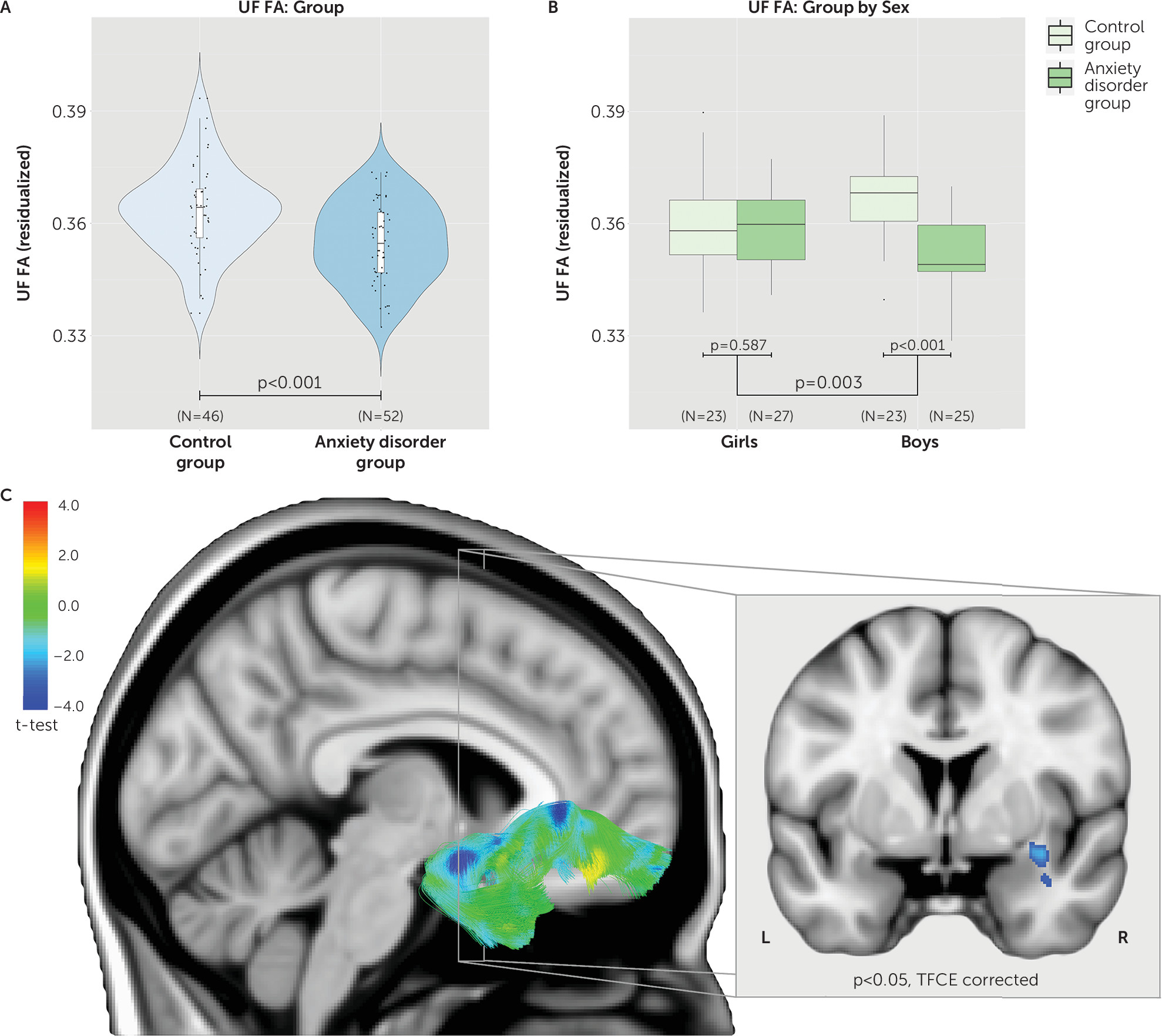
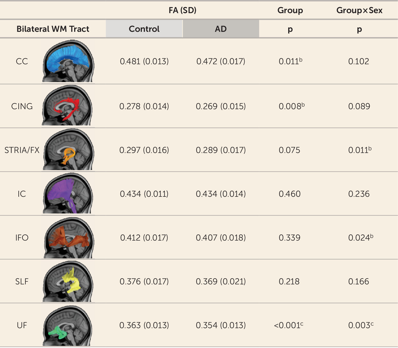
Tract-based analyses were performed on seven white matter pathways, leading to a multiple-comparisons threshold after the Šidák correction of p≤0.0073, and were covaried for age, sex, and site. Results indicated that only the UF tract significantly differed between groups (Figures 1 and 2). Specifically, children with anxiety disorders had reduced UF FA compared with control children (t=3.650, df=92, p<0.001; d=0.73) (see also Figures 1 and 2). In addition, the interaction between group and sex was significant for UF FA (t=3.058, df=92, p=0.003). Post hoc tests indicated that boys with anxiety disorders displayed significantly reduced UF FA compared with control boys (t=4.750, df=44, p<0.001) (see Figure 1), whereas girls with anxiety disorders did not differ from control girls (t=0.547, df=46, p=0.587) (see Figure 1). To further investigate which of these four groups were different from the rest (girls with anxiety disorders, boys with anxiety disorders, control girls, control boys), we ran a contrast analysis. The results indicated that boys with anxiety disorders were different from all three other groups (t=3.937, df=92, p<0.001).
FIGURE 1. Reduced fractional anisotropy (FA) in the uncinate fasciculus (UF) in children with anxiety disordersa
a In panel A, a violin plot of UF FA data residualized for sex, age, and site shows that children with anxiety disorders have significantly lower FA in the UF compared with control children. In panel B, a box plot of significant interaction between group and sex, post hoc analyses indicate no significant difference between healthy girls and girls with anxiety disorders, but a difference between healthy boys and boys with anxiety disorders. UF FA is residualized for age and site. In panel C, whole-brain voxel-based analyses confirm significant differences in UF FA between control children and children with anxiety disorders; test results are shown on the right UF fiber tract. The coronal view shows the extent of significant differences after threshold-free cluster enhancement correction. Cooler colors indicate lower FA values in children with anxiety disorders compared with control children.
FIGURE 2. Group differences in tract fractional anisotropy (FA) in the control children and children with anxiety disordersa
a Weighted mean FA values by tract are listed, with standard deviations. All analyses include age, sex, and site as covariates. AD=anxiety disorder; CC=corpus callosum; CING=cingulum bundle; FX=fornix; IC=internal capsule; IFO=inferior fronto-occipital fasciculus; SLF=superior longitudinal fasciculus; UF=uncinate fasciculus; WM=white matter.
b Statistically significant at uncorrected levels.
c Statistically significant at Šidák-corrected levels.
Other variables that could potentially influence the results include age, comorbid symptoms, and hormonal status. While there was a main effect of age on UF FA (t=2.786, df=92, p=0.006), no significant sex-by-age or group-by-sex-by-age interactions were found (p values >0.28) (see Table S1 and Figure S3 in the online supplement). In relation to anxiety, depression, and ADHD scores, we performed analyses to test for interactions and found that these measures did not affect the findings, such that there were no significant group-by-sex-by-rating interactions (p values >0.375) (see Table S1 in the online supplement). We also performed analyses that examined potential influences of the comorbid categorical diagnoses, ADHD (four girls, two boys) and depression (three girls, no boys). The results remained largely unchanged when these patients were excluded from the analyses (excluding ADHD: group, t=3.592, df=86, p<0.001; group by sex, t=2.502, df=86, p=0.014; excluding major depression: group, t=3.749, df=89, p<0.001; group by sex, t=2.872, df=89, p=0.005). Finally, we tested hormonal status (cortisol, testosterone, and estradiol) in relation to the findings, which demonstrated no significant interactions that could account for the effects (group by sex by hormone level, p values >0.175) (see Table S1).
Diffusivity measures other than FA that were explored, such as MD, XD, and RD, indicated that only UF RD was significantly increased in children with anxiety disorders (t=−2.813, df=92, p=0.006; d=0.56) (see Figure S4 in the online supplement), and the group-by-sex interaction was nominally (uncorrected) significant (t=−2.007, df=92, p=0.048) (see Figure S4). Several of the other six white matter tracts displayed some nominally significant group effects and group-by-sex interactions for FA, MD, XD, and RD (see Figure 2; see also Figure S4), although none were significant after correction for multiple comparisons.

Voxel-Based Analyses of FA

Whole-brain voxel-based analyses explored group differences beyond the a priori hypothesized tracts. The results confirmed the observations from the tract-based analyses, demonstrating significantly reduced FA in multiple locations along the UF tract (p<0.05, threshold-free cluster enhancement [TFCE] corrected; see Figure 1, Table 2, and https://neurovault.org/collections/161/). Areas particularly affected included white matter adjacent to the orbital gyrus and white matter in the bend around the lateral fissure between the temporal and frontal lobes. We also detected group differences in the corpus callosum, internal capsule, and inferior fronto-occipital fasciculus pathways (see Table 2 and https://neurovault.org/collections/161/). In the corpus callosum tract, alterations were mainly localized in the genu (p<0.05, TFCE corrected; see Table 2 and https://neurovault.org/collections/161/). In the left and right internal capsule, significant clusters were found in the area of the postcentral gyrus in the somatosensory cortex (p<0.05, TFCE corrected; see Table 2 and https://neurovault.org/collections/161/). In the inferior fronto-occipital fasciculus tract, significant clusters were mainly located in the anterior inferior portions of the right section of this tract (p<0.05, TFCE corrected; see Table 2 and https://neurovault.org/collections/161/).
TABLE 2. Group differences (control group compared with anxiety disorder group) in voxel-based analyses of whole-brain fractional anisotropy valuesa
Cluster NumberVolume (mm3)LocationHemispherePeak t valuePeak MNI Coordinates
17,000CC/UFLeft, right5.688, 153, 73
2425ICRight4.3111, 89, 140
3290UFRight4.85126, 127, 59
4228CC/IFORight4.53118, 74, 103
5148ICLeft5.1174, 98, 133
610IFORight3.03120, 148, 85
74UFRight3.47129, 124, 45
83CCRight4.35111, 59, 121
a
The table provides an overview of the size and location of significant clusters after threshold-free cluster enhancement correction. CC=corpus callosum; IC=internal capsule; IFO=inferior fronto-occipital fasciculus; MNI=Montreal Neurological Institute; UF=uncinate fasciculus.
Based on results from the tract-based analyses, we also examined the group-by-sex interaction, which did not reveal any clusters passing TFCE correction at a p threshold of 0.05. However, separate analyses performed in girls and boys revealed significant FA reductions in boys with anxiety disorders compared with control boys in regions that overlap with the UF, corpus callosum, and bed nucleus of the stria terminalis, and no such differences in girls (see Figure S5 and Table S2 in the online supplement, and https://neurovault.org/collections/161/).

Discussion

In this study, we examined the microstructural integrity of the UF in unmedicated, preadolescent children with anxiety disorders and also examined potential sex differences. The findings revealed decreased UF FA in boys, and not girls, with anxiety disorders. Tractography analyses of other white matter tracts demonstrated the relative specificity of this finding. The UF is a white matter tract critical for prefrontal–temporal lobe functional integration (38), and the present data suggest that in boys this tract may be linked to the prefrontal-limbic dysregulation that is associated with anxiety disorders (2025). While UF FA differences have been reported in adolescents and adults with anxiety disorders, we are unaware of any reports that have addressed the issue of sexual dimorphism in patients with anxiety disorders. However, there have been inconsistent reports of sex-related UF FA alterations in individuals with high levels of trait anxiety (3941).
Notably, we demonstrated that these white matter alterations are present early in life and in children who have never been exposed to psychotropic medications. In contrast to studies in adult populations with anxiety disorders (20), the results from this study can be interpreted without the potential confounders of prior medication exposure or illness chronicity. These findings provide developmental continuity in relation to the previously reported UF FA reductions in adolescents and adults, suggesting that the UF white matter alterations observed in these older populations may have their origins in childhood.
The UF FA differences that we found in boys with anxiety disorders are intriguing and raise the question as to what underlies this sexual dimorphism. We found no evidence that age or sex hormones could account for this finding. Previous studies in children and adults indicate either no effect or a minimal effect of sex on UF FA (4244). Likewise, studies examining sex steroids in pubertal children have found little association between hormonal levels and white matter volume (45, 46). However, little is known about the specific relation between sex hormones and UF FA. In contrast, age is well known to be associated with FA (42, 43, 47, 48), and even with the relatively constrained age range that we studied, we found that UF FA increased with age. One possibility that could explain the sexual dimorphism reported here is that the developmental trajectory of UF FA could differ between boys and girls. However, we found no significant age-by-sex interactions, which is consistent with other work (43, 44), and we found no age-by-sex-by-group interactions. We also assessed levels of the stress hormone cortisol and found no significant influences of cortisol on UF FA that could account for the UF FA reductions in boys with anxiety disorders, or interactions with sex. Because of the episodic nature of the secretion of steroid hormones, it is possible that more frequent sampling would reveal a relation between steroid hormone levels and UF FA.
Most but not all DTI studies of patients with anxiety disorders report reductions in UF FA as well as other white matter alterations (2025, 30, 31, 49, 50). Inconsistencies in the UF FA findings across studies could be due to a variety of factors, including heterogeneity in diagnostic composition of the sample, sample size, and DTI acquisition and processing. We also note that reduced UF FA is not specific to anxiety disorders, as similar white matter alterations have been observed in some studies of individuals with trait anxiety (3941, 5154) as well as in patients with affective and other psychiatric disorders (55, 56).
Our assessment of multiple diffusivity measures allows for a deeper understanding of the nature of the microstructural alterations found in children with anxiety disorders. In addition to reduced FA, boys with anxiety disorders exhibited increased RD. The combination of decreased FA and increased RD is thought to reflect reduced myelination and/or reduced axonal density (57, 58). Changes in myelination and axonal density are associated with alterations in the speed, timing, and accuracy of information passing through white matter (59). Because the UF is the major tract connecting medial temporal lobe structures such as the amygdala and hippocampus with the prefrontal cortex, alterations in UF structure are likely to alter information flow relevant to emotion regulation (26). Although lesions affecting the UF can alter the regulation of anxiety in primates (6062), the extent to which individual differences in UF myelination influence anxiety-related neuronal signaling is unclear. Of note, a recent study in humans demonstrated that individual differences in UF FA are related to individual differences in cognitive-emotional processing (63).
Although axon myelination is most active early in development, the microstructural integrity of UF continues to increase into adulthood and is one of the last white matter pathways to reach peak FA (43). Myelin-producing oligodendrocytes are highly plastic throughout adulthood, and oligodendrocyte precursor cells are the major proliferating cell type in adult brains (64). Evidence is accumulating that myelination occurs dynamically in response to neural activity and can continue throughout adulthood (59, 65, 66). This white matter plasticity provides a potential mechanism by which targeted therapies could be focused on restoring UF integrity. Interestingly, data in children suggest that aerobic exercise increases white matter integrity (67), and it is well known that exercise has antianxiety effects (68). Our data suggest that early-life interventions directed toward increasing UF integrity could be particularly effective in boys in ameliorating the symptoms of anxiety, and also could have a protective impact on the development of white matter connectivity between brain regions critical for adaptive emotion regulation.
Taken together with other studies (1625), these data provide convergent support for prefrontal cortex–temporal lobe dysfunction, which now extends to childhood anxiety. The findings demonstrate early-life alterations in a key white matter tract involved in conveying information relevant to emotion and anxiety regulation. The findings also point to the importance of considering brain-related sex differences before puberty. The data from this study, along with future studies, have the potential to guide the development of novel treatments focused on restoring the adaptive prefrontal regulation of anxiety. Such early interventions support the possibility of using therapies that could reduce or even prevent long-term chronic psychopathology in the treatment of children with anxiety disorders.

Acknowledgments

The authors thank the HealthEmotions Research Institute, the Waisman Laboratory for Brain Imaging and Behavior, the Lane Neuroimaging Laboratory, Neuroscience Training Program.
Supported by NIH grants R21 MH092581 and Intramural Research Program Project number Z-002781.
Footnote

Dr. Alexander is part owner of Thervoyant. At the time of writing, Dr. Kalin had received honoraria from CME Outfitters, Elsevier, and the Pritzker Consortium; served on scientific advisory boards for Ketamine Clinics of America, Neuronetics, and the Pritzker Neuroscience Consortium; served as co-editor of Psychoneuroendocrinology; and was named, along with Dr. Roseboom, on patents on promoter sequences for corticotropin-releasing factor CRF2alpha and a method of identifying agents that alter the activity of the promoter sequences (7,071,323, 7,531,356), promoter sequences for urocortin II and the use thereof (7,087,385), and promoter sequences for corticotropin-releasing factor binding protein and the use thereof (7,122,650). The other authors report no financial relationships with commercial interests.

Supplementary Material

File (appi.ajp.2018.18040425.ds001.pdf)

References

1.
Kessler RC, Aguilar-Gaxiola S, Alonso J, et al: The global burden of mental disorders: an update from the WHO World Mental Health (WMH) surveys. Epidemiol Psichiatr Soc 2009; 18:23–33
2.
Kessler RC, Ruscio AM, Shear K, et al: Epidemiology of anxiety disorders. Curr Top Behav Neurosci 2010; 2:21–35
3.
Kessler RC, Petukhova M, Sampson NA, et al: Twelve-month and lifetime prevalence and lifetime morbid risk of anxiety and mood disorders in the United States. Int J Methods Psychiatr Res 2012; 21:169–184
4.
MacKinaw-Koons B, Vasey MW: Considering sex differences in anxiety and its disorders across the life span: a construct-validation approach. Appl Prev Psychol 2000; 9:191–209
5.
Mendlowicz MV, Stein MB: Quality of life in individuals with anxiety disorders. Am J Psychiatry 2000; 157:669–682
6.
Merikangas KR, Nakamura EF, Kessler RC: Epidemiology of mental disorders in children and adolescents. Dialogues Clin Neurosci 2009; 11:7–20
7.
Copeland WE, Angold A, Shanahan L, et al: Longitudinal patterns of anxiety from childhood to adulthood: the Great Smoky Mountains Study. J Am Acad Child Adolesc Psychiatry 2014; 53:21–33
8.
Merikangas KR, He JP, Burstein M, et al: Lifetime prevalence of mental disorders in US adolescents: results from the National Comorbidity Survey Replication–Adolescent Supplement (NCS-A). J Am Acad Child Adolesc Psychiatry 2010; 49:980–989
9.
Walkup JT, Albano AM, Piacentini J, et al: Cognitive behavioral therapy, sertraline, or a combination in childhood anxiety. N Engl J Med 2008; 359:2753–2766
10.
Ravindran LN, Stein MB: The pharmacologic treatment of anxiety disorders: a review of progress. J Clin Psychiatry 2010; 71:839–854
11.
Ayling E, Aghajani M, Fouche J-P, et al: Diffusion tensor imaging in anxiety disorders. Curr Psychiatry Rep 2012; 14:197–202
12.
Etkin A, Wager TD: Functional neuroimaging of anxiety: a meta-analysis of emotional processing in PTSD, social anxiety disorder, and specific phobia. Am J Psychiatry 2007; 164:1476–1488
13.
Holzschneider K, Mulert C: Neuroimaging in anxiety disorders. Dialogues Clin Neurosci 2011; 13:453–461
14.
Martin EI, Ressler KJ, Binder E, et al: The neurobiology of anxiety disorders: brain imaging, genetics, and psychoneuroendocrinology. Clin Lab Med 2010; 30:865–891
15.
Williams LE, Oler JA, Fox AS, et al: Fear of the unknown: uncertain anticipation reveals amygdala alterations in childhood anxiety disorders. Neuropsychopharmacology 2015; 40:1428–1435
16.
Etkin A, Prater KE, Hoeft F, et al: Failure of anterior cingulate activation and connectivity with the amygdala during implicit regulation of emotional processing in generalized anxiety disorder. Am J Psychiatry 2010; 167:545–554
17.
Hahn A, Stein P, Windischberger C, et al: Reduced resting-state functional connectivity between amygdala and orbitofrontal cortex in social anxiety disorder. Neuroimage 2011; 56:881–889
18.
Birn RM, Shackman AJ, Oler JA, et al: Evolutionarily conserved prefrontal-amygdalar dysfunction in early-life anxiety. Mol Psychiatry 2014; 19:915–922
19.
Monk CS, Telzer EH, Mogg K, et al: Amygdala and ventrolateral prefrontal cortex activation to masked angry faces in children and adolescents with generalized anxiety disorder. Arch Gen Psychiatry 2008; 65:568–576
20.
Tromp DPM, Grupe DW, Oathes DJ, et al: Reduced structural connectivity of a major frontolimbic pathway in generalized anxiety disorder. Arch Gen Psychiatry 2012; 69:925–934
21.
Phan KL, Orlichenko A, Boyd E, et al: Preliminary evidence of white matter abnormality in the uncinate fasciculus in generalized social anxiety disorder. Biol Psychiatry 2009; 66:691–694
22.
Baur V, Brühl AB, Herwig U, et al: Evidence of frontotemporal structural hypoconnectivity in social anxiety disorder: a quantitative fiber tractography study. Hum Brain Mapp 2013; 34:437–446
23.
Hettema JM, Kettenmann B, Ahluwalia V, et al: Pilot multimodal twin imaging study of generalized anxiety disorder. Depress Anxiety 2012; 29:202–209
24.
Baur V, Hänggi J, Rufer M, et al: White matter alterations in social anxiety disorder. J Psychiatr Res 2011; 45:1366–1372
25.
Liao M, Yang F, Zhang Y, et al: White matter abnormalities in adolescents with generalized anxiety disorder: a diffusion tensor imaging study. BMC Psychiatry 2014; 14:41
26.
Schmahmann JD, Pandya DN, Wang R, et al: Association fibre pathways of the brain: parallel observations from diffusion spectrum imaging and autoradiography. Brain 2007; 130:630–653
27.
Catani M, Howard RJ, Pajevic S, et al: Virtual in vivo interactive dissection of white matter fasciculi in the human brain. Neuroimage 2002; 17:77–94
28.
Lenroot RK, Giedd JN: Sex differences in the adolescent brain. Brain Cogn 2010; 72:46–55
29.
De Bellis MD, Keshavan MS, Beers SR, et al: Sex differences in brain maturation during childhood and adolescence. Cereb Cortex 2001; 11:552–557
30.
Zhang Y, Li L, Yu R, et al: White matter integrity alterations in first episode, treatment-naive generalized anxiety disorder. J Affect Disord 2013; 148:196–201
31.
Liao W, Xu Q, Mantini D, et al: Altered gray matter morphometry and resting-state functional and structural connectivity in social anxiety disorder. Brain Res 2011; 1388:167–177
32.
Birmaher B, Ehmann M, Axelson DA, et al: Schedule for Affective Disorders and Schizophrenia for School-Age Children (K-SADS-PL) for the assessment of preschool children: a preliminary psychometric study. J Psychiatr Res 2009; 43:680–686
33.
RStudio Team: RStudio: Integrated Development for R. Boston, RStudio, 2016
34.
Nakagawa S, Cuthill IC: Effect size, confidence interval, and statistical significance: a practical guide for biologists. Biol Rev Camb Philos Soc 2007; 82:591–605
35.
Sullivan GM, Feinn R: Using effect size, or why the p value is not enough. J Grad Med Educ 2012; 4:279–282
36.
Cohen J: Statistical Power Analysis for the Behavioral Sciences, 2nd ed. Mahway, NJ, Lawrence Erlbaum Associates, 1988
37.
Dorn LD, Kolko DJ, Susman EJ, et al: Salivary gonadal and adrenal hormone differences in boys and girls with and without disruptive behavior disorders: contextual variants. Biol Psychol 2009; 81:31–39
38.
Schmahmann JD, Pandya DN: Fiber Pathways of the Brain. New York, Oxford University Press, 2009
39.
Montag C, Reuter M, Weber B, et al: Individual differences in trait anxiety are associated with white matter tract integrity in the left temporal lobe in healthy males but not females. Neuroscience 2012; 217:77–83
40.
Kim MJ, Brown AC, Mattek AM, et al. The inverse relationship between the microstructural variability of amygdala-prefrontal pathways and trait anxiety is moderated by sex. Front Syst Neurosci 2016; 10:93
41.
Kim MJ, Avinun R, Knodt AR, et al: Neurogenetic plasticity and sex influence the link between corticolimbic structural connectivity and trait anxiety. Sci Rep 2017; 7:10959
42.
Hasan KM, Iftikhar A, Kamali A, et al: Development and aging of the healthy human brain uncinate fasciculus across the lifespan using diffusion tensor tractography. Brain Res 2009; 1276:67–76
43.
Lebel C, Gee M, Camicioli R, et al: Diffusion tensor imaging of white matter tract evolution over the lifespan. Neuroimage 2012; 60:340–352
44.
Schmithorst VJ, Holland SK, Dardzinski BJ: Developmental differences in white matter architecture between boys and girls. Hum Brain Mapp 2008; 29:696–710
45.
Peper JS, Brouwer RM, Schnack HG, et al: Sex steroids and brain structure in pubertal boys and girls. Psychoneuroendocrinology 2009; 34:332–342
46.
Perrin JS, Hervé P-Y, Leonard G, et al: Growth of white matter in the adolescent brain: role of testosterone and androgen receptor. J Neurosci 2008; 28:9519–9524
47.
Lebel C, Beaulieu C: Longitudinal development of human brain wiring continues from childhood into adulthood. J Neurosci 2011; 31:10937–10947
48.
Lebel C, Walker L, Leemans A, et al: Microstructural maturation of the human brain from childhood to adulthood. Neuroimage 2008; 40:1044–1055
49.
Zhang L, Zhang Y, Li L, et al: Different white matter abnormalities between the first-episode, treatment-naive patients with posttraumatic stress disorder and generalized anxiety disorder without comorbid conditions. J Affect Disord 2011; 133:294–299
50.
Brambilla P, Como G, Isola M, et al: White-matter abnormalities in the right posterior hemisphere in generalized anxiety disorder: a diffusion imaging study. Psychol Med 2012; 42:427–434
51.
Westlye LT, Bjørnebekk A, Grydeland H, et al: Linking an anxiety-related personality trait to brain white matter microstructure: diffusion tensor imaging and harm avoidance. Arch Gen Psychiatry 2011; 68:369–377
52.
Baur V, Hänggi J, Jäncke L: Volumetric associations between uncinate fasciculus, amygdala, and trait anxiety. BMC Neurosci 2012; 13:4
53.
Kim MJ, Whalen PJ: The structural integrity of an amygdala-prefrontal pathway predicts trait anxiety. J Neurosci 2009; 29:11614–11618
54.
Modi S, Trivedi R, Singh K, et al: Individual differences in trait anxiety are associated with white matter tract integrity in fornix and uncinate fasciculus: preliminary evidence from a DTI based tractography study. Behav Brain Res 2013; 238:188–192
55.
Sexton CE, Mackay CE, Ebmeier KP: A systematic review of diffusion tensor imaging studies in affective disorders. Biol Psychiatry 2009; 66:814–823
56.
Von Der Heide RJ, Skipper LM, Klobusicky E, et al: Dissecting the uncinate fasciculus: disorders, controversies, and a hypothesis. Brain 2013; 136:1692–1707
57.
Feldman HM, Yeatman JD, Lee ES, et al: Diffusion tensor imaging: a review for pediatric researchers and clinicians. J Dev Behav Pediatr 2010; 31:346–356
58.
Alexander AL, Hurley SA, Samsonov AA, et al: Characterization of cerebral white matter properties using quantitative magnetic resonance imaging stains. Brain Connect 2011; 1:423–446
59.
Sampaio-Baptista C, Johansen-Berg H: White matter plasticity in the adult brain. Neuron 2017; 96:1239–1251
60.
Rudebeck PH, Saunders RC, Prescott AT, et al: Prefrontal mechanisms of behavioral flexibility, emotion regulation, and value updating. Nat Neurosci 2013; 16:1140–1145
61.
Kazama A, Bachevalier J: Selective aspiration or neurotoxic lesions of orbital frontal areas 11 and 13 spared monkeys’ performance on the object discrimination reversal task. J Neurosci 2009; 29:2794–2804
62.
Fox AS, Shelton SE, Oakes TR, et al: Orbitofrontal cortex lesions alter anxiety-related activity in the primate bed nucleus of stria terminalis. J Neurosci 2010; 30:7023–7027
63.
Lapate RC, Rokers B, Tromp DPM, et al: Awareness of emotional stimuli determines the behavioral consequences of amygdala activation and amygdala-prefrontal connectivity. Sci Rep 2016; 6:25826
64.
Dawson MRL, Polito A, Levine JM, et al: NG2-expressing glial progenitor cells: an abundant and widespread population of cycling cells in the adult rat CNS. Mol Cell Neurosci 2003; 24:476–488
65.
Gibson EM, Purger D, Mount CW, et al: Neuronal activity promotes oligodendrogenesis and adaptive myelination in the mammalian brain. Science 2014; 344:1252304
66.
Scholz J, Klein MC, Behrens TEJ, et al: Training induces changes in white-matter architecture. Nat Neurosci 2009; 12:1370–1371
67.
Chaddock-Heyman L, Erickson KI, Holtrop JL, et al: Aerobic fitness is associated with greater white matter integrity in children. Front Hum Neurosci 2014; 8:584
68.
Broman-Fulks JJ, Berman ME, Rabian BA, et al: Effects of aerobic exercise on anxiety sensitivity. Behav Res Ther 2004; 42:125–136

Information & Authors

Information

Published In

Go to American Journal of Psychiatry
American Journal of Psychiatry
Pages: 208 - 216
PubMed: 30654645

History

Received: 15 April 2018
Revision received: 5 October 2018
Accepted: 22 October 2018
Published online: 18 January 2019
Published in print: March 01, 2019

Keywords

  1. Anxiety Disorder-Generalized
  2. Biological Markers

Authors

Details

Do P.M. Tromp, Ph.D.
The Department of Psychiatry (Tromp, Williams, Oler, Roseboom, Rogers, Alexander, Kalin), the Neuroscience Training Program (Tromp, Kalin), the Department of Medical Physics (Alexander), and the HealthEmotions Research Institute (Tromp, Williams, Oler, Roseboom, Kalin), University of Wisconsin, Madison; the Department of Psychology and the California National Primate Research Center, University of California, Davis (Fox); and the Section on Development and Affective Neuroscience, NIMH, Bethesda, Md. (Benson, Pine).
Lisa E. Williams, Ph.D.
The Department of Psychiatry (Tromp, Williams, Oler, Roseboom, Rogers, Alexander, Kalin), the Neuroscience Training Program (Tromp, Kalin), the Department of Medical Physics (Alexander), and the HealthEmotions Research Institute (Tromp, Williams, Oler, Roseboom, Kalin), University of Wisconsin, Madison; the Department of Psychology and the California National Primate Research Center, University of California, Davis (Fox); and the Section on Development and Affective Neuroscience, NIMH, Bethesda, Md. (Benson, Pine).
Andrew S. Fox, Ph.D.
The Department of Psychiatry (Tromp, Williams, Oler, Roseboom, Rogers, Alexander, Kalin), the Neuroscience Training Program (Tromp, Kalin), the Department of Medical Physics (Alexander), and the HealthEmotions Research Institute (Tromp, Williams, Oler, Roseboom, Kalin), University of Wisconsin, Madison; the Department of Psychology and the California National Primate Research Center, University of California, Davis (Fox); and the Section on Development and Affective Neuroscience, NIMH, Bethesda, Md. (Benson, Pine).
Jonathan A. Oler, Ph.D.
The Department of Psychiatry (Tromp, Williams, Oler, Roseboom, Rogers, Alexander, Kalin), the Neuroscience Training Program (Tromp, Kalin), the Department of Medical Physics (Alexander), and the HealthEmotions Research Institute (Tromp, Williams, Oler, Roseboom, Kalin), University of Wisconsin, Madison; the Department of Psychology and the California National Primate Research Center, University of California, Davis (Fox); and the Section on Development and Affective Neuroscience, NIMH, Bethesda, Md. (Benson, Pine).
Patrick H. Roseboom, Ph.D.
The Department of Psychiatry (Tromp, Williams, Oler, Roseboom, Rogers, Alexander, Kalin), the Neuroscience Training Program (Tromp, Kalin), the Department of Medical Physics (Alexander), and the HealthEmotions Research Institute (Tromp, Williams, Oler, Roseboom, Kalin), University of Wisconsin, Madison; the Department of Psychology and the California National Primate Research Center, University of California, Davis (Fox); and the Section on Development and Affective Neuroscience, NIMH, Bethesda, Md. (Benson, Pine).
Gregory M. Rogers, Ph.D.
The Department of Psychiatry (Tromp, Williams, Oler, Roseboom, Rogers, Alexander, Kalin), the Neuroscience Training Program (Tromp, Kalin), the Department of Medical Physics (Alexander), and the HealthEmotions Research Institute (Tromp, Williams, Oler, Roseboom, Kalin), University of Wisconsin, Madison; the Department of Psychology and the California National Primate Research Center, University of California, Davis (Fox); and the Section on Development and Affective Neuroscience, NIMH, Bethesda, Md. (Benson, Pine).
Brenda E. Benson, Ph.D.
The Department of Psychiatry (Tromp, Williams, Oler, Roseboom, Rogers, Alexander, Kalin), the Neuroscience Training Program (Tromp, Kalin), the Department of Medical Physics (Alexander), and the HealthEmotions Research Institute (Tromp, Williams, Oler, Roseboom, Kalin), University of Wisconsin, Madison; the Department of Psychology and the California National Primate Research Center, University of California, Davis (Fox); and the Section on Development and Affective Neuroscience, NIMH, Bethesda, Md. (Benson, Pine).
Andrew L. Alexander, Ph.D.
The Department of Psychiatry (Tromp, Williams, Oler, Roseboom, Rogers, Alexander, Kalin), the Neuroscience Training Program (Tromp, Kalin), the Department of Medical Physics (Alexander), and the HealthEmotions Research Institute (Tromp, Williams, Oler, Roseboom, Kalin), University of Wisconsin, Madison; the Department of Psychology and the California National Primate Research Center, University of California, Davis (Fox); and the Section on Development and Affective Neuroscience, NIMH, Bethesda, Md. (Benson, Pine).
Daniel S. Pine, M.D.
The Department of Psychiatry (Tromp, Williams, Oler, Roseboom, Rogers, Alexander, Kalin), the Neuroscience Training Program (Tromp, Kalin), the Department of Medical Physics (Alexander), and the HealthEmotions Research Institute (Tromp, Williams, Oler, Roseboom, Kalin), University of Wisconsin, Madison; the Department of Psychology and the California National Primate Research Center, University of California, Davis (Fox); and the Section on Development and Affective Neuroscience, NIMH, Bethesda, Md. (Benson, Pine).
Ned H. Kalin, M.D. [email protected]
The Department of Psychiatry (Tromp, Williams, Oler, Roseboom, Rogers, Alexander, Kalin), the Neuroscience Training Program (Tromp, Kalin), the Department of Medical Physics (Alexander), and the HealthEmotions Research Institute (Tromp, Williams, Oler, Roseboom, Kalin), University of Wisconsin, Madison; the Department of Psychology and the California National Primate Research Center, University of California, Davis (Fox); and the Section on Development and Affective Neuroscience, NIMH, Bethesda, Md. (Benson, Pine).

Notes

Send correspondence to Dr. Kalin ([email protected]).

Funding Information

NIH Clinical Center10.13039/100000098: R21 MH092581, Z-002781

Metrics & Citations

Metrics

Citations

Export Citations

If you have the appropriate software installed, you can download article citation data to the citation manager of your choice. Simply select your manager software from the list below and click Download.

For more information or tips please see 'Downloading to a citation manager' in the Help menu.

Format
Citation style
Style
Copy to clipboard

View Options

View options

PDF/EPUB

View PDF/EPUB

Get Access

Login options

Already a subscriber? Access your subscription through your login credentials or your institution for full access to this article.

Personal login Institutional Login Open Athens login
Purchase Options

Purchase this article to access the full text.

PPV Articles - American Journal of Psychiatry

PPV Articles - American Journal of Psychiatry

Not a subscriber?

Subscribe Now / Learn More

PsychiatryOnline subscription options offer access to the DSM-5-TR® library, books, journals, CME, and patient resources. This all-in-one virtual library provides psychiatrists and mental health professionals with key resources for diagnosis, treatment, research, and professional development.

Need more help? PsychiatryOnline Customer Service may be reached by emailing [email protected] or by calling 800-368-5777 (in the U.S.) or 703-907-7322 (outside the U.S.).

Media

Figures

Other

Tables

Share

Share

Share article link

Share