Abstract

Objective:

Neuroimaging research has revealed that structural brain alterations are common across broad diagnostic families of disorders rather than specific to a single psychiatric disorder. Such overlap in the structural brain correlates of mental disorders mirrors already well-documented phenotypic comorbidity of psychiatric symptoms and diagnoses, which can be indexed by a general psychopathology or p factor. The authors hypothesized that if general psychopathology drives the convergence of structural alterations common across disorders, then 1) there should be few associations unique to any one diagnostic family of disorders, and 2) associations with the p factor should overlap with those for the broader diagnostic families.

Methods:

Analyses were conducted on structural MRI and psychopathology data collected from 861 members of the population-representative Dunedin Multidisciplinary Health and Development Study at age 45.

Results:

Study members with high scores across three broad diagnostic families of disorders (externalizing, internalizing, thought disorder) exhibited highly overlapping patterns of reduced global and widely distributed parcel-wise neocortical thickness. Study members with high p factor scores exhibited patterns of reduced global and parcel-wise neocortical thickness nearly identical to those associated with the three broad diagnostic families.

Conclusions:

A pattern of pervasively reduced neocortical thickness appears to be common across all forms of mental disorders and may represent a transdiagnostic feature of general psychopathology. As has been documented with regard to symptoms and diagnoses, the underlying brain structural correlates of mental disorders may not exhibit specificity, and the continued pursuit of such specific correlates may limit progress toward more effective strategies for etiological understanding, prevention, and intervention.
The search for a structural basis of psychopathology in the brain has historically been dominated by case-control studies in which comparisons are made between groups of individuals with or without a specific psychiatric diagnosis (1). While such studies have reported a multitude of structural brain differences between case and control subjects, they have generally failed to identify specific differences in brain structure that are unique to one diagnosis. On the contrary, the results from these studies generally reveal structural features of the brain that are highly conserved across many disorders (e.g., reference 2). For example, a meta-analysis of neuroimaging data from 15,892 individuals across six categorical disorders identified transdiagnostic structural deficits within attentional and cognitive control networks (3). A second meta-analysis of data from 14,027 patients identified transdiagnostic structural deficits in multiple cortical regions across eight categorical disorders (4). Thus, it appears that categorical disorders may not systematically differ in their patterns of associated structural brain alterations.
A similar convergence has been documented in research on psychiatric nosology, where accumulating evidence demonstrates that sets of disorders or symptoms predictably co-occur and can be captured within broader families of disorders (5, 6). For example, depression and anxiety emerge in the same individuals and comprise the internalizing family; and antisocial behavior and drug abuse emerge in the same individuals and comprise the externalizing family. Recent work has shown that disorganized thoughts, delusional beliefs, hallucinations, obsessions, and compulsions emerge in the same individuals over time and comprise the thought disorder family (7). In addition, a single general psychopathology factor, often called the p factor (7), has been identified that robustly captures the shared variance across these three broader families of common mental disorders (810). It is thus possible that the aforementioned convergence of structural brain alterations across categorical disorders reflects the pervasive co-occurrence of psychiatric disorders in individuals, which can be indexed by the p factor. Indeed, a recent study reported global gray matter volume reductions not only across categorical disorders but also with higher general psychopathology (11).
In the present study, we hypothesized that if general psychopathology drives the convergence of structural alterations common across disorders, then 1) there should be few associations unique to any one of the three diagnostic families of disorders, and 2) associations with the p factor should overlap with those for the three diagnostic families. We tested our hypotheses through variability in two structural features of the neocortex—cortical thickness and surface area—derived from high-resolution structural MRI data collected from members of the Dunedin Study, which has followed a population-representative birth cohort for five decades. Our focus on the neocortex reflects both the preponderance of previous transdiagnostic neuroimaging findings in cortical regions (3, 4) and the preferential role of cortical circuits in supporting higher-order integrative and executive processes, in which dysfunctions are hypothesized to be a core feature of general psychopathology (10).

Methods

Study Design and Population

Participants are members of the Dunedin Multidisciplinary Health and Development Study, a longitudinal investigation of health and behavior in a representative birth cohort. Participants (N=1,037; 91% of eligible births; 52% male) were born between April 1972 and March 1973 in Dunedin, New Zealand, and participated in the first assessment at age 3 (12). The cohort represented the full range of socioeconomic status in the general population of New Zealand’s South Island, and as adults the cohort matched the New Zealand National Health and Nutrition Survey on key adult health indicators (e.g., body mass index, smoking, general-practice physician visits) and the New Zealand Census of citizens of the same age on educational attainment. The cohort is primarily white (93%), matching South Island demographics (12). Assessments were carried out at birth and at ages 3, 5, 7, 9, 11, 13, 15, 18, 21, 26, 32, 38, and, most recently (completed in April 2019) 45. The relevant ethics committees approved each phase of the study, and informed consent was obtained from all participants. Of 1,037 study members in the original cohort, 997 were still alive at age 45, and 938 took part in the age-45 assessment. Of these, 875 (441 of them [50.4%] male) underwent scanning. Scanned study members did not differ from other living study members on childhood socioeconomic status or childhood IQ, nor on the p factor (see Figure S1 in the online supplement).

Assessment of Psychopathology

We have previously described the structure of psychopathology from age 18 up to age 38 (7); here we use the models extended to include the age-45 data (see the online supplement for details). To examine the structure of psychopathology, we used ordinal measures that represented the number of the observed symptoms associated with each disorder assessed repeatedly from age 18 to age 45 (see Figure S2 in the online supplement). Using confirmatory factor analysis, we tested two standard models (10): a correlated-factors model and a hierarchical or bifactor model. Using a correlated-factors model (see Figure S3, model A, in the online supplement), we tested three factors representing externalizing disorders (with loadings from attention deficit hyperactivity disorder, conduct disorder, and alcohol, cannabis, tobacco, and other drug dependence), internalizing disorders (with loadings from major depressive episode, generalized anxiety disorder, fears/phobias, posttraumatic stress disorder, and eating disorders), and thought disorders (with loadings from obsessive-compulsive disorder, mania, and schizophrenia). The model fit the data well (see Table S1 in the online supplement), confirming that three correlated factors (i.e., internalizing, externalizing, thought disorder) explain well the structure of the disorder symptoms.
A hierarchical or bifactor model (see Figure S3, model B, in the online supplement) tested the hypothesis that symptom measures reflect both general psychopathology and three narrower styles of psychopathology. General psychopathology (labeled p) is represented by a factor that directly influences all of the diagnostic symptom factors. In addition, styles of psychopathology are represented by three factors, each of which influences a smaller subset of the symptom items. For example, alcohol symptoms load jointly on the general psychopathology factor and on the externalizing style factor. The specific factors represent the constructs of externalizing, internalizing, and thought disorders apart from general psychopathology. After identifying a Heywood case—an estimated variance that was negative for one of the lower-order disorder/symptom factors (specifically mania)—we respecified the model accordingly (see Figure S3, model B′). This model fit the data well (see Table S1 in the online supplement). The p factor captured how cohort members differ from each other in the variety and persistence of many different kinds of disorders over the adult life course (see Figure S4 in the online supplement). Cohort members with higher p scores experienced a greater variety of psychiatric disorders from early adolescence to midlife (r=0.77).

MRI Data Acquisition and Processing

Scanning was performed in a Siemens 3-T Skyra scanner equipped with a 64-channel head/neck coil at the Pacific Radiology imaging center in Dunedin. High-resolution T1-weighted images were collected, as well as three-dimensional fluid-attenuated inversion recovery (FLAIR) images and a gradient echo field map (see the online supplement for details).
Structural MRI data were analyzed using the Human Connectome Project (HCP) minimal preprocessing pipeline (see the online supplement for details). For each study member, the mean cortical thickness and surface area were extracted from each of the 360 cortical areas in the HCP-MPP1.0 parcellation (13). Outputs of the minimal preprocessing pipeline were visually checked for accurate surface generation by examining each study member’s myelin map, pial surface, and white matter boundaries. Of the 875 study members for whom data were available, four were excluded because of major incidental findings or previous injuries (e.g., large tumors or extensive damage to the brain or skull), nine because of missing T2-weighted or field map scans, and one because of poor surface mapping, yielding 861 data sets for analyses.

Statistical Analysis

All analyses were conducted in R, version 3.4.1 (14). For expository purposes, we scaled study members’ scores on the three psychopathology factors (internalizing, externalizing, and thought disorders) from the correlated-factor model, as well as p, to a mean of 100 and a standard deviation of 15. We used ordinary least squares regression to test associations between each of the three psychopathology factors from the correlated-factor model, as well as p, and average cortical thickness and total surface area. Next, we used ordinary least squares regression to predict cortical thickness and surface area in each of the 360 parcels from the scheme described above (13). We used a false discovery rate procedure to independently correct for multiple comparisons across the 360 tests conducted for p factor scores as well as internalizing, externalizing, and thought disorder factor scores (15). Sex was included as a covariate in all analyses. Total surface area and average cortical thickness were not included as covariates in any of the parcel-based analyses because we were interested in examining specific rather than relative regional associations with the psychopathology factor scores as well as regional contributions to general cortex-wide effects. We also conducted exploratory whole-brain analyses of gray matter volume using voxel-based morphometry paralleling those described above for our surface-based measures to facilitate comparison with previous research (see the online supplement for details).

Network Enrichment Analyses

In light of the pervasive transdiagnostic pattern of reduced neocortical thickness associated with general psychopathology identified in our primary analyses described above, we conducted secondary analyses to test whether the strength of the parcel-wise associations between p factor scores and cortical thickness were evenly distributed across the cortex or enriched in heteromodal association cortices including frontoparietal and default mode networks (16, 17). Specifically, we tested whether the standardized betas describing the parcel-wise associations between p factor scores and cortical thickness corresponded with a gradient that situates heteromodal association cortices at one end of a spectrum and unimodal sensory and somatomotor cortices at the other (18). To test correspondence between the two maps, we first parcellated the connectivity gradient into the 360 HCP-MMP1.0 parcellation by taking the mean of each parcel. This parcellated gradient was then correlated with the parcel-wise standardized betas for the association between p factor scores and cortical thickness. To determine significance, we compared this value to a null distribution generated by spin permutation testing (19, 20), in which each of the gradient and standardized beta maps was randomly spherically rotated 1,000 times and correlated with the other map. Results were considered significant at p<0.05.

Results

Cortical Thickness

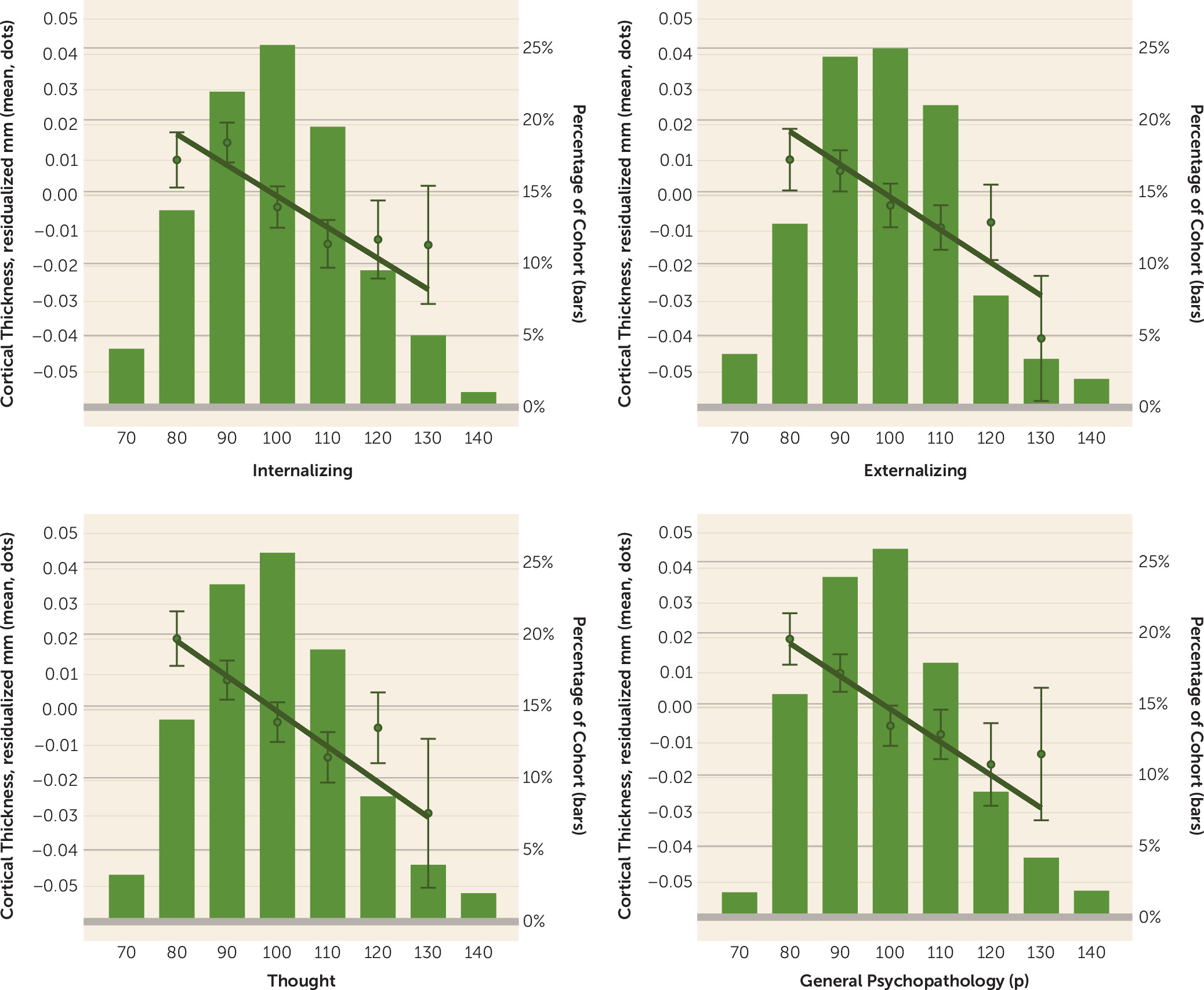
Global cortical thickness was normally distributed across all study members (mean=2.556 mm, SD=0.089). Each of the three psychopathology factors from the correlated-factor model was equally and similarly associated with global cortical thickness (Figure 1). Study members with high internalizing scores had thinner global neocortex (β=−0.156; 95% CI=−0.223, −0.089; p<0.0001), as did study members with high externalizing scores (β=−0.164; 95% CI=−0.232, −0.097; p<0.0001) and those with high thought disorder scores (β=−0.169; 95% CI=−0.234, −0.103; p<0.0001). Consistent with evidence pointing to similar negative associations between each of the three psychopathology factors and global neocortical thickness, study members with high scores on the transdiagnostic p factor also exhibited reduced mean neocortical thickness (β=−0.159; 95% CI=−0.224, −0.093; p<0.0001) (Figure 1).
FIGURE 1. Association of higher scores across the three broad diagnostic families of disorders and general psychopathology as captured by the p factor with thinner average neocortexa
a The regression coefficients reported in the text are based on the full distribution of factor scores. The circles and bars show the mean scores and standard errors, respectively, of individuals in each p factor score group; these groups have been clumped solely for graphing purposes (with group size >50). The values on the x-axis indicate the midpoint of the bins: (<75, 75 to <85, 85 to <95, 95 to <105, 105 to <115, 115 to <125, 125 to <135, and ≥135. The histograms depict the distribution of factor scores.
We also conducted several post hoc analyses. First, we examined the potential confounding effects of childhood socioeconomic status, medical disease, and psychoactive medication use at age 45 as well as image quality. These analyses revealed that all of the above associations were robust to the inclusion of these covariates (see Figure S5 in the online supplement). Second, we determined relative effect sizes of global cortical thickness using traditional case-control analyses by comparing four common diagnostic categories observed in the Dunedin Study—depression, anxiety, substance abuse, and schizophrenia—against healthy control subjects (the 147 control subjects in these analyses represent study members who have never met diagnostic criteria for any of the mental disorders assessed in the Dunedin Study until age 45; that is, they have enduring mental health [21]). Consistent with our factor-based analyses above, all of these case-control analyses revealed decreased global cortical thickness as a feature of diagnosis (see Figure S6 in the online supplement). Not surprisingly, the effect was largest for those diagnosed with schizophrenia, which falls at the extreme end of high p factor scores. Of note, these effect sizes were larger than those observed for the psychopathology factor scores, because they involve comparisons between extreme groups (i.e., diagnosed case subjects compared with healthy control subjects who have never met diagnostic criteria for a mental disorder).
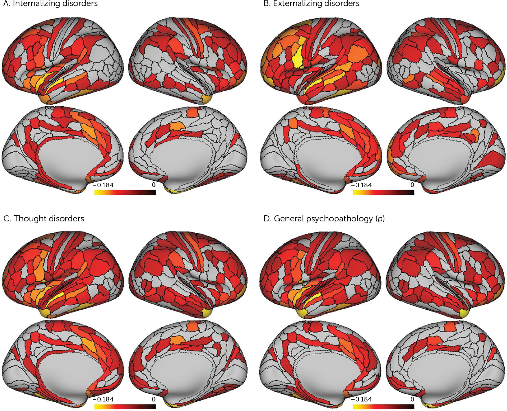
Parcel-wise analyses of 360 neocortical areas revealed that high scores on each of the three psychopathology factors were associated with widely distributed patterns of reduced regional cortical thickness (Figure 2A-C). High internalizing scores were associated with significant reductions in cortical thickness in 150 parcels, high externalizing scores with 171 parcels, and high thought disorder scores with 202 parcels (see Figure S7 in the online supplement for standardized betas and 95% confidence intervals for all 360 parcels for each factor). Consistent with the global effects reported above, parcel-wise analyses revealed that high p factor scores also were associated with widely distributed reductions in cortical thickness (Figure 2D). High p factor scores were associated with significant reductions in cortical thickness in 174 parcels (see Figure S7 in the online supplement). Direct contrasts of the parcel-wise cortical thickness associations with the p factor and the internalizing, externalizing, and thought disorder families of disorders revealed overlaps of 77%, 75%, and 99%, respectively.
FIGURE 2. Association of pervasive and highly overlapping patterns of thinner regional neocortex with higher scores across the three broad diagnostic families of disorders and general psychopathology as captured by the p factora
a Statistical parametric maps from parcel-wise analyses are shown to illustrate significant negative associations between cortical thickness and internalizing scores, externalizing scores, thought disorder scores, and p factor scores. All associations shown are false discovery rate corrected. Color bars reflect effect sizes (standardized betas).
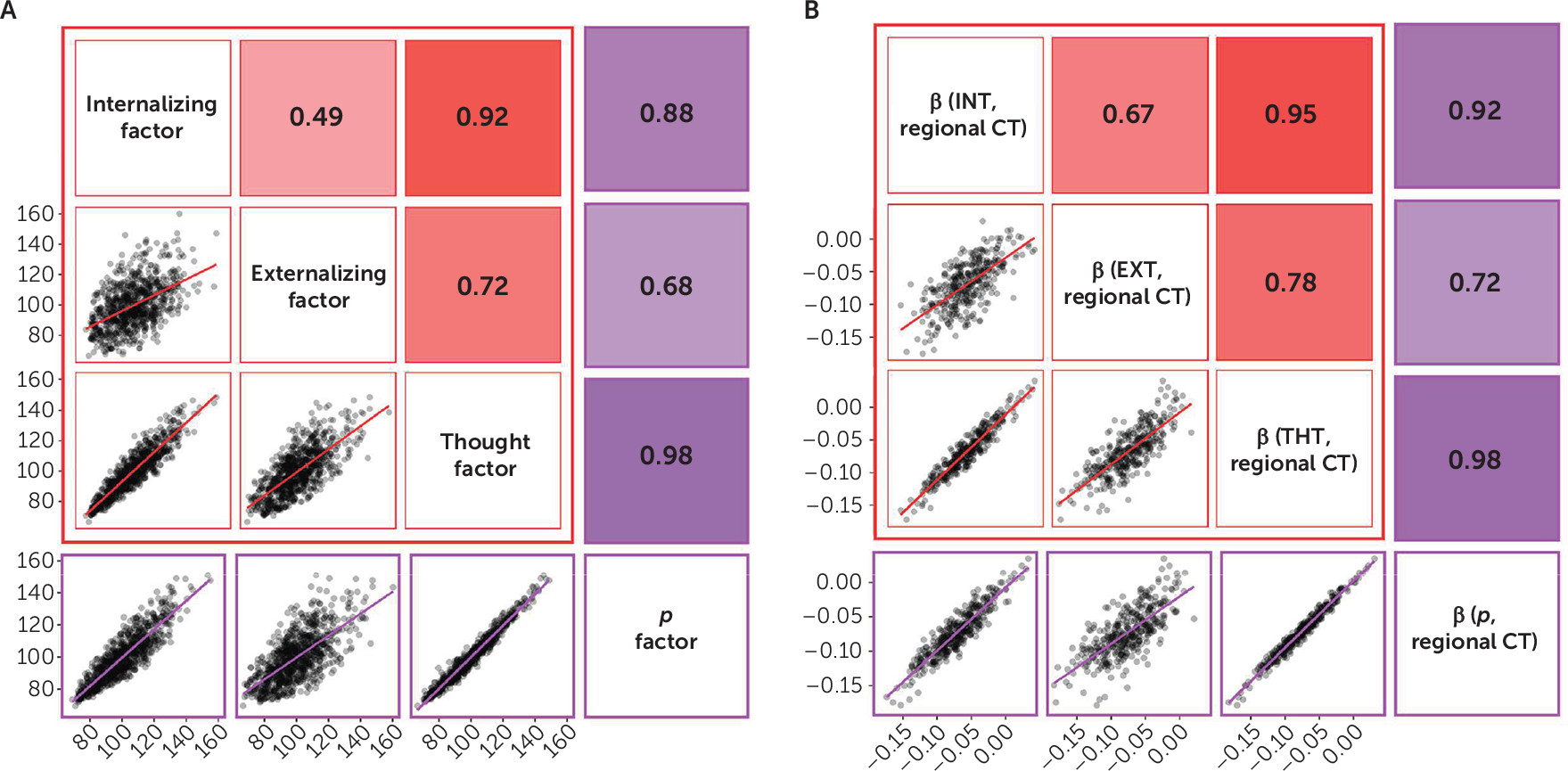
There were virtually no parcel-wise associations exhibiting reduced cortical thickness that were specific to any of the three broad diagnostic families of disorders (see Figure S8 in the online supplement). The pervasive and nonspecific nature of reduced neocortical thickness was further evident when we compared cortical thickness across all 360 parcels in relation to internalizing, externalizing, and thought disorder scores, as well as the p factor. The high correlations in Figure 3 (r values range from 0.67 to 0.95) show that those parcels exhibiting reduced cortical thickness among study members with high scores on one psychopathology factor (e.g., internalizing) were also reduced among study members with high scores on the other psychopathology factors (e.g., externalizing, thought disorder). This nonspecificity is well captured by evidence that the cortical thickness of the parcels that is reduced among study members with high scores on any of the three psychopathology factors is also reduced among study members with high p factor scores (r values range from 0.72 to 0.98).
FIGURE 3. Highly conserved and pervasive pattern of thinner neocortex across the three broader diagnostic families and the p factor mirrors relationships between the phenotypes themselvesa
a Correlations between scores for the three factors from the correlated-factors model (red boxes) and between these three factors and the p factor from the bifactor model (purple boxes) across all 861 study members are shown in panel A, and those between the standardized betas representing the parcel-wise cortical thinning associated with each factor across all 360 parcels are shown in panel B. The matrix cells below the diagonal show scatterplots of the associations. The matrix cells above the diagonal show their correlations expressed as Pearson’s r. The red and purple regression lines illustrate the slopes of associations between each pair of measures.

Surface Area

Global surface area was normally distributed across all study members (mean=185,472.1 mm2, SD=16,347.1). In contrast to the pervasive patterns of reduced cortical thickness found in study members with higher scores across all factors, a significant reduction in global surface area was present only in study members who scored higher on the externalizing factor (β=−0.062, CI=−0.117, −0.007; p=0.026). There were no significant associations between global surface area and scores on either the internalizing (β=−0.029, CI=−0.084, 0.026; p=0.304) or thought disorder factors (β=−0.047, CI=−0.100, 0.007; p=0.087) or the p factor (β=−0.044; CI=−0.098, 0.010; p=0.108). Parcel-wise surface areas were not significantly associated with internalizing, externalizing, thought disorder, or p factor scores. The lack of meaningful parcel-wise associations was further evident on visual examination of the standardized betas and 95% confidence intervals for all 360 parcels (see Figure S9 in the online supplement).

Network Enrichment

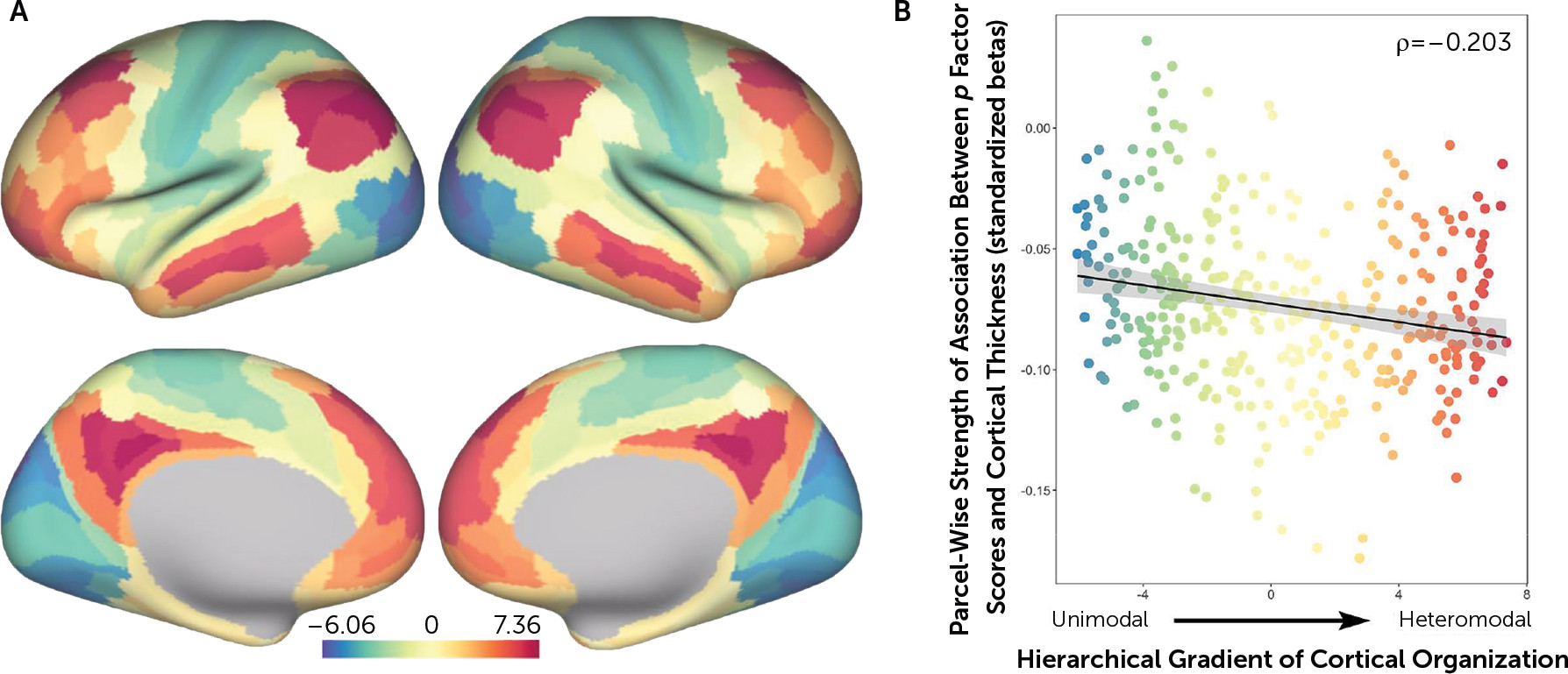
Given that high p factor scores were associated with a pervasive transdiagnostic pattern of reduced neocortical thickness, we conducted exploratory analyses to test whether larger than expected associations between p factor scores and reduced cortical thickness were present in heteromodal association cortices including the frontoparietal and default mode networks. Spatial permutation testing revealed a significant spatial correspondence between the strength of parcel-wise associations between cortical thickness and p factor scores (i.e., standardized betas) and a cortical gradient spanning from heteromodal association cortices on one end to unimodal sensory and somatomotor cortices on the other end (Figure 4). Specifically, parcels encompassing heteromodal association cortices tended to have larger negative associations between cortical thickness and p factor scores than parcels encompassing unimodal sensory and somatomotor cortices (Spearman’s rho=−0.203, p=0.049).
FIGURE 4. Magnitude of parcel-wise associations between p factor scores and cortical thickness across cortical networksa
a A cortical gradient spanning from heteromodal association cortices, including frontoparietal and default mode networks, on one end to unimodal sensory and somatomotor cortices on the other end is shown in panel A. As shown in panel B, spatial permutation testing revealed that parcels encompassing heteromodal association cortices tended to have larger negative associations (standardized betas) between cortical thickness and p factor scores than parcels encompassing unimodal sensory and somatomotor cortices (Spearman’s rho=−0.203, p=0.049).

Discussion

Our analyses suggest that there is little specificity in the brain structural correlates of mental disorders. Rather, a nonspecific and pervasive pattern of thinner neocortex appears to be a transdiagnostic feature of general psychopathology as indexed by the p factor. Consistent with our hypotheses that general psychopathology (p) drives the convergence of structural alterations common across disorders, we found 1) very few associations unique to the internalizing, externalizing, or thought disorder diagnostic families and 2) that associations with the p factor highly overlapped with those for the three diagnostic families. The pervasive and transdiagnostic nature of these associations is consistent with studies revealing that most structural brain differences are not unique to categorical mental disorders but rather are shared across disorders (3, 4, 11).
The present findings help to refine the interpretation of recent studies that have identified widely distributed reductions in neocortical gray matter volumes associated with higher general psychopathology (10, 11, 22). Specifically, our findings suggest that these previously reported associations may be driven by reduced neocortical thickness and not surface area, which together comprise gray matter volume. Unlike the broad mapping of reduced global and parcel-wise cortical thickness onto psychopathology, there were no significant associations with parcel-wise neocortical surface area. While there was a significant association with reduced global surface area, this was restricted to higher scores only on the externalizing factor. This is consistent with previous research showing that externalizing disorders tend to load least strongly on the p factor (7, 23). As we did not hypothesize this effect, and it could arise from chance, we refrain from extensive discussion. It raises the question of whether this specific association reflects early-life vulnerability to externalizing disorders as a result of differences in the developing brain (e.g., due to genetics or maltreatment) or a result of harm done to the brain from an externalizing lifestyle (e.g., due to drug use, sexually transmitted diseases, or violent injuries, including concussions) (24, 25). Regardless, the effect sizes associated with either global or parcel-wise surface area, irrespective of statistical significance, were an order of magnitude smaller than those observed for associations with global and parcel-wise cortical thickness.
Thus, the general pattern of associations suggests that reduced cortical thickness and not surface area is a transdiagnostic feature of mental disorders reflected in general psychopathology. Consistent with this pattern, a recent cross-disorder genome-wide association study identified four common variants predicted to affect the function of radial glia and interneurons critical for the development of cortical layers, which are reflected in cortical thickness (26). That said, the cytological and histological basis of cortical thickness remains unclear. MRI-derived estimates of cortical thickness do accurately reflect the width of the cortical mantle as determined on postmortem examination (27). Differences in cortical thickness, however, are unlikely to reflect numbers of neurons or loss of neurons over time (28). Rather, the thickness of the cortex likely reflects a combination of neuron size (i.e., degree of shrinkage) and dendritic arborization (i.e., degree of branching, spine density).
A thinner neocortex has been associated with a host of negative outcomes across the lifespan. For example, a thinner neocortex has been associated with lower intelligence in midlife (29) and is a feature of older brain age relative to chronological age, which is accompanied by greater cognitive impairment (30). A thinner cortex has also been observed in categorical mental disorders spanning the internalizing, externalizing, and thought disorder diagnostic families (e.g., 3133). Meanwhile, accelerated thinning has been associated with worsening daily functioning and other symptoms in Alzheimer’s disease (34, 35). Our secondary analyses of network enrichment are relevant here as they highlight preferential associations between higher p factor scores and reduced cortical thickness of parcels falling within heteromodal association cortices, including the frontoparietal and default mode networks, supporting higher cognitive processes and executive functions (36). Thus, while a pervasive pattern of reduced neocortical thickness may be a transdiagnostic feature of general psychopathology, it is possible that specific reductions in heteromodal association cortices (i.e., frontoparietal and default mode networks) may feature in the disordered form and content of thought hypothesized to represent the core of the p factor (10, 37). This is consistent with previous transdiagnostic research indicating alterations within frontoparietal and default mode networks supporting executive control and self-referential processes across diagnostic categories (2, 4, 36, 37). Further evidence comes from parallel neuroimaging studies implicating structural alterations within a cerebello-thalamo-cortical circuit supporting executive functions in the expression of the p factor (3840).
One limitation of this study is the availability of only a single, cross-sectional assessment of brain structure in midlife. Unfortunately, we have only recently been able to collect neuroimaging data in the Dunedin Study and we are unable to examine temporal order in the data. It remains to be determined whether a higher burden of psychopathology contributes to a pervasively thinner neocortex or if a pervasively thinner cortex contributes to a higher burden of psychopathology. Longitudinal collection of neuroimaging data beginning in early childhood and continuing throughout life is needed to address this question. Comparisons with other cross-sectional studies, as well as longitudinal data, are necessary to determine the extent to which the patterns observed here, at age 45, generalize across development or the extent to which associations between psychopathology and brain structure change across the lifespan. Second, while the Dunedin Study cohort is a population-representative birth cohort free of the selection biases often present in neuroimaging research, it is a predominantly white cohort born in the 1970s in one part of the world; therefore, results will require replication in other samples from other ethnic groups and countries.
Conclusions

Analyses of data collected from a large population-representative birth cohort in midlife revealed that a pervasively thinner neocortex is a transdiagnostic feature of mental disorders reflecting general psychopathology as indexed by the p factor. In addition to furthering the utility of the p factor in capturing the shared phenotypic features of common mental disorders, our findings reinforce the value of a transdiagnostic approach, ideally including explicit modeling of general psychopathology, in future neuropsychiatric research. More broadly, our findings underscore the possibility that a continued search for specificity among mental disorders may be not only elusive but also likely counterproductive in addressing existing gaps in etiology, treatment, and prevention research.

Acknowledgments

The authors thank members of the Advisory Board for the Dunedin Neuroimaging Study, Dunedin Study members, unit research staff, Pacific Radiology staff, and study founder Phil Silva, Ph.D., University of Otago.

Supplementary Material

File (appi.ajp.2020.19090934.ds001.pdf)

References

1.
Mazziotta JC, Toga AW, Frackowiak RSJ: Brain Mapping: The Disorders. San Diego, Academic Press, 2000
2.
Barch DM: The neural correlates of transdiagnostic dimensions of psychopathology. Am J Psychiatry 2017; 174:613–615
3.
Goodkind M, Eickhoff SB, Oathes DJ, et al: Identification of a common neurobiological substrate for mental illness. JAMA Psychiatry 2015; 72:305–315
4.
Sha Z, Wager TD, Mechelli A, et al: Common dysfunction of large-scale neurocognitive networks across psychiatric disorders. Biol Psychiatry 2019; 85:379–388
5.
Kessler RC, Chiu WT, Demler O, et al: Prevalence, severity, and comorbidity of 12-month DSM-IV disorders in the National Comorbidity Survey Replication. Arch Gen Psychiatry 2005; 62:617–627
6.
Krueger RF, Markon KE: Reinterpreting comorbidity: a model-based approach to understanding and classifying psychopathology. Annu Rev Clin Psychol 2006; 2:111–133
7.
Caspi A, Houts RM, Belsky DW, et al: The p factor: one general psychopathology factor in the structure of psychiatric disorders? Clin Psychol Sci 2014; 2:119–137
8.
Lahey BB, Applegate B, Hakes JK, et al: Is there a general factor of prevalent psychopathology during adulthood? J Abnorm Psychol 2012; 121:971–977
9.
Lahey BB, Krueger RF, Rathouz PJ, et al: A hierarchical causal taxonomy of psychopathology across the life span. Psychol Bull 2017; 143:142–186
10.
Caspi A, Moffitt TE: All for one and one for all: mental disorders in one dimension. Am J Psychiatry 2018; 175:831–844
11.
Kaczkurkin AN, Park SS, Sotiras A, et al: Evidence for dissociable linkage of dimensions of psychopathology to brain structure in youths. Am J Psychiatry 2019; 176:1000–1009
12.
Poulton R, Moffitt TE, Silva PA: The Dunedin Multidisciplinary Health and Development Study: overview of the first 40 years, with an eye to the future. Soc Psychiatry Psychiatr Epidemiol 2015; 50:679–693
13.
Glasser MF, Coalson TS, Robinson EC, et al: A multi-modal parcellation of human cerebral cortex. Nature 2016; 536:171–178
14.
R Core Team: R: A language and environment for statistical computing. Vienna, R Foundation for Statistical Computing, 2017 (https://www.R-project.org/)
15.
Benjamini Y, Hochberg Y: Controlling the false discovery rate: a practical and powerful approach to multiple testing. J R Stat Soc B 1995; 57:289–300
16.
Mesulam MM: From sensation to cognition. Brain 1998; 121:1013–1052
17.
Buckner RL, DiNicola LM: The brain’s default network: updated anatomy, physiology, and evolving insights. Nat Rev Neurosci 2019; 20:593–608
18.
Margulies DS, Ghosh SS, Goulas A, et al: Situating the default-mode network along a principal gradient of macroscale cortical organization. Proc Natl Acad Sci USA 2016; 113:12574–12579
19.
Alexander-Bloch AF, Shou H, Liu S, et al: On testing for spatial correspondence between maps of human brain structure and function. Neuroimage 2018; 178:540–551
20.
Váša F, Bullmore ET, Patel AX: Probabilistic thresholding of functional connectomes: application to schizophrenia. Neuroimage 2018; 172:326–340
21.
Schaefer JD, Caspi A, Belsky DW, et al: Enduring mental health: prevalence and prediction. J Abnorm Psychol 2017; 126:212–224
22.
Snyder HR, Hankin BL, Sandman CA, et al: Distinct patterns of reduced prefrontal and limbic grey matter volume in childhood general and internalizing psychopathology. Clin Psychol Sci 2017; 5:1001–1013
23.
Laceulle OM, Vollebergh WAM, Ormel J: The structure of psychopathology in adolescence: replication of a general psychopathology factor in the TRAILS study. Clin Psychol Sci 2015; 3:850–860
24.
Durazzo TC, Tosun D, Buckley S, et al: Cortical thickness, surface area, and volume of the brain reward system in alcohol dependence: relationships to relapse and extended abstinence. Alcohol Clin Exp Res 2011; 35:1187–1200
25.
Kaag AM, Crunelle CL, van Wingen G, et al: Relationship between trait impulsivity and cortical volume, thickness, and surface area in male cocaine users and non-drug using controls. Drug Alcohol Depend 2014; 144:210–217
26.
Schork AJ, Won H, Appadurai V, et al: A genome-wide association study of shared risk across psychiatric disorders implicates gene regulation during fetal neurodevelopment. Nat Neurosci 2019; 22:353–361
27.
Scholtens LH, de Reus MA, van den Heuvel MP: Linking contemporary high resolution magnetic resonance imaging to the von Economo legacy: a study on the comparison of MRI cortical thickness and histological measurements of cortical structure. Hum Brain Mapp 2015; 36:3038–3046
28.
Morrison JH, Hof PR: Life and death of neurons in the aging brain. Science 1997; 278:412–419
29.
Schnack HG, van Haren NEM, Brouwer RM, et al: Changes in thickness and surface area of the human cortex and their relationship with intelligence. Cereb Cortex 2015; 25:1608–1617
30.
Liem F, Varoquaux G, Kynast J, et al: Predicting brain-age from multimodal imaging data captures cognitive impairment. Neuroimage 2017; 148:179–188
31.
van Haren NEM, Schnack HG, Cahn W, et al: Changes in cortical thickness during the course of illness in schizophrenia. Arch Gen Psychiatry 2011; 68:871–880
32.
Zhao K, Liu H, Yan R, et al: Altered patterns of association between cortical thickness and subcortical volume in patients with first episode major depressive disorder: a structural MRI study. Psychiatry Res Neuroimaging 2017; 260:16–22
33.
Shaw P, Lerch J, Greenstein D, et al: Longitudinal mapping of cortical thickness and clinical outcome in children and adolescents with attention-deficit/hyperactivity disorder. Arch Gen Psychiatry 2006; 63:540–549
34.
Donovan NJ, Wadsworth LP, Lorius N, et al: Regional cortical thinning predicts worsening apathy and hallucinations across the Alzheimer disease spectrum. Am J Geriatr Psychiatry 2014; 22:1168–1179
35.
Marshall GA, Lorius N, Locascio JJ, et al: Regional cortical thinning and cerebrospinal biomarkers predict worsening daily functioning across the Alzheimer’s disease spectrum. J Alzheimers Dis 2014; 41:719–728
36.
Menon V: Large-scale brain networks and psychopathology: a unifying triple network model. Trends Cogn Sci 2011; 15:483–506
37.
Elliott ML, Romer A, Knodt AR, et al: A connectome-wide functional signature of transdiagnostic risk for mental illness. Biol Psychiatry 2018; 84:452–459
38.
Romer AL, Knodt AR, Houts R, et al: Structural alterations within cerebellar circuitry are associated with general liability for common mental disorders. Mol Psychiatry 2018; 23:1084–1090
39.
Romer AL, Knodt AR, Sison ML, et al: Replicability of structural brain alterations associated with general psychopathology: evidence from a population-representative birth cohort. Mol Psychiatry (Epub ahead of print, December 3, 2019)
40.
Moberget T, Alnæs D, Kaufmann T, et al: Cerebellar gray matter volume is associated with cognitive function and psychopathology in adolescence. Biol Psychiatry 2019; 86:65–75

Information & Authors

Information

Published In

Go to American Journal of Psychiatry
American Journal of Psychiatry
Pages: 174 - 182
PubMed: 32600153

History

Received: 10 September 2019
Revision received: 25 February 2020
Accepted: 30 March 2020
Published online: 30 June 2020
Published in print: February 01, 2021

Keywords

  1. Neuroimaging
  2. Cortical Thickness
  3. General Psychopathology
  4. Transdiagnostic
  5. p Factor

Authors

Details

Adrienne L. Romer, Ph.D.
Laboratory of NeuroGenetics (Romer, Elliott, Knodt, Sison, Hariri), Department of Psychology and Neuroscience (Romer, Knodt, Houts, Moffitt, Caspi, Hariri), Department of Psychiatry and Behavioral Sciences (Moffitt, Caspi), and Center for Genomic and Computational Biology (Moffitt, Caspi), Duke University, Durham, N.C.; Dunedin Multidisciplinary Health and Development Research Unit, Department of Psychology, University of Otago, Dunedin, New Zealand (Ireland, Ramrakha, Poulton); Christchurch Radiology Group, Christchurch, New Zealand (Keenan); Department of Medicine, University of Otago, Christchurch (Melzer); New Zealand Brain Research Institute, Christchurch (Melzer); Social, Genetic, and Developmental Psychiatry Research Center, Institute of Psychiatry, Psychology, and Neuroscience, King’s College London (Moffitt, Caspi); and McLean Hospital/Harvard Medical School, Belmont, Mass. (Romer).
Maxwell L. Elliott, M.A.
Laboratory of NeuroGenetics (Romer, Elliott, Knodt, Sison, Hariri), Department of Psychology and Neuroscience (Romer, Knodt, Houts, Moffitt, Caspi, Hariri), Department of Psychiatry and Behavioral Sciences (Moffitt, Caspi), and Center for Genomic and Computational Biology (Moffitt, Caspi), Duke University, Durham, N.C.; Dunedin Multidisciplinary Health and Development Research Unit, Department of Psychology, University of Otago, Dunedin, New Zealand (Ireland, Ramrakha, Poulton); Christchurch Radiology Group, Christchurch, New Zealand (Keenan); Department of Medicine, University of Otago, Christchurch (Melzer); New Zealand Brain Research Institute, Christchurch (Melzer); Social, Genetic, and Developmental Psychiatry Research Center, Institute of Psychiatry, Psychology, and Neuroscience, King’s College London (Moffitt, Caspi); and McLean Hospital/Harvard Medical School, Belmont, Mass. (Romer).
Annchen R. Knodt, M.S.
Laboratory of NeuroGenetics (Romer, Elliott, Knodt, Sison, Hariri), Department of Psychology and Neuroscience (Romer, Knodt, Houts, Moffitt, Caspi, Hariri), Department of Psychiatry and Behavioral Sciences (Moffitt, Caspi), and Center for Genomic and Computational Biology (Moffitt, Caspi), Duke University, Durham, N.C.; Dunedin Multidisciplinary Health and Development Research Unit, Department of Psychology, University of Otago, Dunedin, New Zealand (Ireland, Ramrakha, Poulton); Christchurch Radiology Group, Christchurch, New Zealand (Keenan); Department of Medicine, University of Otago, Christchurch (Melzer); New Zealand Brain Research Institute, Christchurch (Melzer); Social, Genetic, and Developmental Psychiatry Research Center, Institute of Psychiatry, Psychology, and Neuroscience, King’s College London (Moffitt, Caspi); and McLean Hospital/Harvard Medical School, Belmont, Mass. (Romer).
Maria L. Sison, B.S.
Laboratory of NeuroGenetics (Romer, Elliott, Knodt, Sison, Hariri), Department of Psychology and Neuroscience (Romer, Knodt, Houts, Moffitt, Caspi, Hariri), Department of Psychiatry and Behavioral Sciences (Moffitt, Caspi), and Center for Genomic and Computational Biology (Moffitt, Caspi), Duke University, Durham, N.C.; Dunedin Multidisciplinary Health and Development Research Unit, Department of Psychology, University of Otago, Dunedin, New Zealand (Ireland, Ramrakha, Poulton); Christchurch Radiology Group, Christchurch, New Zealand (Keenan); Department of Medicine, University of Otago, Christchurch (Melzer); New Zealand Brain Research Institute, Christchurch (Melzer); Social, Genetic, and Developmental Psychiatry Research Center, Institute of Psychiatry, Psychology, and Neuroscience, King’s College London (Moffitt, Caspi); and McLean Hospital/Harvard Medical School, Belmont, Mass. (Romer).
David Ireland, Ph.D.
Laboratory of NeuroGenetics (Romer, Elliott, Knodt, Sison, Hariri), Department of Psychology and Neuroscience (Romer, Knodt, Houts, Moffitt, Caspi, Hariri), Department of Psychiatry and Behavioral Sciences (Moffitt, Caspi), and Center for Genomic and Computational Biology (Moffitt, Caspi), Duke University, Durham, N.C.; Dunedin Multidisciplinary Health and Development Research Unit, Department of Psychology, University of Otago, Dunedin, New Zealand (Ireland, Ramrakha, Poulton); Christchurch Radiology Group, Christchurch, New Zealand (Keenan); Department of Medicine, University of Otago, Christchurch (Melzer); New Zealand Brain Research Institute, Christchurch (Melzer); Social, Genetic, and Developmental Psychiatry Research Center, Institute of Psychiatry, Psychology, and Neuroscience, King’s College London (Moffitt, Caspi); and McLean Hospital/Harvard Medical School, Belmont, Mass. (Romer).
Renate Houts, Ph.D.
Laboratory of NeuroGenetics (Romer, Elliott, Knodt, Sison, Hariri), Department of Psychology and Neuroscience (Romer, Knodt, Houts, Moffitt, Caspi, Hariri), Department of Psychiatry and Behavioral Sciences (Moffitt, Caspi), and Center for Genomic and Computational Biology (Moffitt, Caspi), Duke University, Durham, N.C.; Dunedin Multidisciplinary Health and Development Research Unit, Department of Psychology, University of Otago, Dunedin, New Zealand (Ireland, Ramrakha, Poulton); Christchurch Radiology Group, Christchurch, New Zealand (Keenan); Department of Medicine, University of Otago, Christchurch (Melzer); New Zealand Brain Research Institute, Christchurch (Melzer); Social, Genetic, and Developmental Psychiatry Research Center, Institute of Psychiatry, Psychology, and Neuroscience, King’s College London (Moffitt, Caspi); and McLean Hospital/Harvard Medical School, Belmont, Mass. (Romer).
Sandhya Ramrakha, Ph.D.
Laboratory of NeuroGenetics (Romer, Elliott, Knodt, Sison, Hariri), Department of Psychology and Neuroscience (Romer, Knodt, Houts, Moffitt, Caspi, Hariri), Department of Psychiatry and Behavioral Sciences (Moffitt, Caspi), and Center for Genomic and Computational Biology (Moffitt, Caspi), Duke University, Durham, N.C.; Dunedin Multidisciplinary Health and Development Research Unit, Department of Psychology, University of Otago, Dunedin, New Zealand (Ireland, Ramrakha, Poulton); Christchurch Radiology Group, Christchurch, New Zealand (Keenan); Department of Medicine, University of Otago, Christchurch (Melzer); New Zealand Brain Research Institute, Christchurch (Melzer); Social, Genetic, and Developmental Psychiatry Research Center, Institute of Psychiatry, Psychology, and Neuroscience, King’s College London (Moffitt, Caspi); and McLean Hospital/Harvard Medical School, Belmont, Mass. (Romer).
Richie Poulton, Ph.D.
Laboratory of NeuroGenetics (Romer, Elliott, Knodt, Sison, Hariri), Department of Psychology and Neuroscience (Romer, Knodt, Houts, Moffitt, Caspi, Hariri), Department of Psychiatry and Behavioral Sciences (Moffitt, Caspi), and Center for Genomic and Computational Biology (Moffitt, Caspi), Duke University, Durham, N.C.; Dunedin Multidisciplinary Health and Development Research Unit, Department of Psychology, University of Otago, Dunedin, New Zealand (Ireland, Ramrakha, Poulton); Christchurch Radiology Group, Christchurch, New Zealand (Keenan); Department of Medicine, University of Otago, Christchurch (Melzer); New Zealand Brain Research Institute, Christchurch (Melzer); Social, Genetic, and Developmental Psychiatry Research Center, Institute of Psychiatry, Psychology, and Neuroscience, King’s College London (Moffitt, Caspi); and McLean Hospital/Harvard Medical School, Belmont, Mass. (Romer).
Ross Keenan, M.B.Ch.B
Laboratory of NeuroGenetics (Romer, Elliott, Knodt, Sison, Hariri), Department of Psychology and Neuroscience (Romer, Knodt, Houts, Moffitt, Caspi, Hariri), Department of Psychiatry and Behavioral Sciences (Moffitt, Caspi), and Center for Genomic and Computational Biology (Moffitt, Caspi), Duke University, Durham, N.C.; Dunedin Multidisciplinary Health and Development Research Unit, Department of Psychology, University of Otago, Dunedin, New Zealand (Ireland, Ramrakha, Poulton); Christchurch Radiology Group, Christchurch, New Zealand (Keenan); Department of Medicine, University of Otago, Christchurch (Melzer); New Zealand Brain Research Institute, Christchurch (Melzer); Social, Genetic, and Developmental Psychiatry Research Center, Institute of Psychiatry, Psychology, and Neuroscience, King’s College London (Moffitt, Caspi); and McLean Hospital/Harvard Medical School, Belmont, Mass. (Romer).
Tracy R. Melzer, Ph.D.
Laboratory of NeuroGenetics (Romer, Elliott, Knodt, Sison, Hariri), Department of Psychology and Neuroscience (Romer, Knodt, Houts, Moffitt, Caspi, Hariri), Department of Psychiatry and Behavioral Sciences (Moffitt, Caspi), and Center for Genomic and Computational Biology (Moffitt, Caspi), Duke University, Durham, N.C.; Dunedin Multidisciplinary Health and Development Research Unit, Department of Psychology, University of Otago, Dunedin, New Zealand (Ireland, Ramrakha, Poulton); Christchurch Radiology Group, Christchurch, New Zealand (Keenan); Department of Medicine, University of Otago, Christchurch (Melzer); New Zealand Brain Research Institute, Christchurch (Melzer); Social, Genetic, and Developmental Psychiatry Research Center, Institute of Psychiatry, Psychology, and Neuroscience, King’s College London (Moffitt, Caspi); and McLean Hospital/Harvard Medical School, Belmont, Mass. (Romer).
Terrie E. Moffitt, Ph.D.
Laboratory of NeuroGenetics (Romer, Elliott, Knodt, Sison, Hariri), Department of Psychology and Neuroscience (Romer, Knodt, Houts, Moffitt, Caspi, Hariri), Department of Psychiatry and Behavioral Sciences (Moffitt, Caspi), and Center for Genomic and Computational Biology (Moffitt, Caspi), Duke University, Durham, N.C.; Dunedin Multidisciplinary Health and Development Research Unit, Department of Psychology, University of Otago, Dunedin, New Zealand (Ireland, Ramrakha, Poulton); Christchurch Radiology Group, Christchurch, New Zealand (Keenan); Department of Medicine, University of Otago, Christchurch (Melzer); New Zealand Brain Research Institute, Christchurch (Melzer); Social, Genetic, and Developmental Psychiatry Research Center, Institute of Psychiatry, Psychology, and Neuroscience, King’s College London (Moffitt, Caspi); and McLean Hospital/Harvard Medical School, Belmont, Mass. (Romer).
Avshalom Caspi, Ph.D.
Laboratory of NeuroGenetics (Romer, Elliott, Knodt, Sison, Hariri), Department of Psychology and Neuroscience (Romer, Knodt, Houts, Moffitt, Caspi, Hariri), Department of Psychiatry and Behavioral Sciences (Moffitt, Caspi), and Center for Genomic and Computational Biology (Moffitt, Caspi), Duke University, Durham, N.C.; Dunedin Multidisciplinary Health and Development Research Unit, Department of Psychology, University of Otago, Dunedin, New Zealand (Ireland, Ramrakha, Poulton); Christchurch Radiology Group, Christchurch, New Zealand (Keenan); Department of Medicine, University of Otago, Christchurch (Melzer); New Zealand Brain Research Institute, Christchurch (Melzer); Social, Genetic, and Developmental Psychiatry Research Center, Institute of Psychiatry, Psychology, and Neuroscience, King’s College London (Moffitt, Caspi); and McLean Hospital/Harvard Medical School, Belmont, Mass. (Romer).
Ahmad R. Hariri, Ph.D. [email protected]
Laboratory of NeuroGenetics (Romer, Elliott, Knodt, Sison, Hariri), Department of Psychology and Neuroscience (Romer, Knodt, Houts, Moffitt, Caspi, Hariri), Department of Psychiatry and Behavioral Sciences (Moffitt, Caspi), and Center for Genomic and Computational Biology (Moffitt, Caspi), Duke University, Durham, N.C.; Dunedin Multidisciplinary Health and Development Research Unit, Department of Psychology, University of Otago, Dunedin, New Zealand (Ireland, Ramrakha, Poulton); Christchurch Radiology Group, Christchurch, New Zealand (Keenan); Department of Medicine, University of Otago, Christchurch (Melzer); New Zealand Brain Research Institute, Christchurch (Melzer); Social, Genetic, and Developmental Psychiatry Research Center, Institute of Psychiatry, Psychology, and Neuroscience, King’s College London (Moffitt, Caspi); and McLean Hospital/Harvard Medical School, Belmont, Mass. (Romer).

Notes

Send correspondence to Dr. Hariri ([email protected]).

Competing Interests

The authors report no financial relationships with commercial interests.

Funding Information

Supported by National Institute on Aging grants R01AG032282 and R01AG049789 and UK Medical Research Council grant MR/P005918/1. Additional support was provided by the Jacobs Foundation. Dr. Romer and Mr. Elliott received support from the National Science Foundation Graduate Research Fellowship under grants DGE-1106401 and DGE-1644868, respectively. Dr. Melzer received support from a Sir Charles Hercus Career Development Fellowship from the New Zealand Health Research Council (17/039). The Dunedin Multidisciplinary Health and Development Research Unit was supported by the New Zealand Health Research Council and the New Zealand Ministry of Business, Innovation, and Employment.

Metrics & Citations

Metrics

Citations

Export Citations

If you have the appropriate software installed, you can download article citation data to the citation manager of your choice. Simply select your manager software from the list below and click Download.

For more information or tips please see 'Downloading to a citation manager' in the Help menu.

Format
Citation style
Style
Copy to clipboard

View Options

View options

PDF/EPUB

View PDF/EPUB

Login options

Already a subscriber? Access your subscription through your login credentials or your institution for full access to this article.

Personal login Institutional Login Open Athens login
Purchase Options

Purchase this article to access the full text.

PPV Articles - American Journal of Psychiatry

PPV Articles - American Journal of Psychiatry

Not a subscriber?

Subscribe Now / Learn More

PsychiatryOnline subscription options offer access to the DSM-5-TR® library, books, journals, CME, and patient resources. This all-in-one virtual library provides psychiatrists and mental health professionals with key resources for diagnosis, treatment, research, and professional development.

Need more help? PsychiatryOnline Customer Service may be reached by emailing [email protected] or by calling 800-368-5777 (in the U.S.) or 703-907-7322 (outside the U.S.).

Media

Figures

Other

Tables

Share

Share

Share article link

Share