Anxiety, posttraumatic stress disorder (PTSD), and other trauma- and fear-related disorders are debilitating and common, are important causes of suicide, and cost society tens of billions of dollars. Despite their costs, morbidity, and mortality, these disorders may be among the most tractable of psychiatric illnesses from a neuroscience perspective, for several reasons: 1) the shared neural circuitry underlying threat processing across mammals, which allows translational approaches; 2) major advances in the neurobiology of threat and neural plasticity over recent decades; 3) the ability to examine shared intermediate phenotypes of fear processing across species; 4) the shared behavioral and neural mechanisms that underlie extinction and exposure-based therapies; and 5) in at least some cases, such as PTSD, a specific causal event for illness onset (e.g., exposure to an external trauma) that can be utilized for research and intervention. Nonetheless, established new treatments for these disorders have been slow to arise from this knowledge. Modern psychiatry seeks to expand our understanding of the biological mechanisms of fear-, stress-, and trauma-related disorders with the goal of identifying targeted, rationally designed interventions for these devastating disorders, based on advances in biology and neuroscience.
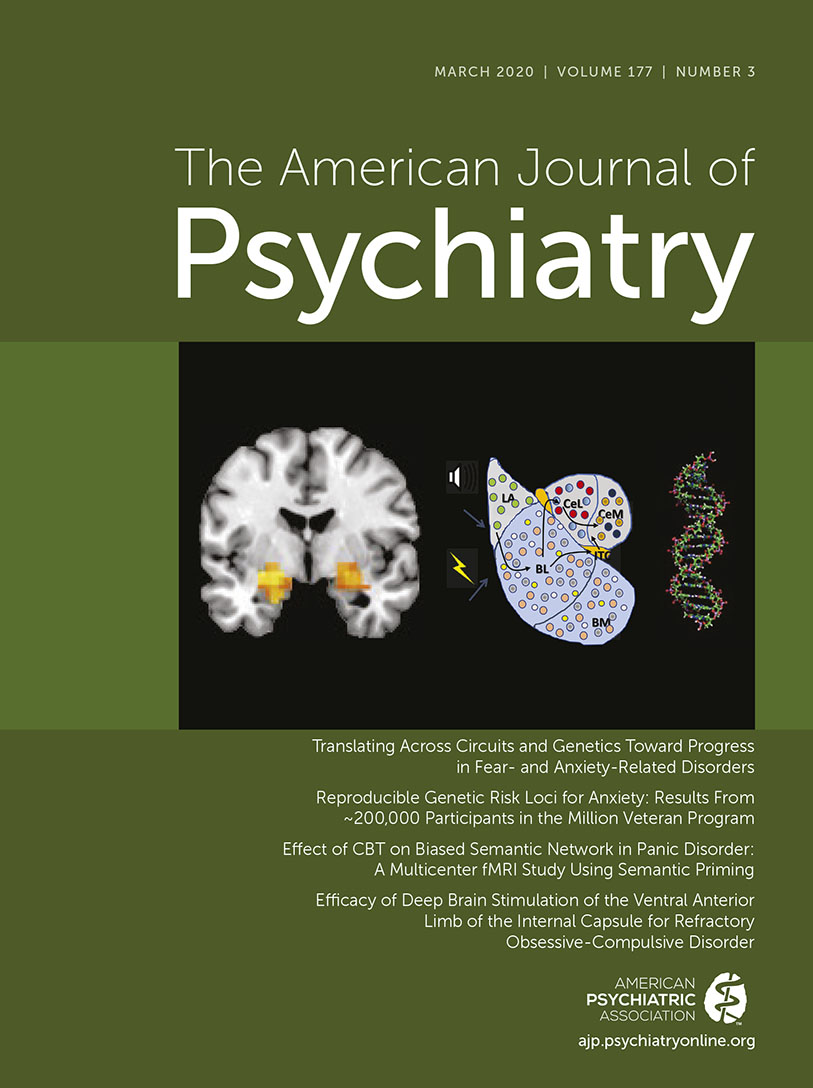
To fully understand fear- and anxiety-related disorders, we need to know what behaviors are affected, which brain regions are involved, and which genes, molecules, and cell types might be involved. The prefrontal cortex, the hippocampus, the insula, and the amygdala are often associated with fear and threat processing (
1–
4). In particular, different amygdala subregions and specific cell populations are being determined to have distinct roles in fear and extinction processing (
5–
12). Furthermore, large-scale international consortia and collaborations have allowed genome-wide association studies (GWASs) of hundreds of thousands of individuals with PTSD and anxiety disorders, confirming their heritability and shared polygenic risk with other severe psychiatric disorders, as well as pointing to novel genes and pathways while also confirming their association with well-known molecular pathways (
13–
16).
We are at a unique time in history, having an embarrassment of riches with new tools for understanding dynamic human brain activity, with powerful combinatorial machine learning approaches, and with enormous biological data sets, from hundreds of thousands of subjects with genome-wide genomics data to single-cell sequencing on millions of cells across brain regions and with causal tools for precise neural circuit manipulation. It is hoped that such promising new approaches will allow integration of amygdala-based microcircuits of threat processing across animal model systems (
1,
2) with postmortem human amygdala (and other related circuits) tissue (e.g., PsychENCODE) (
17) and with large-scale consortium initiatives, such as the Psychiatric Genomics Consortium for PTSD (
18), the Army Study to Assess Risk and Resilience in Servicemembers (Army STARRS) (
19), the Advancing Understanding of Recovery After Trauma (AURORA) Study (
20), and the Million Veteran Program (
21), discussed below. Such progress would allow integration of cell-type-specific regulation of behavior with genomic and biomarker discovery samples from human cohorts.
What is not yet clear is how to integrate the avalanche of new data and findings with our current understanding of the neural circuits of fear and threat processing. Furthermore, how are these circuits functionally and even structurally altered by interventions, from exposure therapy to targeted neurotherapeutics to neurobiologically targeted pharmacology? Such future approaches may allow for rational identification and testing of novel target compounds and biologicals as potential interventions, which can then be efficiently brought from bench to bedside, leading to the next generation of more powerful treatments for PTSD, anxiety, and other fear- and trauma-related disorders.
Intersection Of Neural Circuitry Of Threat And Extinction In Fear- And Anxiety-Related Disorders
Much of the translational progress in understanding mechanisms of anxiety- and fear-related disorders comes from the intersection of decades of work in understanding the biology of mammalian fear or threat conditioning, based on more than a century of work since Pavlov initially worked out the basic rules underlying classical conditioning theory (
22). In the past few decades, groundbreaking work, primarily in rodents, has informed our understanding of the role of the amygdala, modulated by the medial prefrontal cortex (mPFC), hippocampus, thalamus, and insula, among other structures, in underlying the neural mechanisms of behavioral Pavlovian threat conditioning (
4,
5,
7,
9,
23–
27). Importantly, this work and the principal conserved brain regions involved have been well replicated across species in nonhuman primates (
28–
31). Finally, many studies have demonstrated that Pavlovian fear conditioning and its correlated neural mechanisms are dysregulated in human anxiety- and fear-related disorders such as PTSD (
32–
41).
The past decade of progress has focused on more precise dissection of the behavioral, neural, and genetic components underlying fear-related disorders. Behaviorally, it is important to note that fear memory itself can be broken down further into its component parts. During the minutes to hours (to possibly days) following trauma exposure, the fear/trauma memory remains in a labile state, called the consolidation period (
42–
44). A number of exciting areas of inquiry suggest that new pharmacotherapeutic and psychotherapeutic approaches can be initiated that may inhibit the emotional component of fear memory consolidation without markedly affecting the explicit memory formation (
45–
48).
After consolidation of the initial aversive memory, multiple behavioral and cognitive pathways (with component underlying neural circuitry) contribute to healthy recovery from the initial fear or trauma memory (even if the initial triggering events are unknown, as may be the case in panic, obsessive-compulsive disorder [OCD], and phobias) (
2,
46,
47,
49,
50). Processes such as sensitization on reexposure to cues, generalization of context and cue triggers, avoidance of reminder triggers, reinstatement following new threats, and renewal of fear and anxiety behaviors in different contexts following treatment are all associated with increased anxiety- and fear-related symptoms and further comorbidity. In contrast, extinction of fear, discrimination of cues and contexts, and actively facing reminder cues and contexts (allowing natural extinction to occur) are all processes that support recovery. We hope that by understanding multiple components of fear memory modulation in humans and mice, a number of novel, powerful, and targeted treatment and intervention approaches may become possible (
46,
47,
51).
Extinction Of Fear As An Example Of Translational Success
The symptoms of PTSD, panic, and other fear- and anxiety-related disorders can be explained, at least in part, as an inability to inhibit learned fear even during times and contexts that are safe. Notably, multiple studies have demonstrated that fear inhibition or extinction is impaired across PTSD and other anxiety-related disorders (
23,
35,
39–
41,
52–
59). Some of the earliest reports of naturalistic observation following trauma exposure suggested that a failure to naturally recover or to learn to inhibit trauma-based fear reactions is central to these disorders (
60). Other work has demonstrated that amygdala activation is high in individuals with PTSD and that there are delays in inhibition or extinction of fear. Thus, deficits in inhibiting or extinguishing fear are considered a crucial central intermediate phenotype in PTSD and related anxiety- and fear-related disorders.
The observation of failure to extinguish as an important construct in fear- and anxiety-related disorders is exciting for several reasons. From a clinical perspective, exposure-based therapy techniques—which are a central element of cognitive-behavioral therapy (CBT) and its many more specific versions, including cognitive processing therapy, eye movement desensitization and reprocessing, skill training in affective regulation, and of course prolonged exposure therapy—are among the best empirically supported treatments across the range of fear- and anxiety-related disorders. Increasingly, the elements that underscore the emotional learning process of extinction of threat behaviors are appreciated to be the same elements underlying the efficacy of exposure to feared cues and contexts. This provides for a relatively straightforward target for intervention, in which neural circuitry and behavioral psychology intersect in a predictable, reliable, and robust manner.
The Promise Of Fear- And Anxiety Disorders As Translational “Wins” For Psychiatry
Notably, the Research Domain Criteria project (RDoC) of the National Institute of Mental Health (
61) chose “negative valence systems” and, specifically, the constructs of acute threat (fear), potential threat (anxiety), and sustained threat (chronic stress) as among the first RDoC templates. This was in part due to the progress in understanding the neurocircuitry of fear, threat, and aversive behaviors across levels of analyses and model systems. The translation of extinction of fear across species and interventions provides a great example.
Pavlov initially defined extinction of conditioned fear in his classic studies (
22). Decades later, historical leaders in the field such as Rescorla and Bouton provided behavioral evidence that extinction of a prior fear memory did not lead to its forgetting or erasure, but rather to an apparent new memory trace representing fear memory inhibition (
62–
65). In the early 1990s, Michael Davis’s lab at Yale University initially demonstrated the critical observation that extinction of fear required active new amygdala-dependent synaptic plasticity (
66,
67). In the past two decades, a considerable amount of additional research has examined cellular and molecular mechanisms of fear inhibition and extinction. Substantial data now support a variety of mechanisms underlying synaptic plasticity as critical for new extinction learning. For example, brain-derived neurotrophic factor (BDNF) and its receptor TrkB have been shown to be required for memory formation related to fear and its extinction (
68–
71). Furthermore, following the demonstration that extinction memories required
N-methyl-
d-aspartate (NMDA)–dependent plasticity, we demonstrated that a partial agonist at the NMDA receptor,
d-cycloserine, enhances the consolidation of extinction of fear in rodents (
72) and began to translate this finding to enhancement of extinction paradigms in humans (
73). This finding has now been replicated repeatedly in rodents and humans (
74–
81).
Notably, additional studies that failed to replicate the
d-cycloserine effect, along with a number of more recent negative findings, have now begun to be better understood with the observation that NMDA-dependent plasticity enhances both memory reconsolidation and memory extinction (
43,
82,
83). These two opposing processes of strengthening versus inhibiting preexisting memory traces always appear to be at odds on reactivation of a prior fearful memory. This is particularly interesting with clinical translation, given the long-standing observation that reexposure is not always successful, and a variety of tools have been implemented clinically to try to ensure and improve extinction processes over sensitization or reconsolidation processes during memory exposure. The use of cognitive enhancers to improve extinction is now an accepted translational paradigm from bench to bedside and is considered a paradigm-shifting approach. However, this approach has the limitation that both BDNF- and NMDA-dependent plasticity support both extinction and fear learning. Next-generation approaches, as discussed later, hope to identify cell populations or microcircuits, based on translational neuroscience, that are valence-specific and support fear inhibition or extinction and not fear acquisition, expression, or reconsolidation. Targeting cells and circuits, either pharmacologically or neurotherapeutically, that directly enhance extinction or block reconsolidation could provide a true intersection of targeted intervention, combining neurobiological insights with psychotherapeutic tools to drive specific memories.
New Findings And Potential Translational Relevance
Memory Representation, Priming, Generalization, And Anxiety Disorders
In addition to the amygdala, the anterior cingulate cortex (ACC) is one region involved in associative memory formation, conflict and aversive memory expression, and PTSD and anxiety disorders (
34,
84,
85). A study reported in this issue of the
Journal by Yang et al. (
86) examines both the behavioral and semantic representation, as well as neural activation of the ACC, in panic-related representations and exposure therapy in panic disorder. From a translational perspective, this work is exciting in a number of ways. While the concept of priming is primarily a human psychology construct, behavioral studies in model systems have long demonstrated the role of “preparedness” for a threat and overactivation of a threat network, via generalization of cues and contexts that are more likely to contribute to a threat response, despite a relatively low actual probability of danger. Extinction of cues (which appears to depend on the hippocampus/amygdala and mPFC/ACC) in large part is context based, and successful extinction often leads to generalization of the extinction contexts as well (
50,
87). Thus, it is exciting and promising that exposure-based CBT for panic disorder results in decreased priming and, possibly, increased generalization of extinguished cues (with decreased negative valence), along with decreased brain network activity corresponding to such processes.
Data-Driven Approaches For Brain Activity-Based Subtyping In PTSD
Recent work has also examined unbiased data-driven approaches to understanding brain activity patterns that may underlie different biologically distinct forms of anxiety and fear-related disorders. In another article in this issue, Maron-Katz et al. (
88) examine resting-state functional MRI (fMRI) and dynamic brain activity to detect subtypes of PTSD. In parallel with neuroimaging, new analytic approaches with high-density EEG are allowing unsurpassed collection of real-time brain activity in a low-cost fashion that might be more feasible to disseminate. Toll et al. (
89) further demonstrate progress in biotyping in PTSD using EEG in this issue of the
Journal. In addition to the possibility of novel pathways and subgrouping tools, it is interesting that among the primary fMRI findings in the Maron-Katz et al. study (
88) was the identification of visual and sensorimotor findings important in PTSD. As noted by the authors, although abnormalities in frontoparietal connectivity and activity in PTSD case subjects have been reported in several studies, abnormalities in connectivity and activity of sensorimotor and visual regions have been less often seen. Importantly, the translational literature would suggest that these areas are highly likely to be involved in the aftermath of trauma exposure. In fact, in mice, repeated auditory tone–paired foot shocks have been shown to increase voxel-based morphometry gray matter density, likely a result of increased spine and dendritic spine density, in the auditory cortex, sensorimotor regions, and amygdala (
90–
93). These data, combined with a robust literature on structural representation of memory engrams across a large range of primary and secondary sensory areas, suggest that while the amygdala, dorsal ACC, and hippocampus may be “hubs” for memory formation, the actual encoding and sensitization of trauma is likely distributed across the full network encoding the memory (
2,
94–
96). Thus, in addition to the important and hopefully soon-to-be-clinically-utilized tools of EEG- and MRI-based biotyping for anxiety disorders, understanding the networks that may be differentially regulated in these biotypes will expand our neuroscience understanding of risk and resilience.
Deep Brain Stimulation of Specific Limbic Circuits for OCD Treatment
The “holy grail” of a neural circuit model of anxiety disorders would be to utilize the mechanistic understanding of neural circuitry for direct intervention and treatment. Denys et al. (
97), in a study reported in this issue, add to the growing literature suggesting the effectiveness and safety of deep brain stimulation (DBS) in treatment-refractory OCD. Of note, their target—the anterior limb of the internal capsule—is a critical white matter juncture involved in amygdala, ventromedial PFC, and nucleus accumbens (NAcc) connectivity that has been demonstrated across animal model systems to mediate aspects of anxiety and repetitive behaviors (
98–
101). Furthermore, amygdala-striatal circuit activation has been shown to decrease long-term fear and threat processing in model systems, and the NAcc and broader striatum have long been associated with avoidance responses in behavioral learning models (
101–
103)—and, notably, avoidance is often among the most refractory of clinical symptoms in treatment of OCD. This literature also brings together interesting findings associating threat/fear/amygdala networks with addiction/appetitive/NAcc networks. Such preclinical work has recently influenced novel approaches to behavioral exposure therapy as well, by actively bringing reward systems online at the culmination of threat exposures, which appears to enhance the efficacy of extinction and exposure for anxiety- and fear-related disorders (
104,
105).
Large-Scale Genomics Discovery of Anxiety-Related Gene Pathways
In addition to interesting neural circuit approaches for discovery and intervention, ongoing work by large groups of investigators, including the Psychiatric Genomics Consortium Anxiety and PTSD work groups, the UK Biobank, and the Million Veteran Program, have worked to gather data on hundreds of thousands of individuals for large-scale discovery of the genetic architecture underlying anxiety- and fear-related disorders. The latest of these groundbreaking studies is published in this issue by Levey et al. (
106), who examine the largest GWAS of anxiety to date. Notably, with fear- and anxiety-related disorders having approximately twice the incidence in females as males, the authors’ identification of the estrogen receptor gene (
ESR1) as one of the genes robustly associated with anxiety symptoms was particularly exciting. Additionally, they identified the
CRHR1 gene (Corticotropin Releasing Hormone Receptor 1) in gene-based association analyses. This is particularly exciting given the previous findings that the locus—including single-nucleotide polymorphisms associated with gene expression within the
CRHR1 locus (among a number of other genes)—was a top hit meeting genome-wide significance for hyperarousal symptoms in PTSD and alcohol intake in previous recent publications from the same group (
21,
107). This work provides new insights into genetic risk mechanisms underpinning anxiety and related psychiatric disorders. From a translational perspective, some of these findings are enormously exciting. For example, the role of estrogen in the regulation of fear and fear extinction has been gaining much traction recently, and the underlying mechanisms for
ESR1 functioning in the neural circuits of threat are being identified (
108–
112). Certainly, identification of gene pathways that were previously unknown will lead to a whole array of new biology to integrate within the known neural circuit pathways. Perhaps the most exciting development from these recent large-scale GWASs is that they may also provide some GWAS-level biological support for the role of the hypothalamic-pituitary-adrenal (HPA) axis in fear- and anxiety-related disorders. Since its discovery, corticotropin-releasing hormone (CRH) has been a leading translational target in understanding amygdala-hypothalamic-pituitary and other subcortical regulation of the threat reflex (
113–
118). Preclinically, overexpression of CRH or genetic activation of CRH-expressing neurons leads to increased fear and deficits in fear extinction. Conversely, CRH knockout or silencing these cells has opposite effects (
114,
119–
123).
Of note, given the tremendous amount of support for HPA dysregulation and CRH overactivity in depression, PTSD, and fear-related disorders, antagonists targeting CRHR1 were developed with tremendous excitement. Unfortunately, recent trials of these compounds were negative in depression, social anxiety, and PTSD (
124–
127). The assumption is that the drugs, based on our preclinical model of stress disorders, are not effective in the human disorders and that this may represent a failure of translation, which may still be true. However, an important and yet untested possibility is that at the level of precision of a specific neuropeptide antagonist, there may be either sufficient redundancy in the system that monotherapy is inadequate or, perhaps more likely, biological heterogeneity is such that until the appropriate, perhaps transdiagnostic, biological subtypes of patients with specific dysregulation in their CRH systems are stratified for treatment and the ideal treatment conditions (e.g., baseline or with exposure therapy, etc.) are identified, translation of such biological findings will not be successful. Notably, secondary analysis of the PTSD–CRHR1 antagonist study suggests exactly this—that it may be helpful for targeted patients and targeted intermediate phenotypes (
124,
128).
The unbiased identification of the CRHR1 locus in large-scale anxiety- and fear-related disorder GWASs surely should make the field reexamine the disconnect between the biology and targeted pharmacotherapy trials to date. Importantly, how best to integrate data from many gene pathways, each with small effect size, into appropriate treatment targets, perhaps by targeting hub genes or identifying the correct combination of therapies to target the correct combination of critical genes, remains a challenge across all genome-driven approaches to new therapies.
Gaps In Our Understanding
Discussed above are examples of relative translational successes, or at least areas of clear intersection between neuroscience progress in model systems and human clinical neuroscience and clinical disorders. In contrast, below are outlined some clear examples of where our current large-scale data-driven studies do not yet have clear translation with our current framework for understanding the neurobiology of fear- and anxiety-related disorders.
•
Among the most exciting challenges we currently face is how we can utilize the powerful, large-scale, unbiased, data-driven (e.g., model-free) approaches together with many decades of detailed progress in understanding the neural bases of threat processing and extinction (e.g., model-based).
•
We are at a crossroads where large-scale studies from single-cell RNA sequencing of millions of cells throughout the brain may intersect with GWASs of hundreds of thousands of patients and neural imaging of tens of thousands of patients.
•
What are the best next-generation functional genomic approaches to combine with large-scale GWAS discovery, e.g., genetic signatures to predict gene transcription, cell-type-specific expression from the PsychENCODE project, or other functional genomic tools?
•
What are the right model systems, computational approaches, and mechanistic neurobiological questions to address the enormous complexity?
•
Clearly, animal model systems are needed to understand some level of mammalian circuit organization, in particular at a causal level that cannot be done in humans. Which research questions are most appropriate for mouse, nonhuman primate, or other model systems (e.g., organoids)?
The anxiety and fear-related disorders may be more constrained in their complexity than some other areas of psychiatric disease, because of their evolutionary conservation, relatively well understood circuitry, and at least some translational understanding of our best current therapy approaches. Nonetheless, the problems remain very complex, and integrating all levels of analyses—from genetic to cellular to circuit to behavior—will require sophisticated translational approaches.
Conclusions And Future Directions
While some of the deficits in knowledge described above may seem insurmountable, the neuroscientific understanding of fear and threat processing and the inhibition and extinction of these behaviors has rapidly advanced in recent years. Some ideas on ways to leverage this progress in crossing these knowledge gaps could include the following:
•
Increased translational approaches to understanding circuit dynamics in model systems with constrained behaviors (e.g., EEG/fMRI in humans undergoing fear conditioning and extinction with the same specific behaviors in rodent and nonhuman primate model systems, using concomitant circuit-driven mechanistic tools).
•
Integration of circuit knowledge with genetics and molecular tools; this is currently do-able with single-cell RNA sequencing and cell-specific opto-/chemogenetics in rodents, and methods have now been adapted to nonhuman primates. Combining such a mechanistic cell-type-specific understanding of fear and threat regulation with single-cell RNA sequencing from postmortem human brain (from healthy and diseased subjects, for example) to best understand at what level our molecular and circuit translation will hold.
•
Integration of GWASs and other large-scale “omics” with the single-cell and circuit-level understanding to complete a model of how the genetic contributions to risk intersect within known circuits that underlie risk and recovery from fear- and anxiety-related disorders.
•
Experimental-medicine approaches with further targeting of causal circuits related to fear and threat processing in anxiety disorders with neurotherapeutic (e.g., DBS, targeted transcranial magnetic stimulation [TMS] [e.g., with neuronavigation], etc.), combined with quantitative human data collection, both at the clinical and the fMRI level, but also with EEG, other physiological measures, and more sophisticated quantitative behavioral outcome measures.
•
Use of rationally driven approaches to enhance neural plasticity, combined with well-controlled behaviorally targeted extinction of fear and threat (e.g., with either more directly targeted plasticity enhancers that preferentially drive extinction instead of reconsolidation, or with more precise mechanisms of behavior regulation [e.g., targeted psychotherapy and virtual reality approaches] driving extinction over reconsolidation).
•
Translate the great progress in neural understanding of trauma and fear memory consolidation to specific, adaptive intervention and prevention trials in the early aftermath of trauma, done in a thoughtful, well-powered way with a number of potential mechanistic compounds.
In conclusion, while anxiety-, fear-, and trauma-related disorders are common and costly and significantly raise risk for mortality and morbidity, advances in our understanding of genetics and human neuroscience offer tremendous promise. Data-driven tools will provide much greater unbiased insight into the underlying neural circuits and underlying genomic pathways that both predict outcome and allow for stratification of biological subtypes of these clinically heterogeneous syndromes. Next-generation approaches envision combining targeted psychotherapy—as a means to specifically manipulate the memories that are susceptible to modulation by targeting synaptic plasticity—and pharmacologically or targeted neural circuit–based manipulations such as TMS or DBS. While the road is long, tremendous progress has already been made toward translating across circuits and genetics for progress in fear- and anxiety-related disorders—and, fortunately, the best is yet to come.