Full access
Regular Articles
Published Online: 1 April 2012

Human Medial Forebrain Bundle (MFB) and Anterior Thalamic Radiation (ATR): Imaging of Two Major Subcortical Pathways and the Dynamic Balance of Opposite Affects in Understanding Depression

Publication: The Journal of Neuropsychiatry and Clinical Neurosciences

Abstract

The medial forebrain bundle (MFB), a key structure of reward-seeking circuitry, remains inadequately characterized in humans despite its vast importance for emotional processing and development of addictions and depression. Using Diffusion Tensor Imaging Fiber Tracking (DTI FT) the authors describe potential converging ascending and descending MFB and anterior thalamic radiation (ATR) that may mediate major brain reward-seeking and punishment functions. Authors highlight novel connectivity, such as supero-lateral-branch MFB and ATR convergence, caudally as well as rostrally, in the anterior limb of the internal capsule and medial prefrontal cortex. These anatomical convergences may sustain a dynamic equilibrium between positive and negative affective states in human mood-regulation and its various disorders, especially evident in addictions and depression.
Some of you say, “Joy is greater than sorrow,” and others say, “Nay, sorrow is the greater.” But I say unto you, they are inseparable. Together they come, and when one sits alone with you at your board, remember that the other is asleep upon your bed.
Khalil Gibran (1883–1931)
The medial forebrain bundle (MFB) is an important structure in psychobehavioral functioning.110 In rodents, it represents the structural correlate of the system for appetitive motivation (reward-seeking) and euphoric feelings—a state of positive affective excitement,11,12 rather than sensory pleasure. Hence, in primary-process, affective neuroscience/ethological terms, it has been designated as being confluent with the reward-SEEKING System.12 (Capitalization is a nomenclature convention used to designate primary-process emotional systems defined by coherent responses to localized brain simulation.)11,12 Moreover, the MFB includes many other brain regions beyond the reward-seeking system structures, to which it is interconnected, both rostrally and caudally.

Anatomy of Animal MFB

The anatomical designation of the medial forebrain bundle, coursing through the lateral hypothalamus between the descending fornix columns medially and cerebral peduncles laterally, which connects the olfactory and frontal-cortical apparatus with lateral hypothalamic and medial brainstem regions, goes back to the recognition of such a major system in our reptilian ancestors at the end of the 19th and early 20th century.13 It is now understood that this is a more massive system in creatures with strong olfactory guidance of foraging behavior, but still substantial in primates, and with methodological improvements in visualizing subcomponents, it is known to consist of multiple distinct circuits, for instance, all of the ascending monoamine systems,14 including various serotonin, norepinephrine, and dopamine systems. For instance, the mesolimbic dopamine system, which was visualized first using Nauta’s silver impregnation method, followed by the visualization of the nigrostriatal system with the improved Fink-Heimer procedure. However, a distinct mesocortical component was not evident until the use of the glyoxylic acid fluorescent approach by the Swedish histochemists.15
Moreover, these systems are just the tip of the iceberg, for the whole MFB continuum is peppered with interneurons, many of which manufacture critically important neuropepties for SEEKING, such as orexin16 and many others.17 Perhaps the best modern description of the local anatomy is to be found in Palkovitz and Zaborsky (1979),14 followed by the synthetic description by Nieuwenhuys et al. (1988–1989; 2008).18,19 The critical point is that the MFB is a massively heterogeneous system, and only a few of the neurochemical components have been unambiguously identified with the SEEKING urge, especially dopamine and orexin.13 It is important that most of these subcomponents remain to be well described in humans, and this is the first attempt at an overall anatomical characterization using diffusion tensor imaging (DTI). At this point, we cannot say which aspects of the human MFB correspond to the functional SEEKING system, but clearly this functional system is just part of the overall pathway.
Recently, our group demonstrated the importance of the MFB in affective illness in humans.1,2,10 In contrast to humans, the anatomy of the MFB has been well described in rodents, ever since Le Gros Clark’s classic description.13 However, the true extension and topography of this main affective pathway in humans remains uncertain and so far has only been inferred from animal research.19 In a recent study, we were able to describe the MFB with Diffusion Tensor Imaging Fiber Tracking (DTI FT) for the first time in humans.1 Surprisingly, the human MFB has a second supero-lateral branch (slMFB) that shows a far-reaching connectivity to forebrain and frontal lobe structures, which are important for emotion-processing, parts of which correspond to the trajectory of the dopamine component of the MFB in animals. Until now, the MFB has been aggregated with the anterior thalamic radiation (ATR) system, running in close proximity to each other in the anterior limb of the internal capsule.2025 The ATR is likely part of a different emotional/motivational system, perhaps the so-called PANIC system, which helps to mediate separation distress in preclinical models and sadness in humans,12,26 and might functionally convey affective states opposed to euphoric appetitive motivation, mediated by the MFB.1,10
Because of their spatial proximity but distinctly different psychobehavioral affiliations, such pathways deserve greater attention and understanding. The purpose of this study is to describe the localization and extent of the MFB in humans as accurately as currently possible and to clearly distinguish it from the ATR. Furthermore, the intention was to present the first stereotactic atlas for both structures in the ICBM152 standardized brain space.

Methods

Between January and November 2009, 18 subjects were included in this study, 10 women, 8 men. Subjects were mean age 59 (SD: 14) years, with a range of 24–74 years. All subjects were candidates for deep brain stimulation (DBS) surgery for refractory conditions, according to established guidelines. Diagnoses were Parkinson’s disease (N=12), essential tremor (N=4), and dystonia (N=2). MRI sequences were acquired preoperatively on a routine basis under mild sedation. The retrospective evaluation of the DTI data and their publication were approved by the Institutional Ethics Board of Bonn University (#199/09).

Imaging Studies

MR imaging studies were performed with the patient under mild sedation on a 3T Intera MR Imaging System (Philips Medical Systems, Best, The Netherlands) and comprised 3D T2-weighted TSE, diffusion-weighted spin-echo EPI, and 3D T1-weighted gradient echo (MPRAGE) sequences. All sequences were acquired in axial orientation.
The T2-weighted sequence was acquired with the following parameters: TR: 12,646 msec; TE: 100 msec; FOV: 254; matrix: 176×176; sections: 120; slice thickness: 1.44 mm. It resulted in isotropic 1.44-mm3 voxels; acquisition time was 3:44 minutes.
For DTI, a sensitivity encoding (SENSE) spin-echo, echo-planar imaging (SENSE factor 2.9) pulse sequence with scanning parameters as noted was applied (TR: 13,188 msec; TE: 84 msec; FOV: 256; matrix: 128×128; sections: 70; slice thickness: 2 mm; gradient directions: 32; b-value: 1,000 s/mm2). The sequence resulted in isotropic 2-mm3 voxels; acquisition time was 7:54 minutes.
A T1-weighted MPRAGE-sequence was acquired before and after contrast administration (Gadolinium DTPA) with a SENSE factor of 4 and the following parameters: TR: 8.5 msec; TE: 3.8 msec; α: 8°; FOV: 256; matrix: 256×256; sections: 160; slice thickness: 2 mm; spacing between slices: 1 mm. It resulted in reconstructed 1-mm3 voxels; acquisition time was 4:17 minutes.

Deterministic Fiber Tracking Procedure

All raw DTI data were transferred to a surgical navigation system with the embedded advanced visualization module StealthVizDTI (Medtronic Navigation, Louisville, KY) for DTI-analysis and fiber tracking. Images were eddy-current corrected and realigned for bulk motion that occurred during the scans. Subsequently, we calculated the diffusion tensor, as well as its invariant representations, mean diffusion coefficient (MDC), fractional anisotropy (FA), eigenvectors (EVec), and eigenvalues (EVal). Deterministic fiber tractography was performed with Medtronic's proprietary implementation (StealthViz DTI) of a previously described algorithm by Mori et al.1719 fiber assignment by continuous tracking (FACT). An FA (fractional anisotropy) level of 0.2 was set. Seed density was held at 5.0. Minimal fiber length was set to 20 mm. Maximal directional change of fibers was chosen between 45 and 52 degrees.

MFB

The MFB was tracked using a single region of interest in the ipsilateral ventral tegmental area (VTA), as identified in the T2W high-resolution MRI. We defined a rectangular volume of interest (VOI) box of approximately 5×7×7 mm3 (h/l/w) that was aligned in the respective VTA beginning in an axial cut (ACPC-aligned; Figure Supplement 1 [A]) with reiterative adjustments in the correlated triplanar display. The anterior border was the ipsilateral mamillary body and the mamillo-thalamic tract. Laterally, the VOI box reached the medial border of the substantia nigra (SNr). The VOI box was placed below the anatomical level of the red nucleus (Figure Supplement 1 [A–C]).

ATR

The ATR connects the anterior and dorsomedial thalamic nuclei with the frontal cortex. A rectangular box of approximately 5×5×5 mm3 was set at the level of the anterior thalamic nucleus (ATN). This nucleus was identified in T2W MRI as the consociation of the mamillo-thalamic tract into the thalamus (Figure Supplement 1 [B–C]; blue box).
The resulting fiber tract structures for each individual subject were exported as a 3D-mask representing the outermost hull with the largest possible surface area of each structure.

Image Registration and Elastic Transformation Into Standard Space

The T1W MR data set was first co-registered to FSL’s nonlinear ICBM 152 1-mm brain template by affine transformation with 12 degrees of freedom (df), using FSL-FLIRT (FMRIB; Oxford, UK). After consistency and quality checks, a subsequent elastic registration (FSL-FNIRT) was performed to account for the sometimes large brain abnormalities in our patient cohort (i.e., large ventricle, brain atrophy) and to achieve the best possible match with the standard template ICBM 152. To prevent extreme elastic deformation due to, for example, an enlarged ventricular space, we constrained the elastic deformation matrix by a larger-than-default regularization value lambda (λ) of 30 for the last subsampling (default=8). Irresolvable mismatch of the registration result to the ICBM152 led to exclusion of two patients from the study.
After successful registration, the same affine and nonlinear transformation matrix was applied to the FT-masking data, resulting in a normalized representation of all individual WM-structures in the standard ICBM152 template space.

Statistical Maps From White-Matter Tracts

In a heterogeneous population such as our study cohort, normal and pathological variations in patterns are to be expected. To quantify anatomical and functional properties subject-individual variations must, of necessity, be discarded in order to derive common and general guidelines. For this purpose, all individual nonlinearly registered WM-masks in the ICBM152 space were normalized to a pixel-based probability of having a particular segment or voxel of this tract at a certain location in the standard space ICBM152 coordinate reference. Examples of the resulting variability maps of both MFB and ATR with statistical probability between 0% and 100% are depicted in Figure 1 and Figure 2.
FIGURE 1. Three-Dimensional Depiction of MFB and ATR in the ICBM152 Space
Top left: anterior view; bottom left: right superior and lateral view; mid right: lateral view. Depicted are three-dimensional rendering results of a probability map from a cohort of N=16 with cutoff at 80% (ATR, copper: anterior thalamic radiation; MFB, green: medial forebrain bundle). Legend: imMFB: inferomedial medial forebrain bundle, MFB: medial forebrain bundle, PFC: prefrontal cortex, rcHipp: retrocommissural hippocampus, slMFB: supero-lateral medial forebrain bundle, Thal: thalamus, VTA: ventral tegmental area.
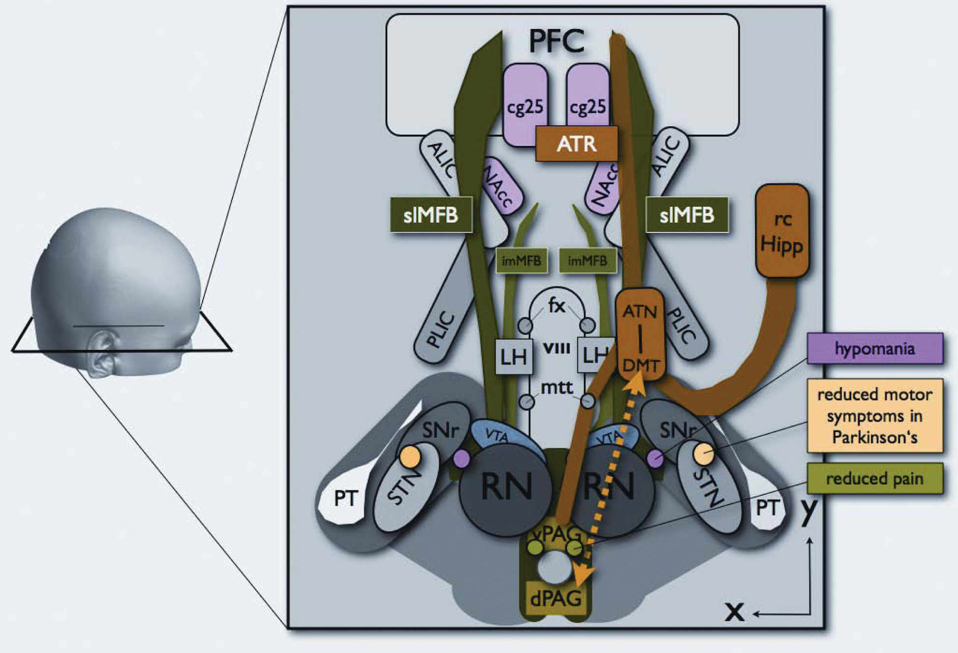
FIGURE 2. Conceptual Sketch According to the DTI Fiber Tracking Results Portraying the Medial Forebrain Bundle (MFB, Green) and the Anterior Thalamic Radiation (ATR, Orange) as Dichotomic Systems. Circles Indicate Deep Brain Stimulation Sites and Effects
The MFB is bipartite with an inferomedial branch (imMFB) running through the lateral third ventricular (VIII) wall to the lateral hypothalamus (LH) and a supero-lateral branch (slMFB) that is distally located in the anterior limb of the internal capsule (ALIC) and connects to the accumbens nucleus (NAcc) and further into the prefrontal cortex (PFC). The ATR system connects Thalamus, prefrontal cortex (PFC), and ventral periaqueductal gray (vPAG) with further connections to the temporomesial region (rcHipp: retrocommissural hippocampus). Further abbreviations: ATN: anterior thalamic nucleus, DM: dorsomedial thalamus, dPAG: dorsal peri-aqueductal gray, fx: fornix, LH: lateral hypothalamus, ot: optic tract, mtt: mamillo-thalamic tract, PAG: periaqueductal gray matter, PLIC: posterior limb of internal capsule, PT: pyramidal tract, RN: red nucleus, STN: subthalamic nucleus, SNr: substantia nigra, VTA: ventral tegmental area.

Individual Appreciation of MFB and ATR Connectivity

Since individual small fiber connections and variations are lost in the statistical evaluation, we performed a case-by-case evaluation of the connectivity of the ATR and MFB to certain brain sites. These results are shown in Table 1 and Table 2.
TABLE 1. Medial Forebrain Bundle (MFB): Individual Connection Analysis From Deterministic Tracking
 p1p2p3p4p5p6p7p8p9p10p11p12p13p14p15p16p17p18
Forceps minor (lateral)++++++++++++++++
OFC++++++++++++++++++++
DLPFC++++++++++++++++++++++++++++++++++++++
VS/NAcc++++ ++++++++++++++++
Pallidum+++++++++++++++++++++++++++++
VC/ALIC (lat)++++++++++++++++++++++++++++++++++++++++++
VC/ALIC (med)+++++++++++++++
Anterior thalamus
Dorsomedial thalamus
Retrocommissural hippocampus
Superior cerebellar peduncle+++++++++++++++++++++++++++++++
Dentate nucleus+++++++++++++++++++++++++++++
Ventral tegmental area++++++++++++++++++++++++++++++++++++++++++++++++++++++
PAG++++++++++++++++++++++++++++++++++++++++++++++++++
Lower brain stem+++++++++++++++++++++++++++++++++++++++++++++++
cg25
Hypothalamus+++++++++++++++++++++++++++++++++++++++++++++++++++
+: low fiber density; ++: medium fiber density; +++: high fiber density.
P: patient; OFC: orbitofrontal cortex; DLPC: dorsolateral prefrontal cortex; VS: ventral striatum; Nacc: nucleus accumbens; VC: ventral capsule; ALIC: anterior limb of internal capsule; PAG: periaqueductal gray; cg25: subgenual cingulate gyrus.
TABLE 2. Anterior Thalamic Radiation (ATR): Individual Connection Analysis From Deterministic Tracking
 p1p2p3p4p5p6p7p8p9p10p11p12p13p14p15p16p17p18
Forceps minor (lateral)+++++++++++++++++++++++++
OFC+++++++++++++++++++++++
DLPFC++++++++++++++++++++++++++++++++++++++
VS / NAcc++  +++++++++++++
Pallidum+++++
VC/ALIC (lat)+++++++++++++++++
VC/ALIC (med)++++++++++++++++++++++++++++++++++++++++++++++++
Anterior thalamus++++++++++++++++++++++++++++++++++++++++++++++++++++++
Dorsomedial thalamus++++++++++++++++++++++++++++++++++++++++++++++++++++++
Retrocommissural hippocampus++++++++++++++++++++++++++++++++++
Superior cerebellar peduncle+++++++++++++++++++
Dentate nucleus++++++++++++++++
Ventral tegmental area+++
Periaqueductal gray+++++++++++++++++
Lower brain stem++++++++++++++
cg25
Hypothalamus
+: low fiber density; ++: medium fiber density; +++: high fiber density.
P: patient; OFC: orbitofrontal cortex; DLPC: dorsolateral prefrontal cortex; VS: ventral striatum; Nacc: nucleus accumbens; VC: ventral capsule; ALIC: anterior limb of internal capsule; PAG: periaqueductal gray; cg25: subgenual cingulate gyrus.

Results

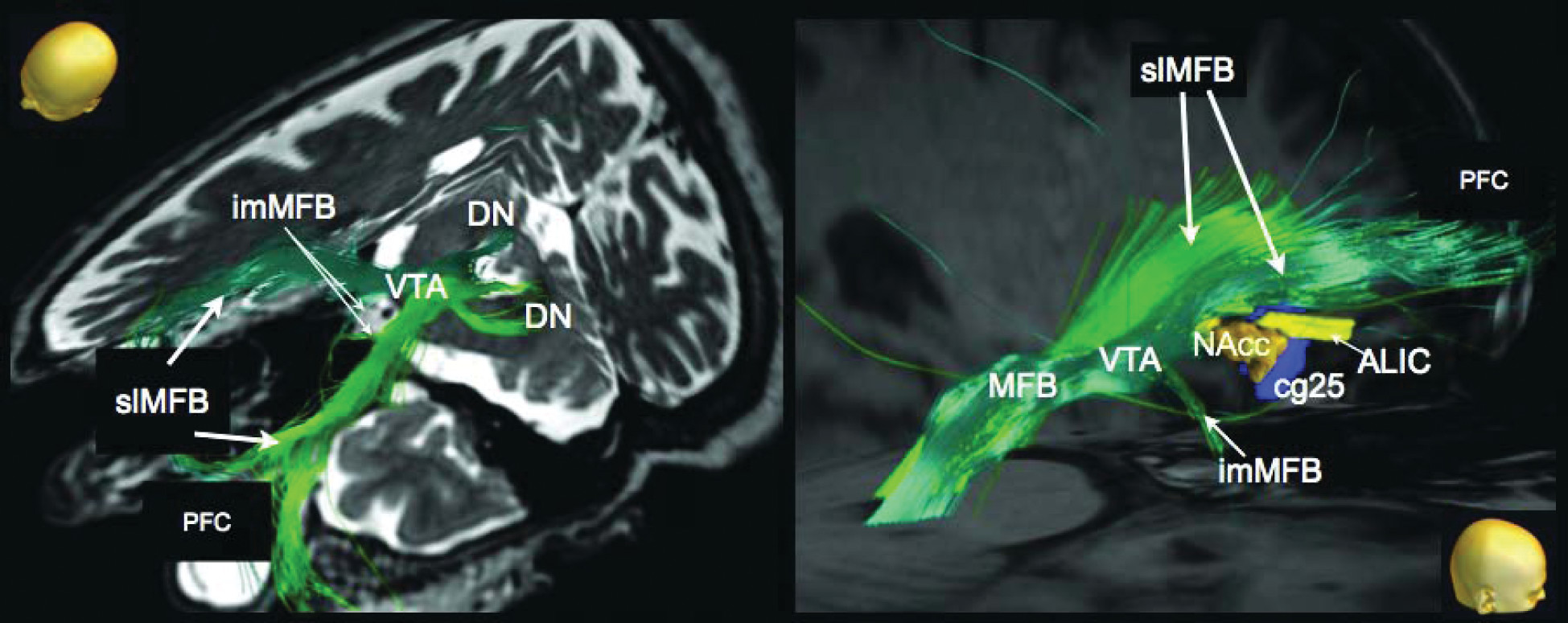
Three-dimensional depiction of the DTI FT results are presented in Figure 3 for a single patient. For clarity, only the MFB is shown.
FIGURE 3. Individual Tracking Result for the Left (Green) and Right (Dark Green)
MFB. left: view from anterolateral and superior left. right: lateral view from right. The MFB traverses the anterior limb of the internal capsule (ALIC) and connects to the accumbens nucleus (NAcc). No direct contact to subgenual region 25 (cg25) could be identified. imMFB: inferomedial medial forebrain bundle, slMFB: supero-lateral medial forebrain bundle, VTA: ventral tegmental area. [3]
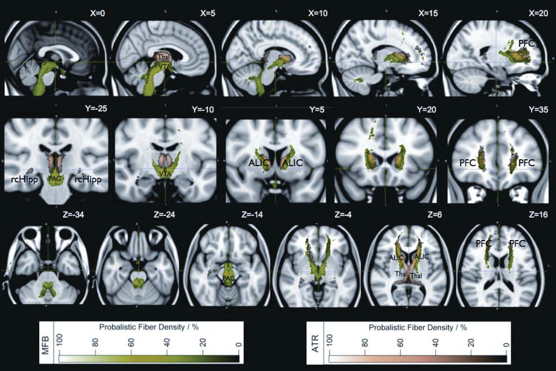
Figure 4 summarizes the two-dimensional projections of the probabilistic atlas templates of the fiber structures MFB and ATR by means of a normalized probability map from 16 subjects registered to the stereotactic space of the ICBM152 standard brain template. The complete extent of MFB and ATR 3D-probabilistic templates of both structures is shown in Figure 1.
FIGURE 4. Probability Map of the MFB (Green) and ATR (Copper) Superimposed onto the ICBM152 Brain Template
top row: sagittal; middle row: coronal; bottom row: axial sections. The color shading (probalistic fiber density) describes the average fiber density over the entire cohort after registration onto ICBM152 per pixel (0% no common overlap; 100% complete and identical overlap). Cutoff for the depiction of MFB and ATR was set to 80% to clearly separate both structures visually. Coordinates (x,y,z) represent the ICBM152 coordinate system. See text (results) for structure description. Legend: ALIC: anterior limb of internal capsule, PFC: prefrontal cortex, rcHipp: retrocommissural hippocampus, Thal: thalamus, VTA: ventral tegmental area). [4]

The Human Medial Forebrain Bundle (MFB)

The MFB, as depicted with DTI FT, is a massive and truly bipartite structure, as described earlier.1 Individual mapping results show that the main trunk splits into two parts that follow distinct directions. Caudal to the VTA, the main trunk connects to the dentate nucleus of the cerebellum, including perhaps Arnold’s bundle59 from where it follows the superior cerebellar peduncle and connects, possibly bidirectionally, to the upper pons, retrorubal area, and the periaqueductal gray (PAG). The location of the bifurcation is the ventral tegmental area (VTA) in the midbrain. From here, an infero-medial branch (imMFB) traces the wall of the third ventricle anteriorly until finally reaching the lateral hypothalamus (LH; Figures 14). The imMFB represents the traditional description of the MFB in the rodent. A second, supero-lateral branch (slMFB), leaves the main trunk in the VTA. This branch originates laterally, undercuts the thalamus, and ascends into the inferior portion of the anterior limb of the internal capsule (ALIC).
In individual deterministic tracking results, the slMFB parallels another fiber structure, the ATR, which always lies strictly medial in the ALIC (Table 1, Table 2). According to the fiber’s variability map, both structures (slMFB and ATR) intermingle in the internal capsule to a certain extent. This is due to inter-individual variability. Despite this, the medial/lateral dichotomy of ATR/MFB in the ALIC is preserved in individual study subjects (cf. Figures 1-3; Table 1, Table 2). Another robustly traceable and reproducible network is the connectivity of the slMFB to the ventral striatum and accumbens nucleus (NAcc; Figure 3). More anteriorly the variability of the slMFB becomes larger depending on interindividual differences in frontal lobe anatomy and size. Therefore, the individual fiber connections do not show up as densely (with smaller probability). As a typical pattern, the fibers fan out medially and laterally to the frontal cortical surface (Figure 3, right). The connections that were seen led to the orbitofrontal cortex (OFC) and dorsolateral prefrontal cortex (DLPFC). In this study, and utilizing our methodology, a direct connection to the white-matter tracts surrounding the subgenual cingulate gyrus (cg25) could not be observed (Table 1).

Anterior Thalamic Radiation (ATR)

The ATR can be visualized as clearly and robustly as the MFB. The ATR connects the dorsomedial thalamus (DM) and the anterior thalamic nucleus (AT) with the prefrontal cortex. It runs strictly medial to the slMFB in the anterior limb of the internal capsule. Further robust connections are seen to the temporo-mesial region (perhaps retrocommissural hippocampus; Figure 1, Figure 4). An obvious connection to the brain stem is made via the mamillo-thalamic tract. A second, less probable connection is seen to the retro-rubral field (also called periaqueductal gray). In the individual analysis (Table 2), the ATR shows strong (very likely) connectivity to the DLPFC. Moderate probable connections are demonstrated to the temporomesial region (retrocommissural hippocampus/amygdala) and the dentate nucleus of the cerebellum. For a complete overview of the ATR connectivity, refer to Table 2 and Figures 1, 2, and 4.

Discussion

The Medial Forebrain Bundle: A Ponto-Frontal Projection Pathway

Textbooks of neuroanatomy typically describe the fronto-pontine tract (Arnold’s bundle) as part of the anterior limb of the internal capsule (ALIC) and as the most anterior and medial fiber structure at the level of the crus cerebri, directly adjacent to the corticospinal tract.13 These classical descriptions focus predominantly on the topography of the anterior limb of the internal capsules and are supported by more recent studies that apply polarized light to identify fiber-tract directions in the anterior limb of the internal capsule and the brainstem on the basis of histological slice sections.2731 However, whether Arnold’s bundle can be found in humans is questioned in the literature, and some authors deny its existence.32 Postmortem examinations of lesioned brains from the frontal-leucotomy era33,34 have demonstrated the existence of descending or ascending tracts that most likely represents Arnold’s bundle in humans. According to these studies, Arnold's bundle originates from the dorsal (superior) and lateral convexity of the prefrontal cortex,20 and not from the agranular parts or the OFC.
However, a recent MRI study in patients after anterior capsulotomy has identified an apparent reciprocal corticopetal pontofrontal projection.21 Classical neuroanatomy does not clearly reflect on a possible bidirectionality of the fronto-pontile projection system (Arnold’s bundle). In this sense, this study extends neuroanatomical knowledge by adding a fiber pathway that is antidromic to Arnold’s bundle. These authors inferred this projection on the basis of early post-axotomy accumulation of axonal lipoproteins that could be identified by MRI. More specifically, accumulation of protein was found in the upper midbrain after lesioning fibers in the anterior capsule in the frontal lobe thus further downstream. This indicates that the cell bodies of this neuronal pathway are situated close to the midbrain (and not in the frontal lobe), indicating a mesencephalo-frontal trajectory. The projection proposed by Hurwitz is clearly extending the classical neuroanatomical description and has not yet been included into newer texts on the neuroanatomy of psychiatry.3,14,3538 Thus, Hurwitz and colleagues first described a reciprocal mesencephalo-frontal pathway in the living human brain. This pontofrontal projection appears to coincide with the supero-lateral branch of the MFB (slMFB) described in our earlier work.1 These results have recently also been replicated by others using the DTI technique in a context of non-motor projections of the cerebellum.59

A Newly-Described Supero-Lateral Branch of the MFB (slMFB)

Anatomically, the MFB in all species that have been studied connects the ventral tegmental area (VTA) with the lateral hypothalamus (LH) and the nucleus accumbens (NAcc);13,19,3942 Le Gros Clark noted that this pathway also connects to the olfactory bulb.13 Nauta had widened the limbic system concept with what he defined as the limbic midbrain. According to his description, the dopamine-containing VTA neurons interface directly with the limbic forebrain via the perifornical region.43 Experimental findings from rodent and primate studies in the last quarter-century list several more pathways of the VTA with basal forebrain structures. These targets are interconnected via the MFB, which serves as the major route of passage along the lateral hypothalamus, including connections to the medial hypothalamus, sublenticular region, lateral and medial preoptic region, diagonal band, septal nuclei, ventral pallidum, and ventral parts of the bed nucleus of the stria terminalis.37
In humans, this pattern of mesolimbic network remains to be described in the classical context of anatomical textbooks. Because of the distinct phylogenetic developmental growth of the brain, the MFB has to follow somewhat distinct trajectories to link up with the aforementioned key limbic structures in man.1 The slMFB described here is ideally placed to serve this role. The slMFB arises out of the trunk of the MFB, and its links with the VTA, NAcc, and ALIC can be well characterized (Figure 3). Our data thus enhance, rather than contradict, established neuroanatomical knowledge in other species.

Clinical Significance

Stereotactic targets in deep brain stimulation (DBS) used for treatment-resistant psychiatric disorders have included cg25 (in depression)44 and, more recently, nucleus accumbens septi (NAcc)9, 45 as well as the anterior limb of the internal capsule (ALIC)20, 46 for depression, and obsessive-compulsive disorder (OCD). The exact neuroanatomical locations for optimal therapeutic effects and the precise mechanism of DBS in these clinical conditions remain unclear,2,44,45 but may include both suppression and activation of neural tissues. For ALIC- and NAcc-DBS, one possible explanation for psychobehavioral benefit is activation of the slMFB,2 which connects to critical higher limbic targets. Stimulation of this system—traditionally called “the brain reward system” —would result in an antidysphoric effect by galvanizing the appetitive motivational SEEKING system that promotes euphoria, accompanied by a motorically-activated, psychologically-energized, and emotionally highly-motivated foraging-planning state.3 (In fact, the brain has several reward systems.11,12) Considering these affective changes, over-activity of the slMFB may promote manic states,1 and, hence, also mood switching in bipolar disorders, whereas the pattern of psychopathology of an expansive mood, enhanced energy, and psychomotor acceleration is consistent with an over-functioning SEEKING system. Such an explanation could also account for the hypomanic states that may complicate DBS subthalamic nucleus (STN) stimulation for Parkinson’s disease, where an incorrectly-placed electrode may inadvertently activate this system.1 Finally, under-functioning of this system could lead to depressive fatigue states, and, as such, suggest a target for future clinical research, given the prevalence of debilitating fatigue syndromes and the limited efficacy of currently available therapies. Indeed, two reported patients treated with anterior capsultomy for depression and OCD developed a severe fatigue syndrome postoperatively. We hypothesize that surgery in those cases might have also severed parts of the slMFB that, as we have shown, lies in close proximity to the ATR in the area of ALIC.
The ATR, in contrast to the MFB, may play a role in depressive illness via the PANIC emotional system that has been revealed by mapping separation-distress calls.11,12 Such brain regions are aroused when humans experience sadness. This is hypothesized to occur because of its presumed connections with cg25, which has been shown to be overactive in depressed patients.47 The horizontal fibers of the ATR are directed to the anterior pole of the frontal lobe, which includes the rostral anterior cingulate gyrus and orbitofrontal cortex.
Earlier work of Herman and Panksepp mapped out the separation-call in guinea pigs, which is directly modulated by endogenous opioids.60 In a human subject, only recently, during a case of anterior thalamic nucleus stimulation for intractable epilepsy, our group was able to repetitively induce acute episodes of depression through activation of deeper electrode contacts located in the ATR (unpublished data).
In rodents, stimulation of several forebrain and anterior diencephalic regions (dorsomedial thalamus, ventral septum, dorsal preoptic area, and bed nucleus of stria terminalis, as part of the extended amygdala) can elicit separation distress. In the light of these facts, it could also be questioned whether the MFB only conveys euphoria. However, with regard to the theorizing of MacLean, it would be reasonable to consider that primary and secondary affect-processing mandates connections of the medial forebrain bundle and anterior thalamic radiation other than the prefrontal cortex. A potential explanation is that ATR and MFB connect at these basal forebrain regions.7,12,61 The relationship of the MFB SEEKING system to these specific areas is not known. It suggests possible areas of interaction that should be investigated in the future.
Deep brain stimulation of the white-matter tracts adjacent to the subgenual cingulate gyrus has been demonstrated to normalize (reduce) pathological hyperactivity of the subgenual cingulate gyrus and adjacent orbitofrontal cortex and is associated with sustained improvement in patients with treatment-resistant depression.21,44 Thus, the MFB and ATR mediate very different, and, possibly, opposing, affective states. As suggested by our in-vivo tracking results, both are located in close proximity within ALIC but are now clearly separable anatomical and, likely, also functional structures. They provide candidate networks for the promotion of opposing emotional states and therefore may be critical for affective equilibrium and disequilibrium.
The anterior thalamic radiation is a structure that has long been targeted for lesioning or DBS in the treatment of psychiatric disorders.2,3,10,21,31,48,49 Why other groups that have applied fiber-tracking in the same brain areas did not describe or identify the slMFB as an individual structure is unclear. We believe that those fibers that we associate with the slMFB have also been seen by other groups, but perhaps were allocated to other tracts.10,2225,50 Indeed, Wakana et al. describe a tracking procedure for the ATR starting with a seed region (ROI) from the frontal lobe that covers the anterior limb of the internal capsule and showed connections to the brain stem that they regarded as atypical for ATR.51 We infer that their ATR has included the slMFB because of the close proximity of slMFB and ATR to their tracking ROI. Figure 5 explains how the ICBM ATR template from the Johns Hopkins University JHU White Matter Tractography Atlas (ATRj)58 might in reality be a combination of ATR and slMFB. The 28 subjects studied for the Johns Hopkins’ white-matter atlas compared with our 16 patients led to quite similar statistical variance of tracking results if one considers that the ATRj indeed composes a superposition of our ATR and slMFB. For a more thorough comparison, refer to Figure 5 and Supplement Figure 2.
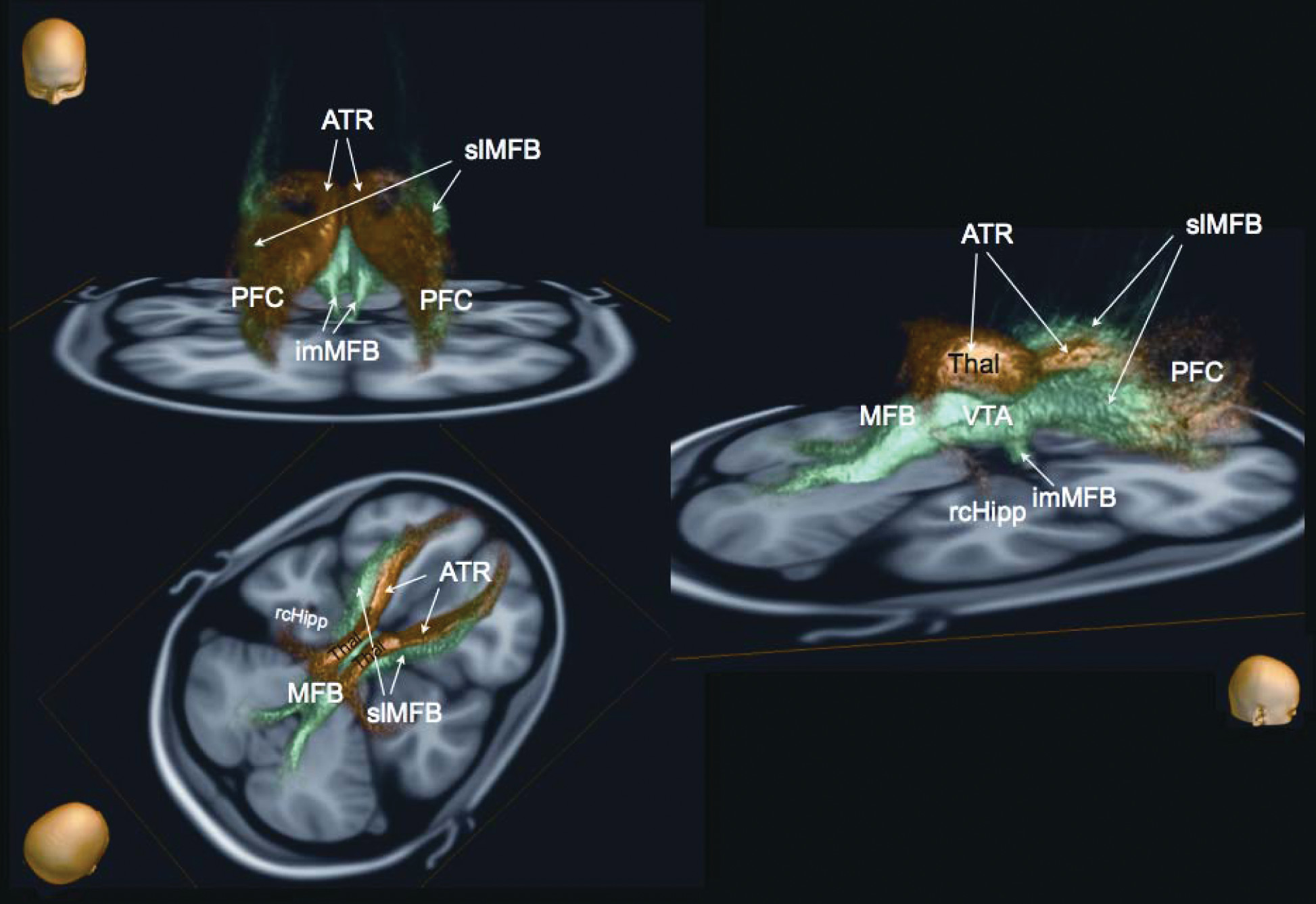
FIGURE 5. Probabilistic Maps of the ATRj (Johns Hopkins University), ATR and slMFB on Standard ICBM152 T1 Template
Top row: coronal view; bottom row: axial view at MNI coordinates x,y,z=(-7.5, 12.0, 3.0).
A: ATRj (left, red), ATR+MFB (right, red), ATR probabilistic (right, yellow hot);
B: ATRj (left, red), ATR+MFB (right, red), ATR probabilistic (right, yellow hot); slMFB probabilistic (right, green)
C: direct comparison of ATRj (left) and combined structure from our ATR and slMFB: ATR+slMFB (right) reveals that ATRj is likely to show both structures together (ATRj=ATR+slMFB).

Limitations

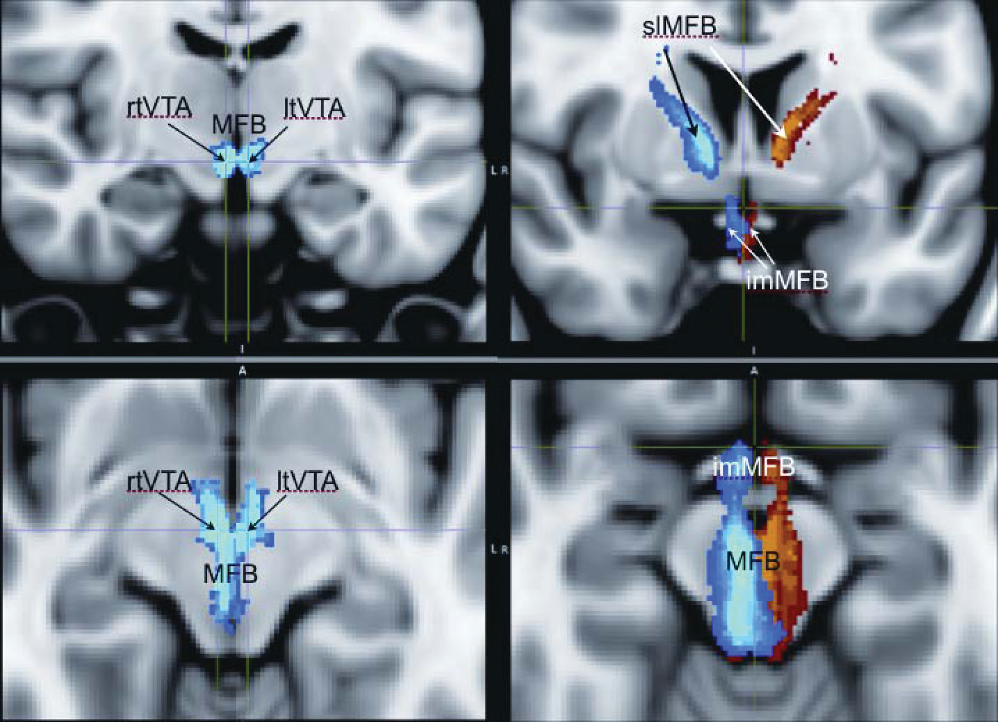
There are several limitations of the deterministic fiber-tracking algorithm (FACT), such as termination of tracking in areas of reduced FA (due to increased intermingling of fibers, approach of the subcortical boundaries, and partial-volume effects), a currently unavoidable ambiguity when attempting to follow the correct connection pathways in areas of dense crossing, kissing, or branching fibers (limitation of single-diffusion tensor model), combined with a relatively low spatial resolution of DTI (about 2-mm isotropic voxel dimension). Nevertheless, our methodology achieved reproducible accuracy and approximates very closely this intrinsic spatial-resolution limit.51 Our method of utilizing DTI scans of patients with movement disorders (Parkinson’s disease, essential tremor, dystonia) to characterize affective pathways might be regarded as problematic because of the uncertainty about any neuropathology that might be linked to these diseases. Although we cannot entirely waive this criticism, however, to our knowledge, there is no valid indication that the pathways under study are anatomically compromised under such neurological conditions. To sustain this hypothesis, a subgroup analysis of Parkinson’s disease (N=12) and tremor/dystonia patients (N=6) was performed (Figure 6) that allowed comparison of resultant fiber tracts between the subgroups and did not identify any differences or disagreements.
FIGURE 6. Subgroup Comparison Between Parkinson's Disease Subjects (N=12; Yellow-Red) and Dystonia-Tremor Group (N=6; Blue)
No difference is seen in this subgroup comparison of slMFB anatomy. Legends: imMFB: inferomedial medial forebrain bundle, ltVTA: left ventral tegmental area, MFB: medial forebrain bundle, rtVTA: right ventral tegmental area, slMFB: supero-lateral medial forebrain bundle.
Given the difficulty of DTI FT in portraying classical anatomy to its full extent,52 caution is required when describing new anatomical findings with this technique, as, for example, in the case of slMFB. However, the lesioning and early MRI study presented by Hurwitz and coworkers considerably strengthens the concept of a fiber tract that runs corticopetally with Arnold’s bundle.21 Older studies from the early frontal leucotomy era also substantiate our findings.2,33,34 More evidence can be found in a thorough study of the literature of inadvertent side effects during DBS. Several authors have reported hypomania (attributed by our own research to MFB activation) for medial and anterior STN stimulation. Furthermore, hypomania was also described for OCD patients in a similar, if not identical, electrode location (STN), and also in ALIC stimulation for OCD.1,5355 Modulation of the same pathway, the slMFB, at different sites (medial STN versus ALIC) thus becomes a reasonable possibility and hence may provide supportive evidence for our anatomical descriptions.
Finally, it should be emphasized that the MFB is a neurochemically heterogeneous bundle, where all ascending biogenic amine systems are represented. Also, immunocytochemical neuropeptide mapping of animal brains, along with receptor distributions, indicates that many neuropeptides, at least a dozen, by our count, are represented along the MFB: angiotensin, atrial natriuretic peptide, corticotropin releasing factor, galanin, neurotensin, Neuropeptide Y, opioid peptides, orexin, oxytocin, thyrotropin-releasing hormone, somatostatin, vasoactive intestinal peptide, and vasopressin.17 Obviously, this level of structural complexity cannot be resolved by DTI, and clarification of the functional complexities requires yet other approaches. How many of these systems contribute to what are here called SEEKING urges is indeterminate at present.

Conclusion

Our studies shed new light on the commonly known anatomy of affective pathways in humans that have traditionally been inferred from human degeneration or animal anatomy studies. We have hereby identified a putative affect-regulating fiber system with DTI that is composed of two distinct fiber pathways, the MFB and the ATR. These pathways, consistent with various clinical studies and our own findings, are concerned with the maintenance of emotional homeostasis and appear to be the anatomical substrate in humans of distinct emotional-action systems, at minimum, the SEEKING and PANIC systems of affective neuroscience.12,26 This knowledge and the ability to visualize these pathways in vivo should greatly facilitate clinical integration with studies of affective circuitries in preclinical animal models.56
So far, the role of MFB in humans as a bipartite fiber system has been greatly underrecognized and misunderstood. From animal studies, it has been known for a long time that the MFB is linked to reward-seeking and appetitive motivation in general. Furthermore, the ATR should be regarded as a separate structure and is probably more likely linked to negative feelings, such as sadness, since the separation–distress circuitry, promoting psychic-pain, traverses this brain region.12,26 MFB and ATR can be clearly distinguished although they converge structurally and conjoin-pass in close proximity to the anterior limb of the internal capsule. The MFB is a central limbic structure and plays an important role in normal functioning and psychopathology. Knowledge about its anatomy may help scientists to generate hypotheses about the subjective psychological side effects of DBS1,53,54 and promote a better understanding of the underlying mechanisms that are essential for defining improved stereotactic targets in the treatment of OCD and depression with DBS.2,10 On the basis of our findings, the MFB may also be implicated in the symptoms of mania and fatigue/depression, both of which deserve further scientific inquiry. Our results utilizing deterministic DTI FT do not contradict, but, instead, enhance the existing classical anatomical concepts: The MFB and ATR are robust and reproducibly-identified structures. We present stereotactic templates, displaying these structures in one place for easy comparison.
In conclusion, we wish to emphasize the anatomical dichotomy of the two presented fiber pathways, suggesting a functional dichotomy that may find its expression in alternating and opposing mood states. These two systems seem to represent two poles of emotional homeostasis, one that allows affective states of joy (MFB), and another, of sadness (ATR), to appear only when one system is highly active while the other is in a lower activation state. Such affective brain systems have long been implicated in the genesis of depression (for a recent full summary and discussion, see Watt and Panksepp57).

Acknowledgments

This paper is dedicated to the memory of Lennart Heimer (1930–2007) and Peter J. Morgane (1927–2010), both of whom were fascinated by the role of the medial forebrain bundle in behavioral and psychological functions.
Dr. Coenen is consultant to Medtronic (Europe, U.S.) and has occasionally received honoraria for lecturing and consultation work. Drs. Coenen and Panksepp acknowledge support from the Hope for Depression Research Foundation and the Institute for Affective Neuroscience.

Supplementary Material

File (jn-11080180_ds001.pdf)

References

1.
Coenen VA, Honey CR, Hurwitz T, et al.: Medial forebrain bundle stimulation as a pathophysiological mechanism for hypomania in subthalamic nucleus deep brain stimulation for Parkinson’s disease. Neurosurgery 2009; 64:1106–1114, discussion 1114–1115
2.
Coenen VA, Schlaepfer TE, Maedler B, et al.: Cross-species affective functions of the medial forebrain bundle: implications for the treatment of affective pain and depression in humans. Neurosci Biobehav Rev 2011; 35:1971–1981
3.
Haber SN, Knutson B: The reward circuit: linking primate anatomy and human imaging. Neuropsychopharmacol Revs 2010; 35:1–23
4.
Heimer L: Anatomy of Neuropsychiatry: The Anatomy of the Basal Forebrain and Its Implication for Neuropsychiatric Illness. Amsterdam, Boston, Heidelberg. London, Academic Press, 2008
5.
Knutson B, Greer SM: Anticipatory affect: neural correlates and consequences for choice. Philos Trans R Soc Lond B Biol Sci 2008; 363:3771–3786
6.
LeDoux JE: Emotion circuits in the brain. Annu Rev Neurosci 2000; 23:155–184
7.
MacLean PD: The Triune Brain in Evolution - Role in Paleocerebral Function. Berlin, Springer, 1990
8.
Schlaepfer TE, Cohen MX, Frick C, et al.: Deep brain stimulation to reward circuitry alleviates anhedonia in refractory major depression. Neuropsychopharmacology 2008; 33:368–377
9.
Schlaepfer TE, Lieb K: Deep brain stimulation for treatment of refractory depression. Lancet 2005; 366:1420–1422
10.
Schoene-Bake JC, Parpaley Y, Weber B, et al.: Tractographic analysis of historical lesion surgery for depression. Neuropsychopharmacology 2010; 35:2553–2563
11.
Panksepp J: Affective consciousness: core emotional feelings in animals and humans. Conscious Cogn 2005; 14:30–80
12.
Panksepp J: Affective Neuroscience: The Foundations of Human and Animal Emotions. Oxford, UK, New York, Oxford University Press, 1998
13.
Le Gros Clark WE: The topography and homologies of the hypothalamic nuclei in man. University Press, 1936
14.
Palkovitz M, Zaborsky L: Neural connections of the hypothalamus, in Handbook of the Hypothalamus: Vol. 1. Anatomy of the Hypothalamus. Edited by Morgane PJ, Panksepp J. New York, Marcel Decker, 1979, pp 379–509
15.
Hattori T: Conceptual history of the nigrostriatal dopamine system. Neurosci Res 1993; 16:239–262
16.
Aston-Jones G, Smith RJ, Sartor GC, et al.: Lateral hypothalamic orexin/hypocretin neurons: a role in reward-seeking and addiction. Brain Res 2010; 1314:74–90
17.
Tohyama M, Takatsuji K: Atlas of Neuroactive Substances and Their Receptors in the Rat. Oxford, UK, Oxford Univ. Press, 1998
18.
Nieuwenhuys R, Veening JG, van Domburg P: Core and paracores; some new chemoarchitectural entities in the mammalian neuraxis. Acta Morphol Neerl Scand 1988-1989-89; 26:131–163
19.
Nieuwenhuys R, Voogd J, van Huijzen C: The Human Central Nervous System. Berlin, Heidelberg, New York, Springer, 2008
20.
Gutman DA, Holtzheimer PE, Behrens TE, et al.: A tractography analysis of two deep brain stimulation white-matter targets for depression. Biol Psychiatry 2009; 65:276–282
21.
Hurwitz TA, Mandat T, Forster B, et al.: Tract identification by novel MRI signal changes following stereotactic anterior capsulotomy. Stereotact Funct Neurosurg 2006; 84:228–235
22.
Mori S: MRI Atlas of Human White Matter. Amsterdam, Boston, Heidelberg, Elsevier, 2005
23.
Mori S, Oishi K, Faria AV: White-matter atlases based on diffusion tensor imaging. Curr Opin Neurol 2009; 22:362–369
24.
Mori S, Oishi K, Jiang H, et al.: Stereotaxic white-matter atlas based on diffusion tensor imaging in an ICBM template. Neuroimage 2008; 40:570–582
25.
Sillery E, Bittar RG, Robson MD, et al.: Connectivity of the human periventricular-periaqueductal gray region. J Neurosurg 2005; 103:1030–1034
26.
Panksepp J: The neurobiology of social loss in animals: some keys to the puzzle of psychic pain in humans, in Social Pain: Neuropsychological and Health Implications of Loss and Exclusion. Edited by, Jensen-Campbell LA, MacDonald G. Washington, DC, American Psychological Association, 2011, pp 11–52
27.
Axer H, Axer M, Krings T, et al.: Quantitative estimation of 3-D fiber course in gross histological sections of the human brain using polarized light. J Neurosci Methods 2001; 105:121–131
28.
Axer H, Berks G, Keyserlingk DG: Visualization of nerve fiber orientation in gross histological sections of the human brain. Microsc Res Tech 2000; 51:481–492
29.
Axer H, Keyserlingk DG: Mapping of fiber orientation in human internal capsule by means of polarized light and confocal scanning laser microscopy. J Neurosci Methods 2000; 94:165–175
30.
Axer H, Leunert M, Mürköster M, et al.: A 3D fiber model of the human brainstem. Comput Med Imaging Graph 2002; 26:439–444
31.
Axer H, Lippitz BE, von Keyserlingk DG: Morphological asymmetry in anterior limb of human internal capsule revealed by confocal laser and polarized light microscopy. Psychiatry Res 1999; 91:141–154
32.
Tredici G, Barajon I, Pizzini G, et al.: The organization of corticopontine fibres in man. Acta Anat (Basel) 1990; 137:320–323
33.
Beck E: The origin, course, and termination of the prefronto-pontine tract in the human brain. Brain 1950; 73:368–391
34.
Meyer, M. A study of efferent connexions of the frontal lobe in the human brain after leucotomy. Brain 1949; 72:265–296
35.
Price JL, Carmichael ST, Drevets WC: Networks related to the orbital and medial prefrontal cortex; a substrate for emotional behavior? Prog Brain Res 1996; 107:523–536
36.
Price JL, Drevets WC: Neurocircuitry of mood disorders. Neuropsychopharmacology 2010; 35:192–216
37.
Zahm DS: The evolving theory of basal forebrain functional-anatomical “macrosystems.” Neurosci Biobehav Rev 2006; 30:148–172
38.
Zald DH, Rauch SL: The Orbitofrontal Cortex. Oxford, New York, Oxford University Press, 2006
39.
Lang J: Lanz Wachsmuth Praktische Anatomie. Kopf. Gehirn- und Augenschädel. Berlin, Heidelberg, Springer, 1979
40.
Lang J: Lanz Wachsmuth Praktische Anatomie. Kopf. Übergeordnete Systeme. Berlin, Heidelberg, New York, Tokyo, Springer, 1979
41.
Standring S: Gray's Anatomy: The Anatomical Basis for Clinical Practice. New York, Elsevier Churchill Livingstone, 2005
42.
Wise RA: Forebrain substrates of reward and motivation. J Comp Neurol 2005; 493:115–121
43.
Nauta WJH: Hypothalamic nuclei and fiber connections, in The Hypothalamus. Edited by, Nauta WJH, Anderson E, Haymaker W. Springfield, IL, Charles C. Thomas, 1969
44.
Lozano AM, Mayberg HS, Giacobbe P, et al.: Subcallosal cingulate gyrus deep brain stimulation for treatment-resistant depression. Biol Psychiatry 2008; 64:461–467
45.
Schlaepfer TE, Fins JJ: Deep brain stimulation and the neuroethics of responsible publishing: when one is not enough. JAMA 2010; 303:775–776
46.
Malone DA, Dougherty DD, Rezai AR, et al.: Deep brain stimulation of the ventral capsule/ventral striatum for treatment-resistant depression. Biol Psychiatry 2009; 65:267–275
47.
Mayberg HS, Brannan SK, Tekell JL, et al.: Regional metabolic effects of fluoxetine in major depression: serial changes and relationship to clinical response. Biol Psychiatry 2000; 48:830–843
48.
Coenen VA, Honey CR: Ablative procedures for depression, in Textbook of Stereotactic and Functional Neurosurgery, Vol 2. Edited by, Lozano AM, Gildenberg P, Tasker RR. Berlin, Heidelberg, Springer-Verlag, 2009, pp 2943–2951
49.
Greenberg BD, Malone DA, Friehs GM, et al.: Three-year outcomes in deep brain stimulation for highly resistant obsessive-compulsive disorder. Neuropsychopharmacology 2006; 31:2384–2393
50.
Zhang W, Olivi A, Hertig SJ, et al.: Automated fiber tracking of human brain white matter using diffusion tensor imaging. Neuroimage 2008; 42:771–777
51.
Wakana S, Jiang H, Nagae-Poetscher LM, et al.: Fiber tract-based atlas of human white-matter anatomy. Radiology 2004; 230:77–87
52.
Bürgel U, Mädler B, Honey CR, et al.: Fiber tracking with distinct software tools results in a clear diversity in anatomical fiber tract portrayal. Cen Eur Neurosurg 2009; 70:27–35
53.
Chang CH, Chen SY, Hsiao YL, et al.: Hypomania with hypersexuality following bilateral anterior limb stimulation in obsessive-compulsive disorder. J Neurosurg 2010; 112:1299–1300
54.
Haq IU, Foote KD, Goodman WK, et al.: A case of mania following deep brain stimulation for obsessive compulsive disorder. Stereotact Funct Neurosurg 2010; 88:322–328
55.
Mallet L, Polosan M, Jaafari N, et al.STOC Study Group: Subthalamic nucleus stimulation in severe obsessive-compulsive disorder. N Engl J Med 2008; 359:2121–2134
56.
Panksepp J: Affective neuroscience of the emotional BrainMind: evolutionary perspectives and implications for understanding depression. Dialogues Clin Neurosci 2010; 12:533–545
57.
Watt D, Panksepp J: Depression: an evolutionarily conserved mechanism to terminate separation-distress? a review of aminergic, peptidergic, and neural network perspectives. (with discussions). Neuro-psychoanalysis 2009; 11:5–104
58.
Hua K, Zhang J, Wakana S, et al.: Tract probability maps in stereotaxic spaces: analyses of white-matter anatomy and tract-specific quantification. Neuroimage 2008; 39:336–347
59.
Engelhardt E, Madeira Moreira D, Laks J: Cerebrocerebellar system and Arnold’s bundle, a tractographic study: preliminary results. Dement Neuropsychol 2010 4(4):293-299.
60.
Herman BH, Panksepp J: Ascending endorphin inhibition of distress vocalization. Science 1981; 211:1060–1062
61.
Panksepp J, Normansell LA, Herman B, et al.: Neural and neurochemical control of the separation distress call, in The Physiological Control of Mammalian Vocalizations. Edited by, Newman JD. New York, Plenum, 1988, pp 263–300

Information & Authors

Information

Published In

Go to The Journal of Neuropsychiatry and Clinical Neurosciences
The Journal of Neuropsychiatry and Clinical Neurosciences
Pages: 223 - 236
PubMed: 22772671

History

Received: 4 August 2011
Revision received: 31 October 2011
Accepted: 14 November 2011
Published online: 1 April 2012
Published in print: Spring 2012

Authors

Details

Volker A. Coenen, M.D.
From the Division of Stereotaxy and MR-based OR - techniques/Department of Neurosurgery (VAC, BM); Department of Radiology/Neuroradiology (HU), Bonn University, Germany; and the Bailey Endowed Chair for Animal Well-Being Science (JP), Washington State University, Pullman, WA; Dept. of Psychiatry, (TAH), University of British Columbia, Vancouver, Canada.
Jaak Panksepp, Ph.D.
From the Division of Stereotaxy and MR-based OR - techniques/Department of Neurosurgery (VAC, BM); Department of Radiology/Neuroradiology (HU), Bonn University, Germany; and the Bailey Endowed Chair for Animal Well-Being Science (JP), Washington State University, Pullman, WA; Dept. of Psychiatry, (TAH), University of British Columbia, Vancouver, Canada.
Trevor A. Hurwitz, M.D.
From the Division of Stereotaxy and MR-based OR - techniques/Department of Neurosurgery (VAC, BM); Department of Radiology/Neuroradiology (HU), Bonn University, Germany; and the Bailey Endowed Chair for Animal Well-Being Science (JP), Washington State University, Pullman, WA; Dept. of Psychiatry, (TAH), University of British Columbia, Vancouver, Canada.
Horst Urbach, M.D.,
From the Division of Stereotaxy and MR-based OR - techniques/Department of Neurosurgery (VAC, BM); Department of Radiology/Neuroradiology (HU), Bonn University, Germany; and the Bailey Endowed Chair for Animal Well-Being Science (JP), Washington State University, Pullman, WA; Dept. of Psychiatry, (TAH), University of British Columbia, Vancouver, Canada.
Burkhard Mädler, Ph.D.
From the Division of Stereotaxy and MR-based OR - techniques/Department of Neurosurgery (VAC, BM); Department of Radiology/Neuroradiology (HU), Bonn University, Germany; and the Bailey Endowed Chair for Animal Well-Being Science (JP), Washington State University, Pullman, WA; Dept. of Psychiatry, (TAH), University of British Columbia, Vancouver, Canada.

Notes

Send correspondence to Volker A. Coenen, M.D.; e-mail: [email protected]

Metrics & Citations

Metrics

Citations

Export Citations

If you have the appropriate software installed, you can download article citation data to the citation manager of your choice. Simply select your manager software from the list below and click Download.

For more information or tips please see 'Downloading to a citation manager' in the Help menu.

Format
Citation style
Style
Copy to clipboard

View Options

View options

PDF/EPUB

View PDF/EPUB

Get Access

Login options

Already a subscriber? Access your subscription through your login credentials or your institution for full access to this article.

Personal login Institutional Login Open Athens login
Purchase Options

Purchase this article to access the full text.

PPV Articles - Journal of Neuropsychiatry and Clinical Neurosciences

PPV Articles - Journal of Neuropsychiatry and Clinical Neurosciences

Not a subscriber?

Subscribe Now / Learn More

PsychiatryOnline subscription options offer access to the DSM-5-TR® library, books, journals, CME, and patient resources. This all-in-one virtual library provides psychiatrists and mental health professionals with key resources for diagnosis, treatment, research, and professional development.

Need more help? PsychiatryOnline Customer Service may be reached by emailing [email protected] or by calling 800-368-5777 (in the U.S.) or 703-907-7322 (outside the U.S.).

Media

Figures

Other

Tables

Share

Share

Share article link

Share