Full access
Windows to the Brain
Published Online: 1 April 2012

The Dynamic Brain: Neuroplasticity and Mental Health

Publication: The Journal of Neuropsychiatry and Clinical Neurosciences

Abstract

This issue's Windows to the Brain series describes the ways in which the adult brain, once thought to be “fixed and stable,” adapts to and recovers from various “brain-altering” conditions. The processes of neurogenesis, migration, and pruning do, in fact, occur in response to both learning and injury. These processes may include synapse-formation, axonal sprouting, and dendritic remodeling, which can be demonstrated with currently-available labeling techniques that highlight newly proliferated cells. Factors such as environmental enrichment, exercise, and learning, and treatment approaches such as ECT and long-term antidepressant administration have been associated with increased neurogenesis; whereas chronic stress, illness, and depression have been correlated with decreased neurogenesis.
Until fairly recently, the adult brain was considered largely fixed and stable.14,15 Although it was accepted that changes occurred in the context of learning and memory, the general consensus was that major processes essential to normal brain development (e.g., generation of new neurons, neuron migration, pruning) ceased once full development was reached. The relationship between physical illness, mental illness, and brain functioning or structure was not heavily considered. However, recent research has led to a major paradigm shift. Most important was the discovery of the birth of new neurons (neurogenesis) in the adult human brain.15 Current perspectives suggest a dynamic brain, physically changed by both internal and external factors.1520 The processes by which the brain is remodeled are collectively referred to as “neuroplasticity.” Neuroplastic changes can be either adaptive or maladaptive. The concept of neuroplasticity is opening the doors to new ways of understanding illness and recovery, as well as how these processes can be utilized to influence and direct outcomes.

Neuroplastic Processes

A variety of processes and mechanisms are included under the umbrella term neuroplasticity.16,17,20 These include the formation of new neurons and glial cells (neurogenesis), as well as formation of new connections and alterations in existing ones through multiple processes (e.g., synapse formation and elimination, dendritic remodeling, axonal sprouting, and pruning). The importance of activity-level or demand in evoking remodeling has been emphasized. Research limitations largely prevent study of these processes directly in humans; thus, what is known has primarily been determined through animal studies. This work provides insight into processes that may be occurring in the human brain.
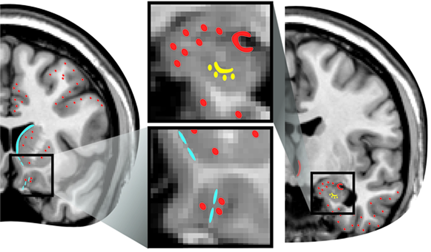
The most surprising and exciting type of neuroplasticity is the actual measurement of birth, migration, maturation, and functional integration of new neurons in the adult brain.35,15 Proliferated or newly-born cells are typically labeled using bromodeoxyuridine (5-bromo-2'-deoxyuridine; BrdU), a synthetic nucleoside that is incorporated into dividing cells in place of thymidine. Once labeled, cells can be subsequently traced through the maturation and integration process. By utilizing animal models, researchers are able to examine patterns and factors related to rates of cell proliferation, maturation, and survival associated with different experimental conditions (e.g., treatment, stress, environmental variables).3 It is now generally accepted that adult neurogenesis occurs in two locations. Neurons born in the subventricular zone (SVZ) adjacent to the caudate are incorporated into the olfactory bulb; those born in the subgranular zone (SGZ) of the dentate gyrus are incorporated into the hippocampus (Figure 1).15,21 Although controversial, there is evidence in adult primates for generation of new neurons in other ventricular regions and incorporation of new neurons into other cortical and subcortical areas (Figure 1).2,510 Rates of survival for newborn cells vary by species and experimental conditions.3 Survivors mature into neurons or glia cells, depending on their location and activity. Growth factors (e.g., brain-derived neurotrophic factor [BDNF]), vascular endothelial growth factor [VEGF], insulin-like growth factor [IGF]) play an important role in regulating the neurogenic process by increasing the rate of cell birth and promoting maturation and survival.2124 Glial cells are believed to play an important role in this process, directly and indirectly, both supporting and regulating the development of new neurons.25,26
Cover and FIGURE 1. It is now generally accepted that adult neurogenesis occurs in two locations in all mammals, including humans.15 Neurons born in the subventricular zone adjacent to the caudate (solid blue area) migrate ventrally, then rostrally (blue dashes), to be incorporated into the olfactory bulb. Neurons born in the subgranular zone of the dentate gyrus (solid yellow area) are incorporated into the dentate gyrus (yellow dots). Although controversial, there is evidence in adult primates for generation of new neurons in other ventricular regions (solid orange areas) and incorporation of new neurons into other cortical and subcortical areas (orange dots).2,610
When confronted with major changes or challenges, the brain can adapt by remodeling and refining existing connections.1720,27 Communication pathways can be strengthened or enhanced by outgrowth of dendrites, axonal sprouting, and increasing or strengthening synaptic connections. Conversely, various factors can contribute to loss of synapses, shrinkage or retraction of dendrites (de-branching), and pruning of axons, thereby reducing communication in those areas. After injury (e.g., stroke, traumatic brain injury), axonal sprouting and pruning can serve to re-establish connections and ultimately restore some functioning.18,20,28 This area of neuroplasticity has particular relevance to humans in the study of cognitive rehabilitation.
Multiple factors have been found to modulate neuroplasticity in animal studies, pointing to internal and external variables that may influence this process in humans, as well. Factors that have been associated with increased neurogenesis include environmental enrichment, exercise, learning, electroconvulsive shock, and chronic administration of antidepressants and other psychotropic medications.15,18,21,24,2934 Reactive neurogenesis (increases after injury) has also been reported, which may be a confounding variable in some animal studies.3,15 Chronic stress, depression, and illness have been associated with suppressed neurogenesis.22,24,3436 All of these factors, as well as altered sensory and motor experiences, appear to modulate dendritic remodeling in a dynamic manner.15,18,2022,24,27,37,38 Considerable variation has been found in all of these neuroplastic processes among species and even between strains.3 This must be borne in mind, particularly when comparing studies or extrapolating from other mammalian lines to humans.
Extensive animal studies have focused on the effects of various types of stress on neuroplastic processes. In considering the effects of both acute and chronic stress, it is essential to differentiate negative (aversive) stress from positive (rewarding) stress.15,34 Although both are associated with increased activation of the hypothalamic–pituitary–adrenal (HPA) axis, they can have opposite effects on measures of structural plasticity (e.g., neurogenesis, dendritic branching, spine number, synapse number) in the hippocampus. As noted previously, multiple animal studies have reported detrimental effects on both behavioral measures and neuroplastic processes associated with chronic negative stress. In contrast, successful coping with intermittent social stress enhanced both spatial learning and hippocampal neurogenesis in adult male squirrel monkeys.39

Neuroplasticity in the Healthy Human Brain

Multiple studies have documented neuroplastic changes in healthy human brains as a result of normal processes, such as learning.16,17,19 Studies using transcranial magnetic stimulation (TMS) to map motor cortex found significantly increased cortical representation with task practice for the involved muscle groups, suggesting increased neural connections to support task performance.16,17 Similar results were found when the task was practiced mentally, suggesting that mental rehearsal alone may produce neuroplastic changes in the brain. Both cross-sectional and longitudinal studies support the induction of neuroplastic changes by musical training.40 A longitudinal study of adults in training to be London taxi drivers reported increased gray-matter volume in posterior hippocampus at the end of training (3–4 years) only in those who passed the stringent qualification examinations.41 A longitudinal study in adults found that the number of stressful life events occurring during the 3-month study period was positively correlated with decreased gray-matter volume in several regions, further supporting the dynamic nature of acquired change in the healthy brain.42
The brain’s ability to efficiently reorganize allocation of its resources to meet demands and compensate for deficits is uniquely illustrated in research utilizing individuals with congenital or acquired absence of a sensory modality. Both blind and deaf individuals often demonstrate superior skills in their remaining senses, as compared with individuals with all senses intact.43,44 Also, areas of brain normally dedicated to the missing sense can be recruited for use by other sensory modalities. Braille reading, for example, has been shown to require participation of visual cortex.17,43,44 Injury to visual cortex or temporary disruption by TMS impairs Braille reading, despite intact somatosensory cortex. The brain’s ability to compensate in these ways can occur rapidly and reversibly, as evidenced by similar cross-modal recruitment in prolonged visual deprivation (e.g., blindfolding) of normal individuals.43 There is growing evidence that sensory-specific areas (e.g., visual cortex, auditory cortex) receive direct (short-latency) inputs from other sensory modalities.43,45 Such heteromodal connections may be the basis for the observed cross-modal compensatory plasticity. This supports the idea of redundancy in the brain, in which there are multiple pre-existing pathways with the potential to sustain similar functions, that are “unmasked” when needed.16,17,43

Neuroplasticity and Neuropsychiatric Disorders
Both animal and human research has provided supportive evidence that chronic stress and some forms of mental illness have deleterious effects on the brain, both structurally and functionally. Numerous studies of various neuropsychiatric disorders have found significant structural differences between patients and healthy individuals.46,47 For example, studies utilizing magnetic resonance imaging (MRI) have reported smaller hippocampal volume in both posttraumatic stress disorder (PTSD) and major depressive disorders (MDD), as well as correlations between volume, symptom severity, and symptom duration.4852 One study reported that veterans with current PTSD symptoms had significantly smaller hippocampal volumes than those who were recovered or who never developed PTSD.53 The authors hypothesized the results may point to a possible reversal of any volume loss among veterans who had recovered or that a smaller hippocampus is a risk factor for chronic, non-remitting PTSD, rather than just for the development of PTSD. Other studies have also found a relationship between hippocampal volume and treatment outcomes, with larger hippocampal volume predictive of positive treatment outcomes and rates of remission.54,55
The cross-sectional design utilized in most studies makes it difficult to determine whether these structural differences were present before the psychiatric illness developed (possible risk factors) or if they are a result of the conditions themselves. This has continued to be an area of debate. For a period of time, it was assumed that stress and mental illness directly caused the observed differences in brain volume. However, a seminal study in pairs of identical twins suggested that smaller hippocampal volume served as a risk factor for developing PTSD, rather than an acquired trait.56,57 Given the conflicting results regarding acquired versus predispositional differences, the field seems to be moving toward a more nuanced, multifaceted understanding of these variables, recognizing that both genetics and environment likely play a role in the etiology and course of mental illness, as well as the associated differences and changes in brain function and structure.52,57 More research is needed, particularly twin and longitudinal studies, in order to clarify the relationship between genetic (i.e., predisposing traits) and environmental (i.e., experience, mental illness, stress) factors.
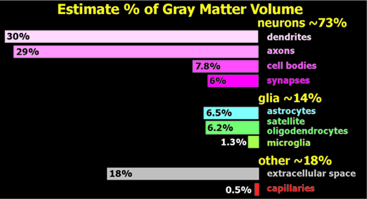
A common, yet simplistic, assumption regarding volumetric changes is that they are directly due to changes in cell quantity (increase or decrease); however, this has not necessarily been well-supported by postmortem studies.11 Recent research suggests that other interpretations may be more accurate. A review of the documented changes in animal models of stress and depression in humans, coupled with the estimated volume fraction of each cortical component, supported the hypothesis that the majority of gray-matter volume changes reported in depression are due to decreased synapses and dendritic regression, rather than a reduction in cell numbers (Figure 2).12
FIGURE 2. Imaging-based studies provide estimates of gray-matter volume associated with particular psychiatric disorders or treatments, but do not allow identification differences at the cellular level.11 Of particular interest is whether reduced neuronal numbers are likely to be present in areas with less gray-matter volume. A recent review provided estimates of the volume fractions occupied by the major constituents of cortex (right).12 Note that much of the cortical volume is occupied by neurons (shades of pink). Their analysis concluded that most gray-matter volume differences reported in depression are likely due to synaptic pruning and dendritic regression, rather than cell loss.

Utilizing Neuroplasticity

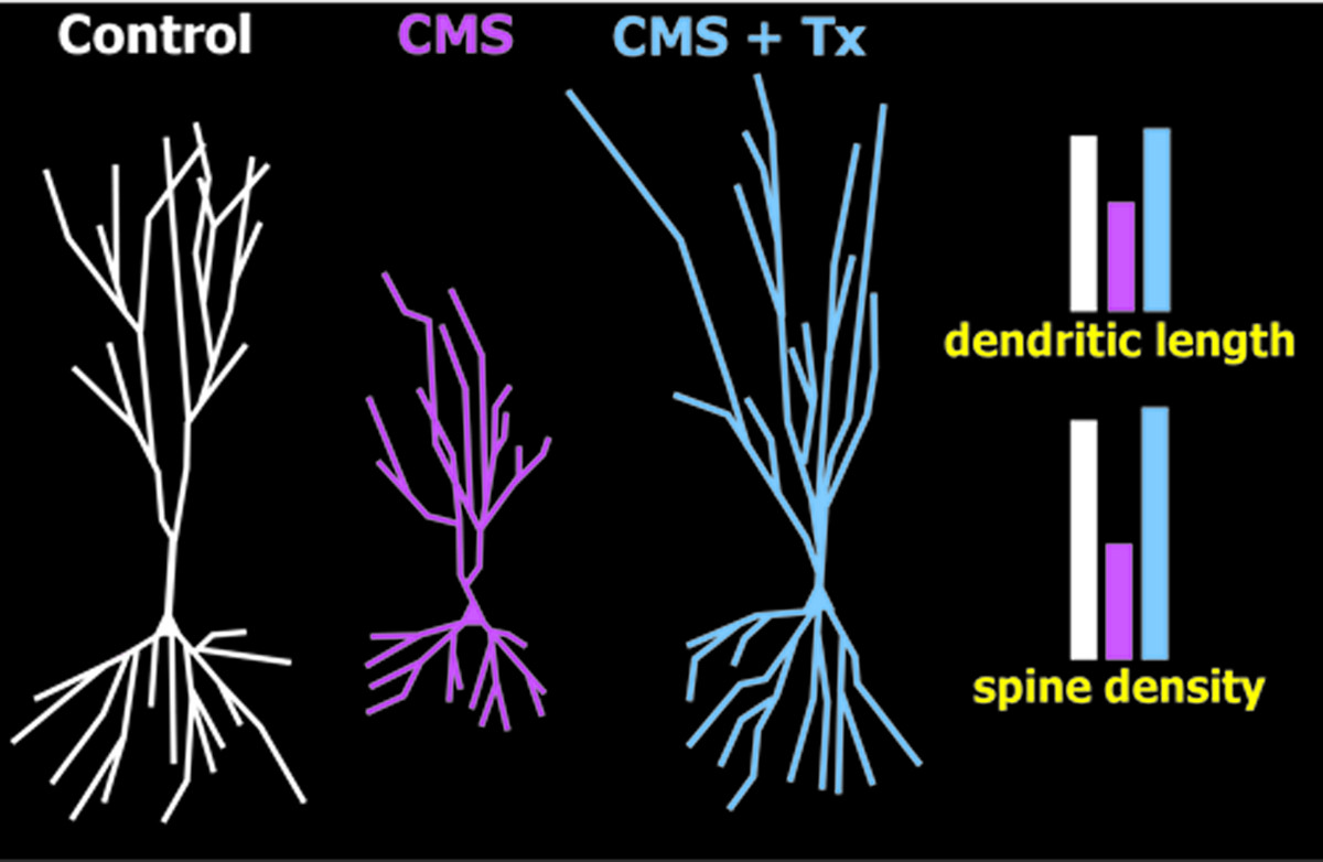
Researchers have begun examining ways to harness neuroplasticity to promote healing and recovery. Although these efforts are still in the beginning stages, there is promising evidence that the dynamic qualities of the brain may play a pivotal role in how one copes with stress and mental illness.58,59 Medications have been shown to affect neuroplasticity in animal models and a few human studies. As noted previously, antidepressant medications can reverse the effects of various types of chronic stress on both behavior and brain structure (Figure 3), although animal studies differ on which aspects of neuroplasticity (e.g., neurogenesis, dendritic remodeling, BDNF levels) are critical for therapeutic efficacy.6062 Several clinical studies have found that successful medication treatment can reverse hippocampal volume deficits in PTSD and depression, although contrary results have also been reported.51,6365 Induced stimulation of the brain focally or generally also effects neuroplasticity. Studies in multiple species, including non-human primates, have shown that electroconvulsive shock increases hippocampal BDNF levels, synaptic density, and neurogenesis.66 A small longitudinal study in patients with depression reported increased hippocampal volume after electroconvulsive therapy (ECT) treatment.67 A case study of a patient with schizophrenia reported that serum levels of BDNF increased during the course of ECT treatment in parallel with symptom improvement.68 Other forms of stimulation (e.g., TMS, deep brain stimulation) have also been utilized in treatment for various conditions by modifying activation patterns in the brain with the intention of improving functioning.59 Exercise, which has been shown to ameliorate behavioral symptoms of stress and enhance hippocampal neuroplasticity in animal models, has also been considered as a potential adjunctive treatment for neuropsychiatric conditions.21,59,6971 Physical activity attenuates many of the harmful effects of stress.70 Reviews of the evidence indicate that exercise can be associated with reduced psychiatric symptoms (particularly of depression [MDD]) and cognitive deficits in multiple conditions (e.g. MDD, schizophrenia, Alzheimer’s dementia).69,71 There is evidence that exercise, used as a supportive treatment, may delay or even prevent disease-onset and progression.71
FIGURE 3. Exposure of rats to 6 weeks of unpredictable chronic mild stress (CMS; pink) induces depressive-like behaviors (e.g., anhedonia, learned helplessness) and multiple detrimental effects in the hippocampus and medial prefrontal cortex (mPFC), including decreases in neurogenesis, dendritic length, and synaptic density, as compared with control conditions (white). Both behavioral and structural deficits can be reversed by administration of antidepressants (Tx) during the final 2 weeks of CMS (CMS + Tx; blue).13 Schematic representations of mPFC neurons under the three conditions illustrate average dendritic changes. The authors of this study noted that these results were independent of neurogenesis, suggesting that restoration of normal dendritic length and synaptic density underlie behavioral recovery.

Conclusions

The brain, once considered to be a fixed and stable organ, is now viewed as dynamic, flexible, and adaptive. Efforts are beginning to focus on ways to harness the plastic qualities of the brain for treatment and recovery. There is much that is still unclear about the relationship between neuroplasticity and mental health. Research capabilities for human studies are limited, so most questions must be addressed by study of animal models. This makes disentangling genetic, environmental, and experiential influences much more challenging. Although there is not yet consensus, it appears the field is moving toward a more multifaceted, nuanced understanding that recognizes the likely contribution of multiple factors, rather than a single explanation. Future research and advances in technology will continue to increase understanding of the human brain and its fascinating abilities and potential.

References

1.
Eriksson PS, Perfilieva E, Björk-Eriksson T, et al.: Neurogenesis in the adult human hippocampus. Nat Med 1998; 4:1313–1317
2.
Gould E: How widespread is adult neurogenesis in mammals? Nat Rev Neurosci 2007; 8:481–488
3.
Bonfanti L, Peretto P: Adult neurogenesis in mammals: a theme with many variations. Eur J Neurosci 2011; 34:930–950
4.
Curtis MA, Kam M, Faull RL: Neurogenesis in humans. Eur J Neurosci 2011; 33:1170–1174
5.
Landgren H, Curtis MA: Locating and labeling neural stem cells in the brain. J Cell Physiol 2011; 226:1–7
6.
Gould E, Reeves AJ, Graziano MS, et al.: Neurogenesis in the neocortex of adult primates. Science 1999; 286:548–552
7.
Bernier PJ, Vinet J, Cossette M, et al.: Characterization of the subventricular zone of the adult human brain: evidence for the involvement of Bcl-2. Neurosci Res 2000; 37:67–78
8.
Bédard A, Cossette M, Lévesque M, et al.: Proliferating cells can differentiate into neurons in the striatum of normal adult monkey. Neurosci Lett 2002; 328:213–216
9.
Bernier PJ, Bedard A, Vinet J, et al.: Newly generated neurons in the amygdala and adjoining cortex of adult primates. Proc Natl Acad Sci USA 2002; 99:11464–11469
10.
Bédard A, Gravel C, Parent A: Chemical characterization of newly generated neurons in the striatum of adult primates. Exp Brain Res 2006; 170:501–512
11.
Couillard-Despres S, Aigner L: In-vivo imaging of adult neurogenesis. Eur J Neurosci 2011; 33:1037–1044
12.
Bennett MR: The prefrontal-limbic network in depression: a core pathology of synapse regression. Prog Neurobiol 2011; 93:457–467
13.
Bessa JM, Ferreira D, Melo I, et al.: The mood-improving actions of antidepressants do not depend on neurogenesis but are associated with neuronal remodeling. Mol Psychiatry 2009; 14:764–773, 739
14.
Cicchetti D, Curtis J. The Developing Brain and Neural Plasticity: Implications for Normality, Psychopatholgy, and Resilience. 2006; 2:1–64
15.
Leuner B, Gould E: Structural plasticity and hippocampal function. Annu Rev Psychol 2010; 61:111–140, C1–C3
16.
Pascual-Leone A, Amedi A, Fregni F, et al.: The plastic human brain cortex. Annu Rev Neurosci 2005; 28:377–401
17.
Warraich Z, Kleim JA: Neural plasticity: the biological substrate for neurorehabilitation. PM R 2010; 2(Suppl 2):S208–S219
18.
Kerr AL, Cheng SY, Jones TA: Experience-dependent neural plasticity in the adult damaged brain. J Commun Disord 2011; 44:538–548
19.
May A: Experience-dependent structural plasticity in the adult human brain. Trends Cogn Sci 2011; 15:475–482
20.
Kolb B, Muhammad A, Gibb R: Searching for factors underlying cerebral plasticity in the normal and injured brain. J Commun Disord 2011; 44:503–514
21.
Yau SY, Lau BW, So KF: Adult hippocampal neurogenesis: a possible way how physical exercise counteracts stress. Cell Transplant 2011; 20:99–111
22.
McEwen BS: Structural plasticity of the adult brain: how animal models help us understand brain changes in depression and systemic disorders related to depression. Dialogues Clin Neurosci 2004; 6:119–133
23.
Taliaz D, Stall N, Dar DE, et al.: Knockdown of brain-derived neurotrophic factor in specific brain sites precipitates behaviors associated with depression and reduces neurogenesis. Mol Psychiatry 2010; 15:80–92
24.
McEwen BS, Eiland L, Hunter RG, et al.: Stress and anxiety: structural plasticity and epigenetic regulation as a consequence of stress. Neuropharmacology 2012; 62:3–12
25.
Ma DK, Ming GL, Song H: Glial influences on neural stem cell development: cellular niches for adult neurogenesis. Curr Opin Neurobiol 2005; 15:514–520
26.
Morrens J, Van Den Broeck W, Kempermann G: Glial cells in adult neurogenesis. Glia 2012; 60:159–174
27.
Chen JL, Nedivi E: Neuronal structural remodeling: is it all about access? Curr Opin Neurobiol 2010; 20:557–562
28.
Yamahachi H, Marik SA, McManus JN, et al.: Rapid axonal sprouting and pruning accompany functional reorganization in primary visual cortex. Neuron 2009; 64:719–729
29.
Castrén E, Rantamäki T: The role of BDNF and its receptors in depression and antidepressant drug action: reactivation of developmental plasticity. Dev Neurobiol 2010; 70:289–297
30.
Kempermann G, Fabel K, Ehninger D, et al.: Why and how physical activity promotes experience-induced brain plasticity. Front Neurosci 2010; 4:189
31.
Bednarczyk MR, Hacker LC, Fortin-Nunez S, et al.: Distinct stages of adult hippocampal neurogenesis are regulated by running and the running environment. Hippocampus 2011; 21:1334–1347
32.
Perera TD, Dwork AJ, Keegan KA, et al.: Necessity of hippocampal neurogenesis for the therapeutic action of antidepressants in adult nonhuman primates. PLoS ONE 2011; 6:e17600
33.
Segi-Nishida E: Exploration of new molecular mechanisms for antidepressant actions of electroconvulsive seizure. Biol Pharm Bull 2011; 34:939–944
34.
Wosiski-Kuhn M, Stranahan AM: Opposing effects of positive and negative stress on hippocampal plasticity over the lifespan. Ageing Res Rev 2011; [Epub ahead of print]
35.
Kubera M, Obuchowicz E, Goehler L, et al.: In animal models, psychosocial stress-induced (neuro)inflammation, apoptosis, and reduced neurogenesis are associated with the onset of depression. Prog Neuropsychopharmacol Biol Psychiatry 2011; 35:744–759
36.
Schoenfeld TJ, Gould E: Stress, stress hormones, and adult neurogenesis. Exp Neurol 2012; 233:12–21
37.
Christoffel DJ, Golden SA, Russo SJ: Structural and synaptic plasticity in stress-related disorders. Rev Neurosci 2011; 22:535–549
38.
Emoto K: Dendrite remodeling in development and disease. Dev Growth Differ 2011; 53:277–286
39.
Lyons DM, Buckmaster PS, Lee AG, et al.: Stress-coping stimulates hippocampal neurogenesis in adult monkeys. Proc Natl Acad Sci USA 2010; 107:14823–14827
40.
Kraus N, Chandrasekaran B: Music training for the development of auditory skills. Nat Rev Neurosci 2010; 11:599–605
41.
Woollett K, Maguire EA: Acquiring “the Knowledge” of London’s layout drives structural brain changes. Curr Biol 2011; 21:2109–2114
42.
Papagni SA, Benetti S, Arulanantham S, et al.: Effects of stressful life events on human brain structure: a longitudinal, voxel-based morphometry study. Stress 2011; 14:227–232
43.
Merabet LB, Pascual-Leone A: Neural reorganization following sensory loss: the opportunity of change. Nat Rev Neurosci 2010; 11:44–52
44.
Ricciardi E, Pietrini P: New light from the dark: what blindness can teach us about brain function. Curr Opin Neurol 2011; 24:357–363
45.
Cappe C, Rouiller EM, Barone P: Multisensory anatomical pathways. Hear Res 2009; 258:28–36
46.
Agarwal N, Port JD, Bazzocchi M, et al.: Update on the use of MR for assessment and diagnosis of psychiatric diseases. Radiology 2010; 255:23–41
47.
Quidé Y, Witteveen AB, El-Hage W, et al.: Differences between effects of psychological versus pharmacological treatments on functional and morphological brain alterations in anxiety disorders and major depressive disorder: a systematic review. Neurosci Biobehav Rev 2012; 36:626–644
48.
Bremner JD, Elzinga B, Schmahl C, et al.: Structural and functional plasticity of the human brain in posttraumatic stress disorder. Prog Brain Res 2008; 167:171–186
49.
Felmingham K, Williams LM, Whitford TJ, et al.: Duration of posttraumatic stress disorder predicts hippocampal grey-matter loss. Neuroreport 2009; 20:1402–1406
50.
McKinnon MC, Yucel K, Nazarov A, et al.: A meta-analysis examining clinical predictors of hippocampal volume in patients with major depressive disorder. J Psychiatry Neurosci 2009; 34:41–54
51.
Malykhin NV, Carter R, Seres P, et al.: Structural changes in the hippocampus in major depressive disorder: contributions of disease and treatment. J Psychiatry Neurosci 2010; 35:337–343
52.
Woon FL, Sood S, Hedges DW: Hippocampal volume deficits associated with exposure to psychological trauma and posttraumatic stress disorder in adults: a meta-analysis. Prog Neuropsychopharmacol Biol Psychiatry 2010; 34:1181–1188
53.
Apfel BA, Ross J, Hlavin J, et al.: Hippocampal volume differences in Gulf War veterans with current versus lifetime posttraumatic stress disorder symptoms. Biol Psychiatry 2011; 69:541–548
54.
Frodl T, Jäger M, Smajstrlova I, et al.: Effect of hippocampal and amygdala volumes on clinical outcomes in major depression: a 3-year prospective magnetic resonance imaging study. J Psychiatry Neurosci 2008; 33:423–430
55.
MacQueen GM, Yucel K, Taylor VH, et al.: Posterior hippocampal volumes are associated with remission rates in patients with major depressive disorder. Biol Psychiatry 2008; 64:880–883
56.
Gilbertson MW, Shenton ME, Ciszewski A, et al.: Smaller hippocampal volume predicts pathologic vulnerability to psychological trauma. Nat Neurosci 2002; 5:1242–1247
57.
Kremen WS, Koenen KC, Afari N, et al.: Twin studies of posttraumatic stress disorder: differentiating vulnerability factors from sequelae. Neuropharmacology 2012; 62:647–653
58.
DeCarolis NA, Eisch AJ: Hippocampal neurogenesis as a target for the treatment of mental illness: a critical evaluation. Neuropharmacology 2010; 58:884–893
59.
Cramer SC, Sur M, Dobkin BH, et al.: Harnessing neuroplasticity for clinical applications. Brain 2011; 134:1591–1609
60.
Hanson ND, Owens MJ, Nemeroff CB: Depression, antidepressants, and neurogenesis: a critical reappraisal. Neuropsychopharmacology 2011; 36:2589–2602
61.
Samuels BA, Hen R: Neurogenesis and affective disorders. Eur J Neurosci 2011; 33:1152–1159
62.
Petrik D, Lagace DC, Eisch AJ: The neurogenesis hypothesis of affective and anxiety disorders: are we mistaking the scaffolding for the building? Neuropharmacology 2012; 62:21–34
63.
Vermetten E, Vythilingam M, Southwick SM, et al.: Long-term treatment with paroxetine increases verbal declarative memory and hippocampal volume in posttraumatic stress disorder. Biol Psychiatry 2003; 54:693–702
64.
Bremner JD, Vermetten E: Neuroanatomical changes associated with pharmacotherapy in posttraumatic stress disorder. Ann N Y Acad Sci 2004; 1032:154–157
65.
Vythilingam M, Vermetten E, Anderson GM, et al.: Hippocampal volume, memory, and cortisol status in major depressive disorder: effects of treatment. Biol Psychiatry 2004; 56:101–112
66.
Bolwig TG: How does electroconvulsive therapy work? theories on its mechanism. Can J Psychiatry 2011; 56:13–18
67.
Nordanskog P, Dahlstrand U, Larsson MR, et al.: Increase in hippocampal volume after electroconvulsive therapy in patients with depression: a volumetric magnetic resonance imaging study. J ECT 2010; 26:62–67
68.
Martinotti G, Ricci V, Di Nicola M, et al.: Brain-derived neurotrophic factor and electroconvulsive therapy in a schizophrenic patient with treatment-resistant paranoid-hallucinatory symptoms. J ECT 2011; 27:e44–e46
69.
Ernst C, Olson AK, Pinel JP, et al.: Antidepressant effects of exercise: evidence for an adult-neurogenesis hypothesis? J Psychiatry Neurosci 2006; 31:84–92
70.
Greenwood BN, Fleshner M: Exercise, stress resistance, and central serotonergic systems. Exerc Sport Sci Rev 2011; 39:140–149
71.
Knöchel C, Oertel-Knöchel V, O’Dwyer L, et al.: Cognitive and behavioural effects of physical exercise in psychiatric patients. Prog Neurobiol 2012; 96:46–68

Information & Authors

Information

Published In

Go to The Journal of Neuropsychiatry and Clinical Neurosciences
The Journal of Neuropsychiatry and Clinical Neurosciences
Pages: 118 - 124
PubMed: 22772660

History

Published online: 1 April 2012
Published in print: Spring 2012

Authors

Details

Jill L. Kays, Psy.D.
Drs. Kays, Taber, and Hurley are affiliated with the Veterans Affairs Mid-Atlantic Mental Illness Research, Education, and Clinical Center, and the W.G. Hefner Veterans Affairs Medical Center in Salisbury, NC. Dr. Hurley is affiliated with the Departments of Psychiatry and Radiology at Wake Forest School of Medicine in Winston-Salem, NC, and the Menninger Dept. of Psychiatry and Behavioral Sciences at Baylor College of Medicine in Houston, TX. Dr. Taber is affiliated with the Division of Biomedical Sciences at the Via College of Osteopathic Medicine in Blacksburg, VA, and the Dept. of Physical Medicine and Rehabilitation at Baylor College of Medicine, Houston, TX.
Robin A. Hurley, M.D.
Drs. Kays, Taber, and Hurley are affiliated with the Veterans Affairs Mid-Atlantic Mental Illness Research, Education, and Clinical Center, and the W.G. Hefner Veterans Affairs Medical Center in Salisbury, NC. Dr. Hurley is affiliated with the Departments of Psychiatry and Radiology at Wake Forest School of Medicine in Winston-Salem, NC, and the Menninger Dept. of Psychiatry and Behavioral Sciences at Baylor College of Medicine in Houston, TX. Dr. Taber is affiliated with the Division of Biomedical Sciences at the Via College of Osteopathic Medicine in Blacksburg, VA, and the Dept. of Physical Medicine and Rehabilitation at Baylor College of Medicine, Houston, TX.
Katherine H. Taber, Ph.D.
Drs. Kays, Taber, and Hurley are affiliated with the Veterans Affairs Mid-Atlantic Mental Illness Research, Education, and Clinical Center, and the W.G. Hefner Veterans Affairs Medical Center in Salisbury, NC. Dr. Hurley is affiliated with the Departments of Psychiatry and Radiology at Wake Forest School of Medicine in Winston-Salem, NC, and the Menninger Dept. of Psychiatry and Behavioral Sciences at Baylor College of Medicine in Houston, TX. Dr. Taber is affiliated with the Division of Biomedical Sciences at the Via College of Osteopathic Medicine in Blacksburg, VA, and the Dept. of Physical Medicine and Rehabilitation at Baylor College of Medicine, Houston, TX.

Notes

Address correspondence to Dr. Robin Hurley, Hefner VA Medical Center, Salisbury, NC; e-mail: [email protected]

Metrics & Citations

Metrics

Citations

Export Citations

If you have the appropriate software installed, you can download article citation data to the citation manager of your choice. Simply select your manager software from the list below and click Download.

For more information or tips please see 'Downloading to a citation manager' in the Help menu.

Format
Citation style
Style
Copy to clipboard

View Options

View options

PDF/EPUB

View PDF/EPUB

Login options

Already a subscriber? Access your subscription through your login credentials or your institution for full access to this article.

Personal login Institutional Login Open Athens login
Purchase Options

Purchase this article to access the full text.

PPV Articles - Journal of Neuropsychiatry and Clinical Neurosciences

PPV Articles - Journal of Neuropsychiatry and Clinical Neurosciences

Not a subscriber?

Subscribe Now / Learn More

PsychiatryOnline subscription options offer access to the DSM-5-TR® library, books, journals, CME, and patient resources. This all-in-one virtual library provides psychiatrists and mental health professionals with key resources for diagnosis, treatment, research, and professional development.

Need more help? PsychiatryOnline Customer Service may be reached by emailing [email protected] or by calling 800-368-5777 (in the U.S.) or 703-907-7322 (outside the U.S.).

Media

Figures

Other

Tables

Share

Share

Share article link

Share