To the Editor: We report on a 22-year-old man who has a 9-year history of glue-sniffing that evolved into a full-blown picture of psychological dependency that was characterized by craving and tolerance, which was subsequently followed by agitation, irritability, and physical struggle. It gradually ended with period of low mood, lack of motivation, and social withdrawal. The disturbance was punctuated by episodes of florid psychosis during intoxication, which included visual hallucination and disorganized behavior. After 6 years of chronic glue-sniffing, there was an insidious onset of bilateral, low-frequency intention tremors involving the upper limbs that gradually worsened and was accompanied by unstable gait, stuttering of speech, and blurring of vision. There was no history to suggest seizure disorder or any infection of the central nervous system. There was a 1-year delay before presentation to medical services due to shame and guilt feelings about coming out of the house because of the physical problems. Mental state examination (MSE) revealed presence of scanning speech and impairment of abstract thinking. He scored 26/30 on Mini-Mental State Exam (MMSE). He had bilateral, persistent, horizontal, pendular nystagmus, and bilateral intention tremors of the hands, broad-based gait, past pointing and dysdiadokinesia of the upper limbs, which were more prominent on the right side. Further examination revealed optic atrophy of the right eye, but sensorimotor functions were normal, and muscle wasting was absent.
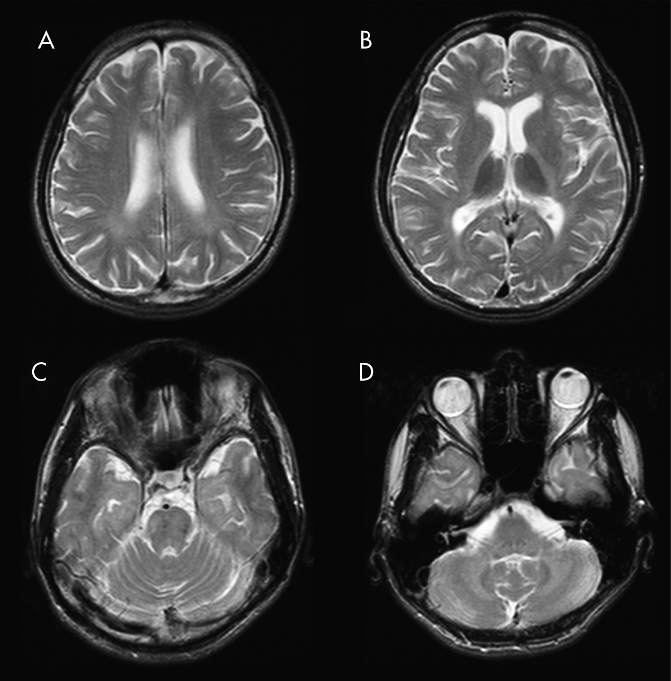
Blood and urine biochemistry that included measurements of serum lactate and ceruloplasmin were negative, as was the screening for other commonly abused substances. Magnetic resonance imaging (MRI) of the brain demonstrated extensive increased T
2 signal in the white matter extending from the centrum semiovale, down the posterior limb of the internal capsule, and through the anterolateral pons, as well as the white matter of the cerebellar hemispheres (
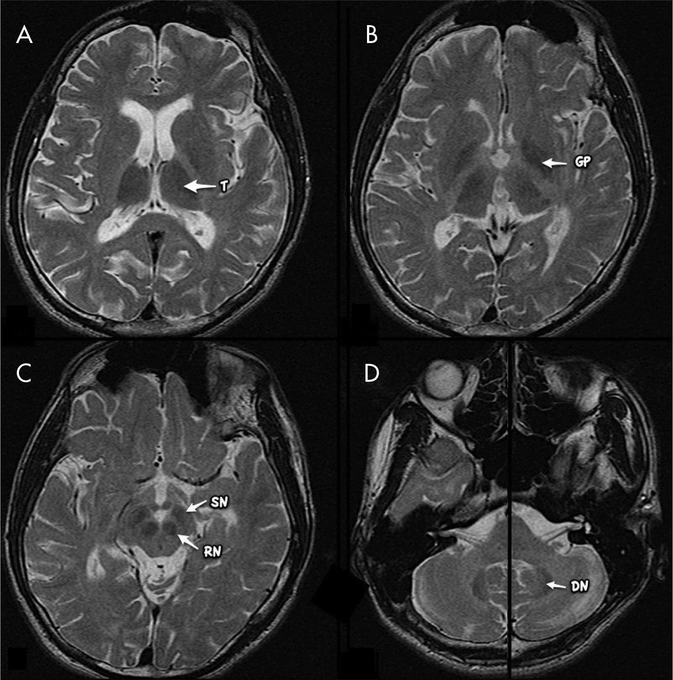
Figure 1). Low signal is seen in the deep gray-matter structures, particularly involving the thalami, globus pallidi, and dentate nuclei, as well as the red nuclei and substantia nigra (
Figure 2). Widespread cerebral and cerebellar volume loss is also present, with marked thinning of the corpus callosum, without T
2 signal change.
Treatment was initiated with propranolol 40 mg bid. This was discontinued after 3 months because of lack of efficacy, and he was started on treatment with amantadine 100 mg nightly, primidone 250 mg nightly, and supplemented with thiamine. Despite 6 months of the above treatments, there has been no improvement. He continues to sniff glue, albeit at a much lower frequency and amount.
Peripheral neuropathy, cerebellar dysfunction, cranial nerve damage, cortical atrophy, encephalopathy, and dementia have all been attributed to result from prolonged and heavy exposure to inhalants.
1,2 N-hexane and toluene adhere to lipid-laden myelin sheaths and neuronal membranes to start the demyelinating process.
3 Cerebellar damage due to toluene was first reported three decades ago, and it commonly presents with tremors and ataxia, which correlate to the extent of sulci enlargement.
1


