Full access
Online Exclusives
Published Online: 10 November 2014

A Neural Circuit Framework for Somatosensory Amplification in Somatoform Disorders

Publication: The Journal of Neuropsychiatry and Clinical Neurosciences

Abstract

Although somatosensory amplification is theorized to serve a critical role in somatization, it remains poorly understood neurobiologically. In this perspective article, convergent visceral-somatic processing is highlighted, and neuroimaging studies in somatoform disorders are reviewed. Neural correlates of cognitive-affective amplifiers are integrated into a neurocircuit framework for somatosensory amplification. The anterior cingulate cortex, insula, amygdala, hippocampal formation, and striatum are some of the identified regions. Clinical symptomatology in a given patient or group may represent dysfunction in one or more of these neurobehavioral nodes. Somatosensory amplification may, in part, develop through stress-mediated aberrant neuroplastic changes and the neuromodulatory effects of inflammation.
Approximately one-third of symptoms reported by patients in primary care and subspecialty settings remain medically unexplained after a complete evaluation.1 The term somatization refers to patients with medically unexplained symptoms that seek medical attention because of suspected disease or individuals reporting symptoms out-of-proportion to their medical condition. Their symptoms commonly include abdominal pain, bloating, dizziness, chest pain, breathlessness, pelvic pain, food intolerance, palpitations, and back pain among others.2 When considering symptoms that have “bothered [the patient] a lot” in the past month, 16%−33% have no demonstrable medical basis.3 Patients with medically unexplained symptoms engage in a disproportionately high rate of medical care utilization, including outpatient visits, hospitalizations, and overall healthcare costs (averaging 4700 USD annually per individual).4
The somatosensory amplification construct has been theorized by Arthur Barsky and colleagues to serve a critical role in the pathophysiology of somatization.5 Somatosensory amplification refers to the tendency to experience a wide range of benign bodily sensations as intrusive, intense, noxious, and disruptive. Several elements are associated with amplification including: 1) a heightened attentional focus on bodily sensations; 2) the tendency to select out certain relatively weak and infrequent sensations; 3) the disposition to react to these sensations with affect and cognitions that intensify them and make them more alarming and distressing.5 Somatosensory amplification has also been linked to alexithymia,6,7 and somatization has been associated with dysphoric-anxious mood and cognitive distortions, particularly pain catastrophizing.8
Neuroimaging techniques applied to somatoform disorder populations and related cognitive-affective neuroscience studies in healthy populations allow for the in vivo detection of neural circuit abnormalities associated with somatization generally, and with somatosensory amplification more specifically. In this perspective article, central nervous system convergence of visceral and somatic processing is first summarized. Thereafter, neuroimaging studies in somatization disorder, undifferentiated somatization disorder, and somatoform pain disorder are reviewed [functional neurological symptom disorder (i.e., conversion disorder) was previously discussed elsewhere9]. Hypothesized cognitive amplifiers of visceral-somatic processing including negative expectation, negative attentional bias, and pain catastrophizing, along with affective modifiers including alexithymia and dysphoric-anxious mood are discussed by integrating neuroimaging findings in healthy populations. Lastly, a neurocircuit model of somatosensory amplification is suggested. We propose that somatosensory amplification involves abnormal interactions among large-scale neural systems mediating visceral-somatic perception, emotional processing/awareness, and cognitive control. Frontolimbic, subcortical, and brainstem structures are particularly linked to the neurocircuitry of abnormal symptom amplification.
The purpose of this article is to provide a theoretical neurocircuit framework through which physician-scientists may begin to understand brain-behavior relationships in somatosensory amplification, rather than to provide a comprehensive review of published studies on somatosensory amplification and somatoform disorders. Articles using functional magnetic resonance imaging (fMRI), single photon-emission computed tomography (SPECT) and positron emission tomography (PET) to investigate somatization disorder, undifferentiated somatoform disorder, and somatoform pain disorder were emphasized. Studies exploring other “functional” disorders including fibromyalgia, chronic fatigue syndrome, and irritable bowel syndrome were largely omitted to limit the discussion to DSM-IV somatoform disorders.

Somatic and Visceral Afferent Processing

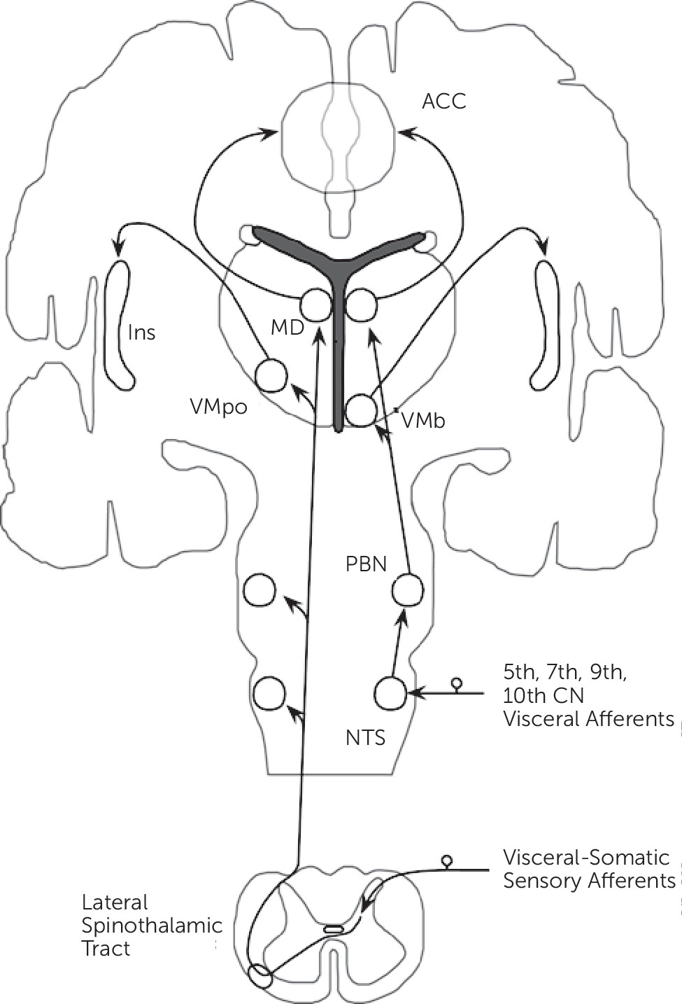
Neurobiological, noninvasive neuroimaging, and neuropathological studies have identified distributed neural networks involved in somatic and visceral sensory processing. This section focuses on sites of information convergence, and mainly details the lamina I spinothalamocortical pathway (Figure 1).10
FIGURE 1. Graphic Depiction of the Lamina I Spinothalamocortical Pathway Demonstrating Convergence of Visceral-Somatic Processing Within the Brainstem, Thalamus, Anterior Cingulate Cortex (ACC), and Insula (Ins)a
a CN=cranial nerve; MD=medial dorsal thalamic nuclei; NTS=nucleus tractus solitarius; PBN=parabrachial nucleus; VMb=basal ventral medial thalamic nuclei; VMpo=ventromedial thalamic nuclei
Small diameter sensory afferents conveying physiological information from tissues of the body (including mechanical, thermal, stress, metabolic, inflammatory, and visceral) terminate in lamina I within the dorsal horn of the spinal cord. Second-order neurons project contralaterally in the spinal cord and ascend as part of the lateral spinothalamic tract. These ascending projections, prior to terminating in the thalamus, project within the brainstem onto the nucleus tractus solitarius (NTS), parabrachial nucleus (PBN), and nucleus cuneiformis (NCF). Visceral afferents from the 5th, 7th, 9th, and 10th cranial nerves terminate on the NTS, which subsequently project onto the PBN. Lamina I spinothalamic afferents synapse topographically onto the posterior part of the ventromedial nucleus (VMpo) and the medial dorsal nucleus (MD) of the thalamus. Afferents from the PBN project onto an adjacent part of the thalamus, the basal ventral medial nucleus (VMb), and the MD nucleus. [Note: conventional spinothalamic afferents synapse ventrolaterally in the thalamus before projecting to primary somatosensory cortex (SI).] Notably, visceral-somatic afferents converge within the brainstem and the MD nucleus of the thalamus.
The VMpo and VMb thalamic nuclei project topographically onto the posterior insula, while the MD nucleus projects to the anterior cingulate cortex (ACC). Based on their afferent connections, the ACC and insula are major cortical sites of visceral-somatic processing and information convergence; the ACC and insula are reciprocally connected and these regions also connect to the orbitofrontal cortex (OFC) and the amygdala.11 Additional components of visceral-somatic processing include SI, secondary somatosensory (SII), posterior parietal, medial and lateral prefrontal cortices, the striatum, hippocampal formation, lateral thalamus, hypothalamus, pituitary, and brainstem structures including the periaqueductal gray (PAG) and rostral ventromedial medulla (RVM). The brainstem structures form part of the descending pain neuromodulatory system, which may either inhibit or amplify visceral-somatic processing within the spinal cord.12 Of note, while differences in the cortical-subcortical processing of visceral and somatic stimuli have been reported,13 evidence suggests considerable cortical-subcortical and brainstem overlap.1417

Neuroimaging Studies in Somatization Disorder, Undifferentiated Somatoform Disorder, and Somatoform Pain Disorder

Functional and structural neuroimaging studies have used fMRI, PET, SPECT, and structural MRI techniques to investigate neurocircuit abnormalities in somatization disorder, undifferentiated somatoform populations, and somatoform pain disorder.
Neuroimaging studies thus far have mainly identified striatal and amygdalar abnormalities in somatization disorder and undifferentiated somatoform disorder. Hakala and colleagues performed the first series of investigations in 10 female patients (six with somatization disorder; four with undifferentiated somatoform disorder) compared with healthy subjects using resting-state, fluorodeoxyglucose-PET, and demonstrated bilateral caudate-putamen hypometabolism in patients.18,19 The same cohort exhibited increased bilateral caudate volumes compared with controls using a manualized MRI tracing technique.20 In addition to the striatum, amygdalar abnormalities have also been characterized. Manualized tracings identified bilateral amygdalar volumetric reductions in 20 women with somatization disorder compared with healthy females.21 Similarly, in an fMRI affectively valenced picture viewing task, 20 mixed somatoform disorder patients (13 undifferentiated, five somatoform pain disorder, two somatization disorder) exhibited decreased left amygdala and right parahippocampal activity during processing of emotionally valenced facial expressions compared with unrecognizable smoothed pictures.22 Less frequently, smaller pituitary volumes were reported in somatization disorder using manualized tracings,23 and bilateral superior temporal and left lateralized postcentral, precentral, inferior parietal, and middle occipital regional SPECT hyperperfusion was identified in patients with undifferentiated somatoform disorder compared with healthy subjects.24
Studies of patients with somatoform pain disorder commonly, though not exclusively, demonstrated increased regional central pain processing activity and reduced prefrontal cortex activity. Several investigations have compared patients with somatoform pain disorder with healthy subjects using pain provocation paradigms. In a fMRI study using a pinprick noxious stimuli task, 17 mixed-gender somatoform pain disorder patients compared with healthy subjects showed increased activity in pain processing regions including the anterior insula, hippocampus, putamen, and thalamus; in addition, somatoform pain disorder patients exhibited increased inferior parietal, temporo-occipital, lateral temporal, ventrolateral prefrontal, and dorsomedial prefrontal regional activity.25 This somatoform pain disorder cohort compared with healthy subjects also demonstrated increased left insula and decreased bilateral temporo-occipital, superior parietal and right OFC activity during exposure to pictures and audio scenes of physical violence. Noxious thermal stimuli delivery was associated with anterior insula, parahippocampal, and amygdalar hyperactivity, along with orbitofrontal/ventromedial prefrontal hypoactivity, in 12 women with somatoform pain disorder compared with healthy subjects.26 Decreased lateral prefrontal and increased right ACC, left thalamus and bilateral brainstem, caudate, and posterior cingulate cortex regional cerebral blood flow were also noted in a SPECT study of 10 patients with somatoform pain disorder compared with healthy subjects.27 In parallel, an automated voxel-based structural MRI whole-brain analysis identified decreased prefrontal (ventromedial/OFC/ACC/middle frontal/superior medial), insula, parahippocampal, inferior temporal, and posterior cingulate cortex gray matter volumes.28 Resting-state functional neuroimaging studies have reported increased brainstem, caudate, thalamus and ACC activity, and decreased lateral prefrontal activity.29,30 In summary, neuroimaging studies suggest enhanced central pain processing activity in patients with somatoform pain disorder, whereas striatal and amygdalar dysfunction have been linked to somatization disorder and undifferentiated somatoform disorder.

Cognitive Amplifiers of Visceral-Somatic Processing

Although few neuroimaging studies have investigated circuit abnormalities in somatization disorder, undifferentiated somatoform disorder and somatoform pain disorder, advances in cognitive-affective neuroscience provide additional important insights into the neurocircuitry of somatosensory amplification. The following section examines the neural correlates of negative expectation/anticipation, negative attentional bias, and pain catastrophizing, cognitive processes implicated in somatosensory amplification.58 These amplifiers converge to limit an individual’s ability to shift cognitive resources away from visceral-somatic perceptions.

Appraisal: Negative Expectation/Anticipation

The hippocampal formation has been linked to negative expectancy effects by the work of Ploghaus and colleagues. Investigators compared behavioral and neural responses in 18 healthy men under two conditions: during condition 1, a visual cue reliably predicted the delivery of moderately intense thermal noxious stimulation; in condition 2, a distinct visual cue predicted the delivery of moderately intense noxious stimuli but was also associated with the possibility of stronger noxious stimulus delivery.31 Observationally, moderately intense stimuli were rated as more intense when preceded by the condition 2 cue. Furthermore, comparisons of the neural responses of moderately intense stimuli signaled by condition 1 and 2 showed increased left hippocampal formation activity (including the parahippocampus) and enhanced functional connectivity between the hippocampal formation, mid-insula and perigenual ACC during the less predictable condition. In a modified version of this paradigm, reduced differential hippocampal activity between high and low anxiety states was associated with increased report of daily physical (somatized) symptoms.32 Hippocampal formation activity also correlated with individual differences in pain threshold sensitivity,33 predicted posterior insula activity during noxious stimuli processing,34 and was associated with negative expectancy effects to opiate analgesia.35
The ACC, insula, and brainstem have also been linked to negative expectancy effects. In a study of 27 mixed-gender healthy subjects exposed to high- and low-intensity thermal stimuli, each predicted by distinct visual cues,36 subjects again rated stimuli as more noxious when preceded by a high-intensity signaling cue. High-intensity stimuli coupled to the higher intensity cue demonstrated increased activity in the caudal ACC and NCF compared with the same stimulus paired to a lower intensity cue. The authors suggested that involvement of the descending pain modulatory system potentially accounted for negative expectation effects. In other investigations, enhanced expectation of thermal stimuli unpleasantness increased ACC and posterior insula activity.37
The nocebo effect, an adverse effect or symptom worsening based on the negative expectation of a given treatment, is a subtype of the negative expectancy effect. In a study of 13 mixed-gender subjects using a within-subject design, thermal pain, and sham acupuncture to induce nocebo hyperalgesia, pain processing regions including the ACC, insula, frontal-parietal operculum, OFC, and hippocampus were hyperactivated.38 Nocebo hyperalgesia has been linked to decreased opioid and dopaminergic activity in the insula, dlPFC, nucleus accumbens, and PAG.39 Overall, studies of negative expectancy effects, including nocebo hyperalgesia, suggest hippocampal formation, ACC, insula and brainstem involvement.

Attentional Bias

Negative attentional bias, the enhanced sensitivity and detection of negatively valenced stimuli, involves the ACC, amygdala, and lateral prefrontal cortex. A series of functional neuroimaging studies evaluated the interactions of hypo-serotonergic brain states, negative attentional bias, and neural activation patterns using an acute tryptophan depletion (ATD) protocol; ATD transiently induces a hypo-serotonergic brain state. Employing an affectively valenced Stroop paradigm to probe emotional interference, 15 healthy women demonstrated delayed color naming during negatively valenced word viewing and increased ACC activity during interference following ATD.40 Studies using ATD paradigms have also shown increased amygdalar/hippocampal activity associated with heightened threat sensitivity41 and low mood42 during hypo-serotonergic brain states. In network analyses, tryptophan depletion altered amygdalar‒ventral ACC/ventrolateral prefrontal cortex connectivity in healthy individuals during an aversive facial viewing task.43
Neural activation patterns to visceral rectal distension following ATD have been specifically studied.44 Twelve healthy females exhibited increased amygdala, rostral ACC, insula, and thalamic activity during rectal balloon stimulation following ATD, along with reduced prefrontal mediated amygdala inhibition. Although not the focus of this article, this pattern of decreased top-down amygdala inhibition during rectal stimulation following ATD in healthy females resembled the functional connectivity pattern of patients with irritable bowel syndrome.
A role for the lateral prefrontal cortex in attentional bias has also been reported.45 Twelve healthy men rated visceral distension as significantly less painful during performance of a 1-back working memory task compared with no distraction.46 Esophageal stimulation during distraction resulted in decreased right dorsal ACC and lateral prefrontal activity. These studies, overall, suggested top-down (ACC, lateral prefrontal) and bottom-up (amygdala, hippocampal formation) mechanisms in the biology of negative attentional bias.

Pain Catastrophizing

Neural correlates of pain catastrophizing, the forecasting of future visceral-somatic experiences as markedly aversive, involve the ACC, insula, and dlPFC. Seminowicz and colleagues examined the effects of pain catastrophizing on central nociceptive processing in 21 mixed-gender healthy subjects using fMRI.47 During mild electrical stimulation of the median nerve, a positive correlation was observed between pain catastrophizing reports and blood-oxygen-level-dependence (BOLD) signal in the right rostral ACC and bilateral insula (including anterior and posterior regions). With moderately intense electrical pain, an inverse correlation was observed between the dlPFC and reported catastrophizing. In addition, preserved integrity of white matter tracts in the external capsule adjacent to the anterior and posterior insula correlated positively with catastrophizing in healthy subjects using diffusion tensor imaging (DTI).48 These findings, while preliminary, linked the ACC, insula, and dlPFC to pain catastrophizing.

Affective Modifiers of Visceral-Somatic Processing

In addition to integrating cognitive amplifiers in the theorized biology of somatosensory amplification, it is also important to discuss affective modifiers including alexithymia and dysphoric/anxious mood.

Alexithymia

Alexithymia is the reduced ability to recognize and verbalize one’s emotions, along with difficulties distinguishing bodily sensations from emotional experiences. Alexithymia been associated with somatosensory amplification,6,7 and alexithymia has been linked to activity in right hemisphere lateralized49 ACC, insula, amygdala, and less frequently posterior cingulate cortex.5062 In an early PET study probing the neural correlates of emotional awareness, ACC metabolism positively correlated with emotional awareness.51 Hypoactivity52,53,58 and less frequently hyperactivity57,61 of the ACC has been observed in patients with alexithymia across a variety of affectively valenced stimuli. Structural ACC abnormalities have also been reported (studies have identified increased,55 decreased,54,60 and a lack of differential ACC volumes56). Sex differences potentially account for these divergent findings; several studies reported that alexithymic men and women exhibited distinct ACC abnormalities.56,57,61 An inverse correlation between alexithymia and right caudate volumes has also been reported,63 suggesting potential ACC-subcortical circuit involvement.
Similar to reduced amygdala and insula activity observed in patients with somatization disorder,22 patients with alexithymia exhibited reduced amygdala and insula activity to extrinsic emotional stimuli. In an fMRI paradigm, 21 mixed-gender healthy subjects viewed masked affectively valenced faces and displayed an inverse correlation between right amygdala activity and reported difficulty identifying feelings59; these findings were independently replicated.61 Decreased bilateral insular responses to emotional stimuli have also been demonstrated in alexithymic populations.54,62 These investigations link reduced amygdala and insula activity, along with ACC functional and structural disturbances, to emotional unawareness in alexithymia.

Negative Emotion

The amplifying effects of negative emotion on visceral-somatic processing may be mediated, in part, through prefrontal, insular, striatal-thalamic, and brainstem activity. Several studies have experimentally induced sad mood to study its effects on aversive sensory processing.6467 Thirty-two healthy women participated in either a sad or neutral mood induction protocol; subjects received painful thermal stimulation during fMRI scanning both before and after mood induction.66 Sad mood was associated with pain amplification and increased bilateral ventrolateral thalamic activity. This finding suggested that dysphoric mood potentially enhanced thalamo-cortical sensory gating to reduce pain thresholds. In a similar paradigm applied to 20 mixed-gender subjects, dysphoric mood induction augmented the perceived unpleasantness of thermal stimuli, and increased subgenual ACC, inferior frontal gyrus, OFC, posterior insula, hippocampal, caudate, and thalamic activity.64 Visceral stimulation using balloon esophageal distention also increased right dorsal ACC, anterior insula and inferior frontal gyrus activity following negative mood induction.65
Anxiety may also amplify visceral-somatic processing. Applying thermal pain, 13 mixed-gender subjects exhibited a positive correlation between rostral ACC/ventromedial prefrontal cortex activity and reported trait anxiety sensitivity; OFC activity correlated with fear of pain.68 Individuals with increased trait anxiety also demonstrated less functional connectivity between the anterior insula and PAG; decreased insula-PAG functional connectivity increased the likelihood of pain perception following a near-threshold stimulus.69 Thus, negative mood may enhance visceral-somatic processing by modulating cortical emotional/sensory processing (ACC, insula, OFC), striatal-thalamic, and cortico-brainstem activity.

A Neural Circuit Model of Somatosensory Amplification

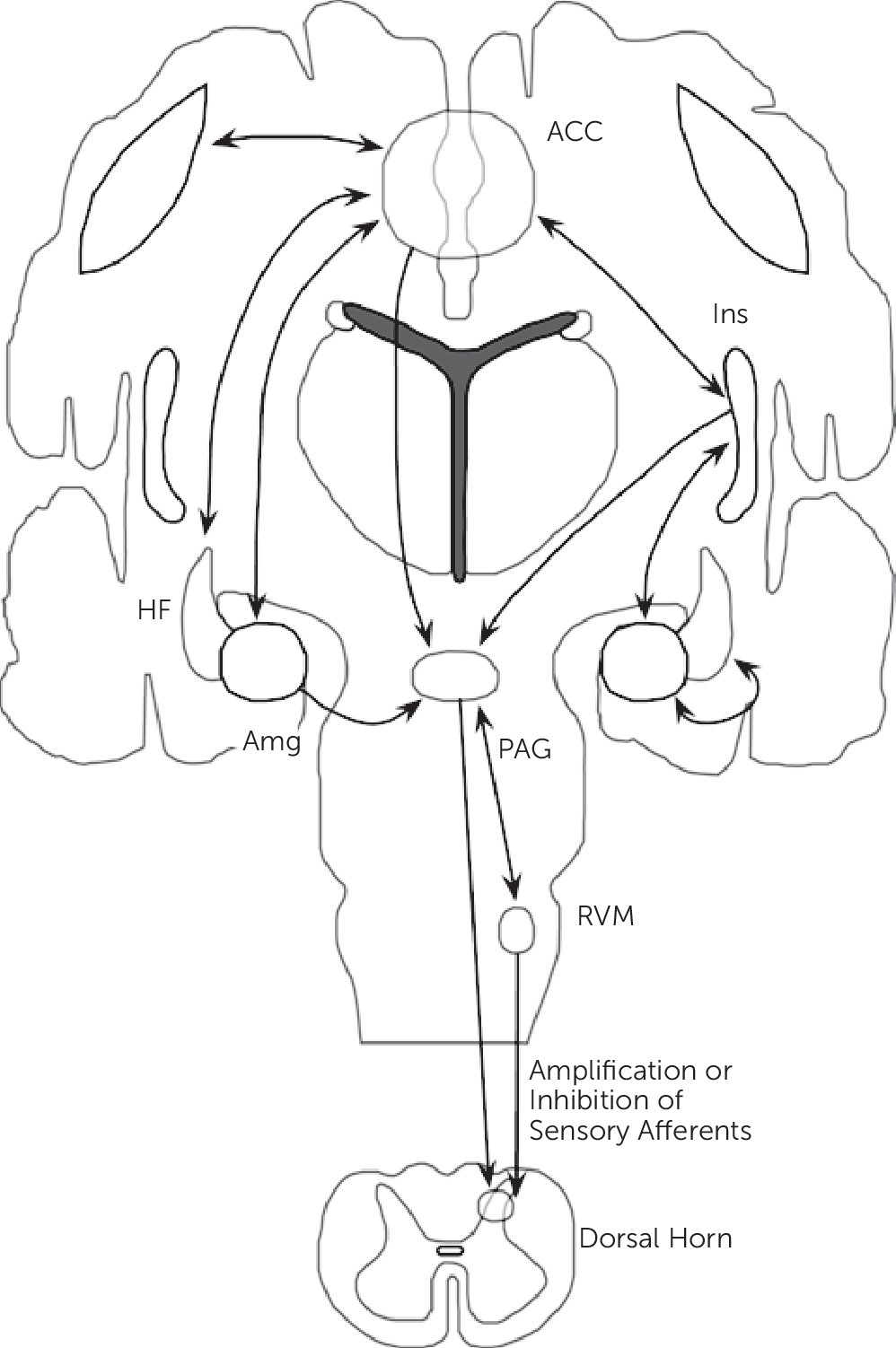
Synthesizing neuroimaging findings across somatization disorder, undifferentiated somatoform disorder, and somatoform pain disorder, and integrating these abnormalities with cognitive-affective neuroscience findings, we propose that aberrant circuit interactions across large-scale neural systems mediating visceral-somatic perception, emotional processing/awareness, and cognitive control serve critical roles in the neurobiology of somatosensory amplification. Important brain regions linked to somatosensory amplification include the ACC, insula, amygdala, hippocampal formation, and striatum among other regions (Figure 2).
FIGURE 2. Suggested Theoretical Neural Framework for Somatosensory Amplification in Somatoform Illnessa
a Aberrant circuit interactions across neural systems mediating visceral-somatic perception, emotional processing/awareness, and cognitive control serve critical roles in the neurobiology of somatosensory amplification. Cortical-subcortical-brainstem-spinal cord interactions are theorized to mediate the amplification of visceral-somatic sensations. Important cognitive processes in somatosensory amplification include: negative expectation bias (anterior cingulate cortex (ACC), orbitofrontal cortex (OFC), insula (Ins), hippocampal formation (HF), and brainstem); negative attentional bias (ACC, amygdala (Amg), dorsolateral prefrontal cortex (dlPFC)); and pain catastrophizing (ACC, dlPFC, Ins). Affective processes linked to somatosensory amplification include alexithymia (ACC, Ins, Amg) and dysphoric-anxious mood (ACC, Ins, OFC). Note: not shown are OFC connections with the ACC, Ins, Amg, HF and PAG (periaqueductal gray). RVM indicates rostral ventromedial medulla.
The lamina I spinothalamic pathway allows for the integration of visceral-somatic information within the brainstem, thalamus, and cortex. Visceral-somatic information converges cortically within the ACC and insula. The ACC has been classically subdivided into a subgenual/pregenual affective component and a dorsal ACC/anterior middle cingulate cortex (aMCC) cognitive component.70 Nociceptive studies across visceral-somatic stimuli elicit robust cingulate gyrus activations; visceral stimuli frequently activate pregenual and dorsal ACC, whereas noxious cutaneous stimuli more robustly activate the MCC. Uniquely positioned for integration is the dorsal ACC/aMCC where affectively ladened information, visceral-somatic processing, motivated behavior, and cognitive control converge. Shackman and colleagues71 recently proposed this region as a critical integrator of negative affect, pain, and cognitive control. ACC-subcortical connections include the nucleus accumbens/ventral caudate, ventral globus pallidus, and MD, and ventral anterior thalamic nuclei; cortico-cortical connections occur between the ACC and the dlPFC, insula, OFC, amygdala, and hippocampal formation.11 ACC/aMCC functional and structural abnormalities have been reported in somatoform pain disorder,27,28 and this region is involved in negative expectation bias,36,38,39 negative attentional bias,40,43,44 alexithymia,5255,57,58,60,61 and negative affect modulation of visceral-somatic processing.64,65,68 Cortical ACC‒dlPFC interactions may mediate negatively valenced, visceral-somatic forecasting associated with catastrophizing,47 and negative attentional bias, whereas ACC‒OFC connections may mediate top-down influences of negative expectation and negative mood hierarchically. Caudate-putamen components of the ACC, dlPFC, and OFC cortical-subcortical circuits potentially relate more specifically to somatization and undifferentiated somatoform disorders.1820
Apart from the ACC, the insula is also likely a critical region for somatosensory amplification. The posterior insula receives somatosensory, nociceptive/thermoceptive, and visceral information from the thalamus. Classic intraoperative electrical stimulation studies of the posterior insula performed by Penfield verified that many visceral sensations including gurgling, burning, rising/rolling sensations, and nausea can be produced through posterior insula activations.72 A.D. Craig has specifically suggested that the posterior insula provides an interoceptive representation of the physiological condition of the body.10 The mid-insula is considered an integrative zone where affectively and motivationally valenced information from the ACC, amygdala, and OFC influence sensory processing. The integration of visceral-somatic, affective, and motivational information converges onto the anterior insula, and together with the ACC, the anterior insula (right>left) has been linked to emotional awareness.73 Interestingly, the insula and ACC share large spindle-shaped neurons, termed von Economo neurons, linked to social-emotional cognition. Differential insular activity occurs in somatization disorder,74 undifferentiated somatoform disorder,74 and somatoform pain disorder,25,26,28,74 and the insula is involved in negative expectation bias,3739 alexithymia,54,62 and the modulation of visceral-somatic processing by negative emotion.64,65,69 Opioid system activity may potentially drive insula and ACC related negative expectation responses within somatoform illness,39 particularly in the context of negative emotion.
Somatosensory amplification may involve a bidirectional pattern of insula and amygdala activity. Patients with somatization disorder, undifferentiated somatoform disorder, and somatoform pain disorder22 exhibit insula and amygdala hypoactivity to external (environmental) emotionally valenced stimuli. Amygdalar and insular hypoactivity was also observed in subjects with alexithymia exposed to extrinsic emotionally valenced stimuli.61,62 Conversely, delivery of self-oriented, bodily-related stimuli (i.e., tactile) increased amygdala and insula activity in patients with somatoform pain disorder,2527,30 and these regions also showed hyperactivity in studies of negative attentional bias.4044 These findings suggest that somatosensory amplification may be partially the result of selective heightened attention and salience for bodily sensations (internal states) and parallel under-processing of external emotionally valenced information. Tryptophan depletion studies suggest serotonergic dysfunction may potentially mediate this bidirectional aberrant activity pattern.
The hippocampal formation (including the parahippocampal gyrus) also demonstrated abnormal activity in somatization disorder,22 undifferentiated somatoform disorder,22 and somatoform pain disorder patients.22,25,26 Similarly, studies of negative expectation bias3135 reported increased hippocampal formation activity. Using the Gray-McNaughton theory,75 which posits a role for the hippocampal formation in responding to aversive information in the context of behavioral conflict (including uncertainty, novelty, and contextual processing), these studies suggest an amplifying role for the hippocampal formation in visceral-somatic processing during uncertainty; this effect may be mediated by enhanced insula and ACC activity.31
The ACC, insula, amygdala, and hippocampal formation are all part of the descending pain modulatory system, which also includes multiple brainstem structures (PAG, NCF, and RVM).12 Reciprocal connections between the ACC and the amygdala, OFC, dlPFC, insula, and hippocampal formation position the ACC as a key cortical modulatory region of PAG activity (both directly and indirectly though ACC-amygdala-PAG connections). Efferent connections between the ACC and the PAG have been described in mammals, though DTI failed to reliably identify human ACC-PAG connections76 suggesting further anatomical clarification is necessary. Nonetheless, the PAG is connected to and modulated by the amygdala, insula and multiple prefrontal cortical regions as well.76 Within the brainstem, the PAG is interconnected with the RVM and NCF. Descending RVM and PAG projections can either amplify or inhibit afferent sensory processing within the dorsal horn of the spinal cord. These cortico-brainstem connections likely play a role in somatosensory amplification and future research will help determine if intrinsic brainstem abnormalities (independent of cortical, top-down influence) are also involved in the pathophysiology of somatosensory amplification.
Having delineated a neurocircuit framework for somatosensory amplification, it is important to emphasize that in a given subject or patient group, dysfunction in distinct nodes or levels of these hierarchical cortical-subcortical-brainstem-spinal cord circuits may occur. One patient may have amplification of visceral-somatic processing driven by enhanced entry-level afferent information within the dorsal horn of the spinal cord or brainstem nuclei, whereas another individual may have amplification driven by cortical-subcortical mediated cognitive and affective processes.

Additional Considerations–Inflammation, Stress-Mediated Neuroplastic Change and Autonomic Imbalance

Although this article focuses on neurocircuit disturbances, it is important to introduce roles for inflammation, aberrant neuroplastic change, and autonomic imbalance in the pathophysiology of somatosensory amplification. Recent research suggests that proinflammatory states modulate frontolimbic circuits to alter affective and cognitive elements of visceral-somatic processing. Chronic interpersonal stress, prevalent in patients with somatoform disorders, has been associated with enhanced inflammatory leukocyte response to microbial challenge,77 and women with elevated levels of circulating cytokines report poorer health despite controlling for physical health and diagnosis.78 Furthermore, pain catastrophizing following noxious stimuli delivery has been associated with interleukin-6 (IL-6) reactivity.79 A series of studies by Harrison and colleagues explored the modifying effects of systemic inflammation on neural activity.80,81 In a double-blind, randomized study using typhoid injection compared with placebo, individuals who received a typhoid injection reported worsening mood and demonstrated increased systemic IL-6 levels. In the typhoid injected group, enhanced subgenual ACC (sgACC) activity was associated with dysphoric mood; decreased functional connectivity of the sgACC to the amygdala, medial prefrontal cortex, and nucleus accumbens was modulated by peripheral IL-6 levels.80 In this same cohort, self-reported fatigue following injection correlated with bilateral mid/posterior insula and left ACC activity.81 These studies suggest a neuromodulatory role for circulating cytokines in the emergence of dysphoric mood and fatigue.
Aberrant neuroplastic change and impaired development following early-life interpersonal stress, including sexual/physical abuse and neglect, are additional elements potentially mediating the emergence of somatic symptoms. Patients with somatoform illness frequently experience early-life trauma. Animal models of chronic stress suggest that the medial prefrontal cortex (including the ACC), hippocampus, and amygdala undergo neuroplastic changes in response to prolonged stress.82 The hippocampal CA3 region and medial prefrontal cortex exhibit dendritic spine density reductions following repeated stress; amygdalar experience-dependent changes, although less well studied, have also been described. Recent large-scale volumetric analyses in human subjects observed ACC, OFC, insula, hippocampal, and caudate gray matter reductions associated with a history of childhood trauma.83 Early-life interpersonal stress also predisposes to insecure adult attachment. Individuals with insecure attachment display amygdala and striatal dysfunction,84 and abnormal hypothalamic-pituitary-adrenal axis function.85 Strikingly, these findings overlap with brain regions theorized to play a role in abnormal somatosensory amplification.
An imbalance of the autonomic nervous system has also been reported in somatoform disorders.8690 Rief and colleagues characterized elevated morning heart rates and cortisol levels, along with persistent heart rate increases (failed habituation) during an emotionally valenced word viewing task in patients with prominent somatization compared with healthy controls90; a similar cohort of patients exhibited persistently elevated heart rates during rest following completion of an attentional task.89 Patients with somatization disorder displayed increased heart rate and decreased baroreceptor sensitivity during autonomic testing.86 Patients with somatoform symptoms also exhibited reduced heart rate variability during affectively valenced facial viewing88 and pain processing paradigms.87 These findings suggest a static imbalance of increased sympathetic and decreased parasympathetic tone. Thayer and colleagues proposed an important role for the central autonomic network (CAN), which includes frontolimbic, insular and brainstem structures, in the pathobiology of autonomic imbalance facilitating noxious psychosomatic experiences.91 Importantly, the CAN converges with the theorized neurocircuitry of somatosensory amplification.

Therapeutic Implications

Having proposed prominent prefrontal dysfunction (i.e., ACC, dlPFC, OFC) in the pathophysiology of somatosensory amplification, several therapeutic implications emerge. Noninvasive and invasive neuromodulation, particularly rapid transcranial magnetic stimulation (rTMS) and deep brain stimulation (DBS), have been investigated in the treatment of neuropsychiatric disorders. Modulation of dlPFC activity, in conjunction with trans-synaptic sgACC modulation, has been suggested as the mechanism for therapeutic efficacy of rTMS in the treatment of major depressive disorder.92 Future investigations in somatoform illness should evaluate dlPFC neuromodulation to potentially improve pain catastrophizing, negative attentional bias, and negative affective disturbances. DBS targeting sensory thalamus and periventricular/periaqueductal gray matter have been used to treat chronic intractable pain syndromes for the past half-century. Interestingly, ventral caudal thalamic stimulation for chronic pain treatment modulated perigenual and dorsal ACC activity,93 and ventral PAG stimulation enhanced parasympathetic activity.94 For patients with disabling, chronic somatoform pain disorders, consideration should be given to investigating the PAG, dorsomedial thalamus, and dorsal ACC as potential therapeutic targets.
Prominent roles for the monoamine and opioid neurotransmitter systems in negative attentional bias, negative expectation, and affective disturbances in somatosensory amplification are consistent with the pharmacologic evidence in somatoform disorders. A meta-analysis of antidepressants (mainly tricyclic and selective serotonin reuptake inhibitors) compared with placebo for the treatment of somatoform pain disorder showed significant decreased pain intensity following antidepressant use.95 A more recently conducted randomized, double-blind placebo-controlled trial of fluoxetine in patients with somatoform pain disorder compared with controls demonstrated drug-related analgesia, with the greatest therapeutic efficacy in patients with comorbid depression.96 Negative expectation, particularly the nocebo effect, has been linked to decreased opioid and dopaminergic neurotransmitter activity,39 and this finding may be particularly noteworthy in the context of an association between severe somatization and opiate drug misuse.
Empirical evidence supports using cognitive-behavioral therapy (CBT) for the treatment of somatoform disorders. Randomized controlled trials investigating the effectiveness of CBT for somatization disorder, undifferentiated somatoform disorder, and somatoform pain disorder have demonstrated improvements in physical symptom severity and level of functioning.97,98 The particular mechanisms utilized in CBT [which include modifying physiological arousal, attention, attributional processes, and cognitive distortions (i.e., catastrophizing)] target many of the theorized cognitive and affective modifiers of visceral-somatic processing in somatosensory amplification.
Emerging data for mindfulness-based techniques (MBT) also suggest some promise in targeting the circuitry of abnormal somatosensory amplification. MBT involve training in specific meditative practices that encourage moment-to-moment, nonjudgmental, nonreactive awareness. Through such training, it has been proposed that a distributed, large-scale network (including the dorsal ACC, dlPFC, and anterior insula among other regions) are functionally recruited to guide therapeutic changes in neural systems underlying catastrophizing and affect-biased attention.99 Interestingly, the most widely cited brain areas of activity and morphological change during and in response to MBT have been the ACC, dlPFC, anterior insula, and hippocampus.99 MBT have been reported to reduce disability pensions in patients with somatization disorder100 and should be further investigated in patients with somatoform illness.

Limitations and Future Directions

There are several important limitations to address regarding this theoretical neural circuit framework for somatosensory amplification. Even though this approach integrates somatoform disorder neuroimaging abnormalities with related findings in cognitive-affective neuroscience, investigations probing cognitive and affective modifiers of visceral-somatic processing in somatoform disorder populations are necessary to validate the proposed model. In addition, there are limited, underpowered somatoform disorder-specific neuroimaging studies to date, and case-control studies with increased sample sizes are needed to ensure the reliability of the framework, and clarify important concepts such as the role of gender in the pathophysiology of somatosensory amplification. Several somatoform disorder visceral-somatic symptoms (i.e., noncardiac chest pain, breathlessness, dizziness) are also understudied using brain imaging techniques and, thus, not fully integrated into the current framework; these symptoms, however, likely map onto the neurocircuitry in topographic fashion with similar influences from cognitive and affective amplifiers. Future research is necessary to also clarify intra and intercircuit functional connectivity patterns and to delineate common and disorder-specific circuit abnormalities across somatoform disorder subtypes. In addition, this framework focuses on central modifiers of visceral-somatic processing, however, primary end organ dysfunction (i.e., abnormal serotonergic transmission in the gut wall) should be explored for potential additive or synergistic roles in the pathophysiology of symptom amplification. Future research will also incorporate genetic-epigenetic influences, and the neuroimaging based systems-level approach taken in this article should be further refined with other systems-level research modalities including electrophysiology techniques (i.e., event-related potentials). Systems-level measures of brain function, which may be associated with complex mental states, are also not necessarily inherently causative of specific abnormal symptoms; this highlights the need for integrative multilevel research investigations in somatosensory amplification. Lastly, while critical neurocircuit regions were identified, it will be necessary to further investigate ACC (subgenual, perigenual, dorsal) and insula (anterior-midposterior) subregion involvement and directionally of abnormal activation patterns in the pathophysiology of somatosensory amplification.
Conclusions

In summary, visceral-somatic physical symptoms are remarkably prevalent in primary care and subspecialty clinics. Somatosensory amplification has been theorized to play a crucial role in the pathophysiology of somatization. In this article, aberrant interactions across neural circuits mediating visceral-somatic perception, emotional processing/awareness, and cognitive control are proposed to serve important roles in the neurobiology of somatosensory amplification. Key sites of abnormal activity include the prefrontal cortex, insula, medial temporal lobe, and striatum within the context of the lamina I spinothalamocortical and descending pain modulatory pathways. At the individual level, activation patterns within hierarchical cortical-subcortical-brainstem-spinal cord circuits may vary. Future diagnoses may be based upon the delineation and classification of distinct patterns of abnormal activity at the individual patient and group level. The emergence of regional neural patterns of dysfunction may potentially be explained, in part, through aberrant neuroplastic change in the context of early-life trauma and the neuromodulatory effects of inflammatory cytokines. Future investigations are needed to refine the proposed theoretical framework and explore therapeutic opportunities to modify neurocircuit activity through psychotherapy, pharmacology, and region-specific neuromodulation.
Acknowledgments

The authors thank Laura Ortiz Terán, M.D., Ph.D. and Benjamin Fuchs, B.A. for their assistance on the illustrations and manuscript comments.

References

1.
Kroenke K: Patients presenting with somatic complaints: epidemiology, psychiatric comorbidity and management. Int J Methods Psychiatr Res 2003; 12:34–43
2.
Rief W, Barsky AJ: Psychobiological perspectives on somatoform disorders. Psychoneuroendocrinology 2005; 30:996–1002
3.
Katon W, Ries RK, Kleinman A: The prevalence of somatization in primary care. Compr Psychiatry 1984; 25:208–215
4.
Smith GR Jr, Monson RA, Ray DC: Patients with multiple unexplained symptoms. Their characteristics, functional health, and health care utilization. Arch Intern Med 1986; 146:69–72
5.
Barsky AJ, Goodson JD, Lane RS, et al: The amplification of somatic symptoms. Psychosom Med 1988; 50:510–519
6.
Jones MP, Schettler A, Olden K, et al: Alexithymia and somatosensory amplification in functional dyspepsia. Psychosomatics 2004; 45:508–516
7.
Nakao M, Barsky AJ, Kumano H, et al: Relationship between somatosensory amplification and alexithymia in a Japanese psychosomatic clinic. Psychosomatics 2002; 43:55–60
8.
Rief W, Hiller W, Margraf J: Cognitive aspects of hypochondriasis and the somatization syndrome. J Abnorm Psychol 1998; 107:587–595
9.
Perez DL, Barsky AJ, Daffner K, et al: Motor and somatosensory conversion disorder: a functional unawareness syndrome? J Neuropsychiatry Clin Neurosci 2012; 24:141–151
10.
Craig AD: How do you feel? Interoception: the sense of the physiological condition of the body. Nat Rev Neurosci 2002; 3:655–666
11.
Heimer L, Van Hoesen GW, Trimble M, et al: Anatomy of Neuropsychiatry: The New Anatomy of the Basal Forebrain and Its Implications for Neuropsychiatric Illness. Burlington, MA, Academic Press, 2007
12.
Tracey I, Mantyh PW: The cerebral signature for pain perception and its modulation. Neuron 2007; 55:377–391
13.
Dunckley P, Wise RG, Aziz Q, et al: Cortical processing of visceral and somatic stimulation: differentiating pain intensity from unpleasantness. Neuroscience 2005; 133:533–542
14.
Dunckley P, Wise RG, Fairhurst M, et al: A comparison of visceral and somatic pain processing in the human brainstem using functional magnetic resonance imaging. J Neurosci 2005; 25:7333–7341
15.
Lotze M, Wietek B, Birbaumer N, et al: Cerebral activation during anal and rectal stimulation. Neuroimage 2001; 14:1027–1034
16.
Strigo IA, Albanese MC, Bushnell MC, et al: Visceral and cutaneous pain representation in parasylvian cortex. Neurosci Lett 2005; 384:54–59
17.
Strigo IA, Duncan GH, Boivin M, et al: Differentiation of visceral and cutaneous pain in the human brain. J Neurophysiol 2003; 89:3294–3303
18.
Hakala M, Karlsson H, Ruotsalainen U, et al: Severe somatization in women is associated with altered cerebral glucose metabolism. Psychol Med 2002; 32:1379–1385
19.
Hakala M, Vahlberg T, Niemi PM, et al: Brain glucose metabolism and temperament in relation to severe somatization. Psychiatry Clin Neurosci 2006; 60:669–675
20.
Hakala M, Karlsson H, Kurki T, et al: Volumes of the caudate nuclei in women with somatization disorder and healthy women. Psychiatry Res 2004; 131:71–78
21.
Atmaca M, Sirlier B, Yildirim H, et al: Hippocampus and amygdalar volumes in patients with somatization disorder. Prog Neuropsychopharmacol Biol Psychiatry 2011; 35:1699–1703
22.
de Greck M, Scheidt L, Bölter AF, et al: Altered brain activity during emotional empathy in somatoform disorder. Hum Brain Mapp 2012; 33:2666–2685
23.
Yildirim H, Atmaca M, Sirlier B, et al: Pituitary volumes are reduced in patients with somatization disorder. Psychiatry Investig 2012; 9:278–282
24.
Koh KB, Kang JI, Lee JD, et al: Shared neural activity in panic disorder and undifferentiated somatoform disorder compared with healthy controls. J Clin Psychiatry 2010; 71:1576–1581
25.
Stoeter P, Bauermann T, Nickel R, et al: Cerebral activation in patients with somatoform pain disorder exposed to pain and stress: an fMRI study. Neuroimage 2007; 36:418–430
26.
Gündel H, Valet M, Sorg C, et al: Altered cerebral response to noxious heat stimulation in patients with somatoform pain disorder. Pain 2008; 137:413–421
27.
Karibe H, Arakawa R, Tateno A, et al: Regional cerebral blood flow in patients with orally localized somatoform pain disorder: a single photon emission computed tomography study. Psychiatry Clin Neurosci 2010; 64:476–482
28.
Valet M, Gündel H, Sprenger T, et al: Patients with pain disorder show gray-matter loss in pain-processing structures: a voxel-based morphometric study. Psychosom Med 2009; 71:49–56
29.
Karibe H, Arakawa R, Tateno A, et al: Regional cerebral blood flow in patients with orally localized somatoform pain disorder: a single photon emission computed tomography study. Psychiatry Clin Neurosci 2010; 64:476–482
30.
Otti A, Guendel H, Henningsen P, et al: Functional network connectivity of pain-related resting state networks in somatoform pain disorder: an exploratory fMRI study. J Psychiatry Neurosci 2013; 38:57–65
31.
Ploghaus A, Narain C, Beckmann CF, et al: Exacerbation of pain by anxiety is associated with activity in a hippocampal network. J Neurosci 2001; 21:9896–9903
32.
Gondo M, Moriguchi Y, Kodama N, et al: Daily physical complaints and hippocampal function: an fMRI study of pain modulation by anxiety. Neuroimage 2012; 63:1011–1019
33.
Ziv M, Tomer R, Defrin R, et al: Individual sensitivity to pain expectancy is related to differential activation of the hippocampus and amygdala. Hum Brain Mapp 2010; 31:326–338
34.
Fairhurst M, Wiech K, Dunckley P, et al: Anticipatory brainstem activity predicts neural processing of pain in humans. Pain 2007; 128:101–110
35.
Bingel U, Wanigasekera V, Wiech K, et al: The effect of treatment expectation on drug efficacy: imaging the analgesic benefit of the opioid remifentanil. Sci Transl Med 2011; 3:1–9
36.
Keltner JR, Furst A, Fan C, et al: Isolating the modulatory effect of expectation on pain transmission: a functional magnetic resonance imaging study. J Neurosci 2006; 26:4437–4443
37.
Sawamoto N, Honda M, Okada T, et al: Expectation of pain enhances responses to nonpainful somatosensory stimulation in the anterior cingulate cortex and parietal operculum/posterior insula: an event-related functional magnetic resonance imaging study. J Neurosci 2000; 20:7438–7445
38.
Kong J, Gollub RL, Polich G, et al: A functional magnetic resonance imaging study on the neural mechanisms of hyperalgesic nocebo effect. J Neurosci 2008; 28:13354–13362
39.
Scott DJ, Stohler CS, Egnatuk CM, et al: Placebo and nocebo effects are defined by opposite opioid and dopaminergic responses. Arch Gen Psychiatry 2008; 65:220–231
40.
Evers EA, van der Veen FM, Jolles J, et al: Acute tryptophan depletion improves performance and modulates the BOLD response during a Stroop task in healthy females. Neuroimage 2006; 32:248–255
41.
Cools R, Calder AJ, Lawrence AD, et al: Individual differences in threat sensitivity predict serotonergic modulation of amygdala response to fearful faces. Psychopharmacology (Berl) 2005; 180:670–679
42.
van der Veen FM, Evers EA, Deutz NE, et al: Effects of acute tryptophan depletion on mood and facial emotion perception related brain activation and performance in healthy women with and without a family history of depression. Neuropsychopharmacology 2007; 32:216–224
43.
Passamonti L, Crockett MJ, Apergis-Schoute AM, et al: Effects of acute tryptophan depletion on prefrontal-amygdala connectivity while viewing facial signals of aggression. Biol Psychiatry 2012; 71:36–43
44.
Labus JS, Mayer EA, Jarcho J, et al: Acute tryptophan depletion alters the effective connectivity of emotional arousal circuitry during visceral stimuli in healthy women. Gut 2011; 60:1196–1203
45.
Browning M, Holmes EA, Murphy SE, et al: Lateral prefrontal cortex mediates the cognitive modification of attentional bias. Biol Psychiatry 2010; 67:919–925
46.
Coen SJ, Aziz Q, Yaguez L, et al: Effects of attention on visceral stimulus intensity encoding in the male human brain. Gastroenterology 2008;135:2065–2074, e2061.
47.
Seminowicz DA, Davis KD: Cortical responses to pain in healthy individuals depends on pain catastrophizing. Pain 2006; 120:297–306
48.
Chen JY, Blankstein U, Diamant NE, et al: White matter abnormalities in irritable bowel syndrome and relation to individual factors. Brain Res 2011; 1392:121–131
49.
Schäfer R, Popp K, Jörgens S, et al: Alexithymia-like disorder in right anterior cingulate infarction. Neurocase 2007; 13:201–208
50.
Mantani T, Okamoto Y, Shirao N, et al: Reduced activation of posterior cingulate cortex during imagery in subjects with high degrees of alexithymia: a functional magnetic resonance imaging study. Biol Psychiatry 2005; 57:982–990
51.
Lane RD, Reiman EM, Axelrod B, et al: Neural correlates of levels of emotional awareness. Evidence of an interaction between emotion and attention in the anterior cingulate cortex. J Cogn Neurosci 1998; 10:525–535
52.
Berthoz S, Artiges E, Van De Moortele PF, et al: Effect of impaired recognition and expression of emotions on frontocingulate cortices: an fMRI study of men with alexithymia. Am J Psychiatry 2002; 159:961–967
53.
Kano M, Fukudo S, Gyoba J, et al: Specific brain processing of facial expressions in people with alexithymia: an H2 15O-PET study. Brain 2003; 126:1474–1484
54.
Borsci G, Boccardi M, Rossi R, et al: Alexithymia in healthy women: a brain morphology study. J Affect Disord 2009; 114:208–215
55.
Gündel H, López-Sala A, Ceballos-Baumann AO, et al: Alexithymia correlates with the size of the right anterior cingulate. Psychosom Med 2004; 66:132–140
56.
Heinzel A, Minnerop M, Schäfer R, et al: Alexithymia in healthy young men: a voxel-based morphometric study. J Affect Disord 2012; 136:1252–1256
57.
Heinzel A, Schäfer R, Müller HW, et al: Increased activation of the supragenual anterior cingulate cortex during visual emotional processing in male subjects with high degrees of alexithymia: an event-related fMRI study. Psychother Psychosom 2010; 79:363–370
58.
Karlsson H, Näätänen P, Stenman H: Cortical activation in alexithymia as a response to emotional stimuli. Br J Psychiatry 2008; 192:32–38
59.
Kugel H, Eichmann M, Dannlowski U, et al: Alexithymic features and automatic amygdala reactivity to facial emotion. Neurosci Lett 2008; 435:40–44
60.
Paradiso S, Vaidya JG, McCormick LM, et al: Aging and alexithymia: association with reduced right rostral cingulate volume. Am J Geriatr Psychiatry 2008; 16:760–769
61.
Pouga L, Berthoz S, de Gelder B, et al: Individual differences in socioaffective skills influence the neural bases of fear processing: the case of alexithymia. Hum Brain Mapp 2010; 31:1469–1481
62.
Reker M, Ohrmann P, Rauch AV, et al: Individual differences in alexithymia and brain response to masked emotion faces. Cortex 2010; 46:658–667
63.
Lee BT, Lee HY, Park SA, et al: Neural substrates of affective face recognition in alexithymia: a functional magnetic resonance imaging study. Neuropsychobiology 2011; 63:119–124
64.
Berna C, Leknes S, Holmes EA, et al: Induction of depressed mood disrupts emotion regulation neurocircuitry and enhances pain unpleasantness. Biol Psychiatry 2010; 67:1083–1090
65.
Coen SJ, Yaguez L, Aziz Q, et al: Negative mood affects brain processing of visceral sensation. Gastroenterology 2009;137:253–261, e251–e252
66.
Wagner G, Koschke M, Leuf T, et al: Reduced heat pain thresholds after sad-mood induction are associated with changes in thalamic activity. Neuropsychologia 2009; 47:980–987
67.
Roy M, Piché M, Chen JI, et al: Cerebral and spinal modulation of pain by emotions. Proc Natl Acad Sci USA 2009; 106:20900–20905
68.
Ochsner KN, Ludlow DH, Knierim K, et al: Neural correlates of individual differences in pain-related fear and anxiety. Pain 2006; 120:69–77
69.
Ploner M, Lee MC, Wiech K, et al: Prestimulus functional connectivity determines pain perception in humans. Proc Natl Acad Sci USA 2010; 107:355–360
70.
Vogt BA: Pain and emotion interactions in subregions of the cingulate gyrus. Nat Rev Neurosci 2005; 6:533–544
71.
Shackman AJ, Salomons TV, Slagter HA, et al: The integration of negative affect, pain and cognitive control in the cingulate cortex. Nat Rev Neurosci 2011; 12:154–167
72.
Shelley BP, Trimble MR: The insular lobe of Reil—its anatamico-functional, behavioural and neuropsychiatric attributes in humans—a review. World J Biol Psychiatry 2004; 5:176–200
73.
Craig AD: How do you feel—now? The anterior insula and human awareness. Nat Rev Neurosci 2009; 10:59–70
74.
de Greck M, Scheidt L, Bölter AF, et al: Multimodal psychodynamic psychotherapy induces normalization of reward related activity in somatoform disorder. World J Biol Psychiatry 2011; 12:296–308
75.
Gray JA, McNaughton N: The neuropsychology of anxiety. Oxford, Oxford University Press, 2000
76.
Hadjipavlou G, Dunckley P, Behrens TE, et al: Determining anatomical connectivities between cortical and brainstem pain processing regions in humans: a diffusion tensor imaging study in healthy controls. Pain 2006; 123:169–178
77.
Miller GE, Rohleder N, Cole SW: Chronic interpersonal stress predicts activation of pro- and anti-inflammatory signaling pathways 6 months later. Psychosom Med 2009; 71:57–62
78.
Lekander M, Elofsson S, Neve IM, et al: Self-rated health is related to levels of circulating cytokines. Psychosom Med 2004; 66:559–563
79.
Edwards RR, Kronfli T, Haythornthwaite JA, et al: Association of catastrophizing with interleukin-6 responses to acute pain. Pain 2008; 140:135–144
80.
Harrison NA, Brydon L, Walker C, et al: Inflammation causes mood changes through alterations in subgenual cingulate activity and mesolimbic connectivity. Biol Psychiatry 2009; 66:407–414
81.
Harrison NA, Brydon L, Walker C, et al: Neural origins of human sickness in interoceptive responses to inflammation. Biol Psychiatry 2009; 66:415–422
82.
Leuner B, Shors TJ: Stress, anxiety, and dendritic spines: what are the connections? Neuroscience 2013; 251:108–119
83.
Dannlowski U, Stuhrmann A, Beutelmann V, et al: Limbic scars: long-term consequences of childhood maltreatment revealed by functional and structural magnetic resonance imaging. Biol Psychiatry 2012; 71:286–293
84.
Vrticka P, Andersson F, Grandjean D, et al: Individual attachment style modulates human amygdala and striatum activation during social appraisal. PLoS One 2008; 3:e2868
85.
Pierrehumbert B, Torrisi R, Glatz N, et al: The influence of attachment on perceived stress and cortisol response to acute stress in women sexually abused in childhood or adolescence. Psychoneuroendocrinology 2009; 34:924–938
86.
Laederach-Hofmann K, Rüddel H, Mussgay L: Pathological baroreceptor sensitivity in patients suffering from somatization disorders: do they correlate with symptoms? Biol Psychol 2008; 79:243–249
87.
Pollatos O, Dietel A, Herbert BM, et al: Blunted autonomic reactivity and increased pain tolerance in somatoform patients. Pain 2011; 152:2157–2164
88.
Pollatos O, Herbert BM, Wankner S, et al: Autonomic imbalance is associated with reduced facial recognition in somatoform disorders. J Psychosom Res 2011; 71:232–239
89.
Rief W, Auer C: Is somatization a habituation disorder? Physiological reactivity in somatization syndrome. Psychiatry Res 2001; 101:63–74
90.
Rief W, Shaw R, Fichter MM: Elevated levels of psychophysiological arousal and cortisol in patients with somatization syndrome. Psychosom Med 1998; 60:198–203
91.
Thayer JF, Brosschot JF: Psychosomatics and psychopathology: looking up and down from the brain. Psychoneuroendocrinology 2005; 30:1050–1058
92.
Fox MD, Buckner RL, White MP, et al: Efficacy of transcranial magnetic stimulation targets for depression is related to intrinsic functional connectivity with the subgenual cingulate. Biol Psychiatry 2012; 72:595–603
93.
Davis KD, Taub E, Duffner F, et al: Activation of the anterior cingulate cortex by thalamic stimulation in patients with chronic pain: a positron emission tomography study. J Neurosurg 2000; 92:64–69
94.
Pereira EA, Lu G, Wang S, et al: Ventral periaqueductal grey stimulation alters heart rate variability in humans with chronic pain. Exp Neurol 2010; 223:574–581
95.
Fishbain DA, Cutler RB, Rosomoff HL, et al: Do antidepressants have an analgesic effect in psychogenic pain and somatoform pain disorder? A meta-analysis. Psychosom Med 1998; 60:503–509
96.
Luo YL, Zhang MY, Wu WY, et al: A randomized double-blind clinical trial on analgesic efficacy of fluoxetine for persistent somatoform pain disorder. Prog Neuropsychopharmacol Biol Psychiatry 2009; 33:1522–1525
97.
Allen LA, Woolfolk RL, Escobar JI, et al: Cognitive-behavioral therapy for somatization disorder: a randomized controlled trial. Arch Intern Med 2006; 166:1512–1518
98.
Zonneveld LN, van Rood YR, Timman R, et al: Effective group training for patients with unexplained physical symptoms: a randomized controlled trial with a non-randomized one-year follow-up. PLoS ONE 2012; 7:e42629
99.
Vago DR, Silbersweig DA: Self-awareness, self-regulation, and self-transcendence (S-ART): a framework for understanding the neurobiological mechanisms of mindfulness. Front Hum Neurosci 2012; 6:296
100.
Fjorback LO, Carstensen T, Arendt M, et al: Mindfulness therapy for somatization disorder and functional somatic syndromes: analysis of economic consequences alongside a randomized trial. J Psychosom Res 2013; 74:41–48

Information & Authors

Information

Published In

Go to The Journal of Neuropsychiatry and Clinical Neurosciences
The Journal of Neuropsychiatry and Clinical Neurosciences
Pages: e40 - e50
PubMed: 25716493

History

Received: 29 July 2013
Revision received: 27 November 2013
Accepted: 6 January 2014
Published ahead of print: 10 November 2014
Published in print: Winter 2015
Published online: 26 February 2015

Authors

Details

David L. Perez, M.D.
From the Dept. of Psychiatry, Brigham and Women’s Hospital, Boston, MA (DLP, AJB, DRV, GB, DAS);. Dept. of Neurology, Brigham and Women’s Hospital, Boston, MA (DLP); Functional Neuroimaging Laboratory, Brigham and Women’s Hospital, Boston, MA (DLP, DRV DAS); Dept. of Psychiatry, Beth Israel Deaconess Medical Center, Boston, MA (DLP); and Harvard Medical School, Boston, MA (DLP, AJB, DRV, GB, DAS).
Arthur J. Barsky, M.D.
From the Dept. of Psychiatry, Brigham and Women’s Hospital, Boston, MA (DLP, AJB, DRV, GB, DAS);. Dept. of Neurology, Brigham and Women’s Hospital, Boston, MA (DLP); Functional Neuroimaging Laboratory, Brigham and Women’s Hospital, Boston, MA (DLP, DRV DAS); Dept. of Psychiatry, Beth Israel Deaconess Medical Center, Boston, MA (DLP); and Harvard Medical School, Boston, MA (DLP, AJB, DRV, GB, DAS).
David R. Vago, Ph.D.
From the Dept. of Psychiatry, Brigham and Women’s Hospital, Boston, MA (DLP, AJB, DRV, GB, DAS);. Dept. of Neurology, Brigham and Women’s Hospital, Boston, MA (DLP); Functional Neuroimaging Laboratory, Brigham and Women’s Hospital, Boston, MA (DLP, DRV DAS); Dept. of Psychiatry, Beth Israel Deaconess Medical Center, Boston, MA (DLP); and Harvard Medical School, Boston, MA (DLP, AJB, DRV, GB, DAS).
Gaston Baslet, M.D.
From the Dept. of Psychiatry, Brigham and Women’s Hospital, Boston, MA (DLP, AJB, DRV, GB, DAS);. Dept. of Neurology, Brigham and Women’s Hospital, Boston, MA (DLP); Functional Neuroimaging Laboratory, Brigham and Women’s Hospital, Boston, MA (DLP, DRV DAS); Dept. of Psychiatry, Beth Israel Deaconess Medical Center, Boston, MA (DLP); and Harvard Medical School, Boston, MA (DLP, AJB, DRV, GB, DAS).
David A. Silbersweig, M.D.
From the Dept. of Psychiatry, Brigham and Women’s Hospital, Boston, MA (DLP, AJB, DRV, GB, DAS);. Dept. of Neurology, Brigham and Women’s Hospital, Boston, MA (DLP); Functional Neuroimaging Laboratory, Brigham and Women’s Hospital, Boston, MA (DLP, DRV DAS); Dept. of Psychiatry, Beth Israel Deaconess Medical Center, Boston, MA (DLP); and Harvard Medical School, Boston, MA (DLP, AJB, DRV, GB, DAS).

Notes

Send correspondence to Dr. Perez; e-mail: [email protected]

Metrics & Citations

Metrics

Citations

Export Citations

If you have the appropriate software installed, you can download article citation data to the citation manager of your choice. Simply select your manager software from the list below and click Download.

For more information or tips please see 'Downloading to a citation manager' in the Help menu.

Format
Citation style
Style
Copy to clipboard

View Options

View options

PDF/EPUB

View PDF/EPUB

Get Access

Login options

Already a subscriber? Access your subscription through your login credentials or your institution for full access to this article.

Personal login Institutional Login Open Athens login
Purchase Options

Purchase this article to access the full text.

PPV Articles - Journal of Neuropsychiatry and Clinical Neurosciences

PPV Articles - Journal of Neuropsychiatry and Clinical Neurosciences

Not a subscriber?

Subscribe Now / Learn More

PsychiatryOnline subscription options offer access to the DSM-5-TR® library, books, journals, CME, and patient resources. This all-in-one virtual library provides psychiatrists and mental health professionals with key resources for diagnosis, treatment, research, and professional development.

Need more help? PsychiatryOnline Customer Service may be reached by emailing [email protected] or by calling 800-368-5777 (in the U.S.) or 703-907-7322 (outside the U.S.).

Media

Figures

Other

Tables

Share

Share

Share article link

Share