To the Editor: Transient global amnesia (TGA) is a syndrome characterized by abrupt onset of anterograde amnesia accompanied by repetitive questioning. Affected patients show no signs of impaired cognitive functioning and have no focal deficits.
1 TGA shows complete recovery in less than 24 hours.
2 Although various pathophysiologic explanations have been offered, its etiology remains largely unknown.
1 We hereby report a case that is found rarely in the literature.
Case Report
A 58-year-old man arrived at his office at 9.00 a.m. and was found to be confused, asking strange questions and responding inappropriately. He was immediately brought to the emergency department by his colleagues for a medical opinion. A history revealed that the man had been talking to himself, saying that his son was in Chandigarh and that he had to fetch an apple box for him. When asked to whom it was he was talking, he was unable to reply satisfactorily. In the hospital, he was unable to recall the sequence of events that happened on the same day and kept asking, “What has happened? Why I have been brought to the hospital?” Information obtained from the patient’s family members indicated that he had no history of similar episode, seizure disorder, fever, vomiting, neck stiffness, giddiness, weakness, numbness, or abnormal sensation in any part of the body. In addition, there was no history of difficulty in speech, double vision, loss of consciousness, hearing of voices, migraine, abnormal taste, or smell. The patient had no significant medical illness, including diabetes, hypertension, varicose veins, heart disease, nor was he taking any medication.
The patient was afebrile, with a blood pressure of 110/70 mmHg, pulse rate of 80 bpm, which was regular and good volume. Cardiovascular, chest, and abdominal examinations were within the normal limits. A CNS examination revealed confusion and disorientation to time with impaired immediate and recent memory but intact remote memory. The patient’s speech was effortless, fluent, and appropriate to educational level, and he executed commands correctly and completely. No difficulty in naming, reading, or writing was noted. Cranial nerve, motor and sensory systems, coordination, gait, and stance examinations were all within normal limits.
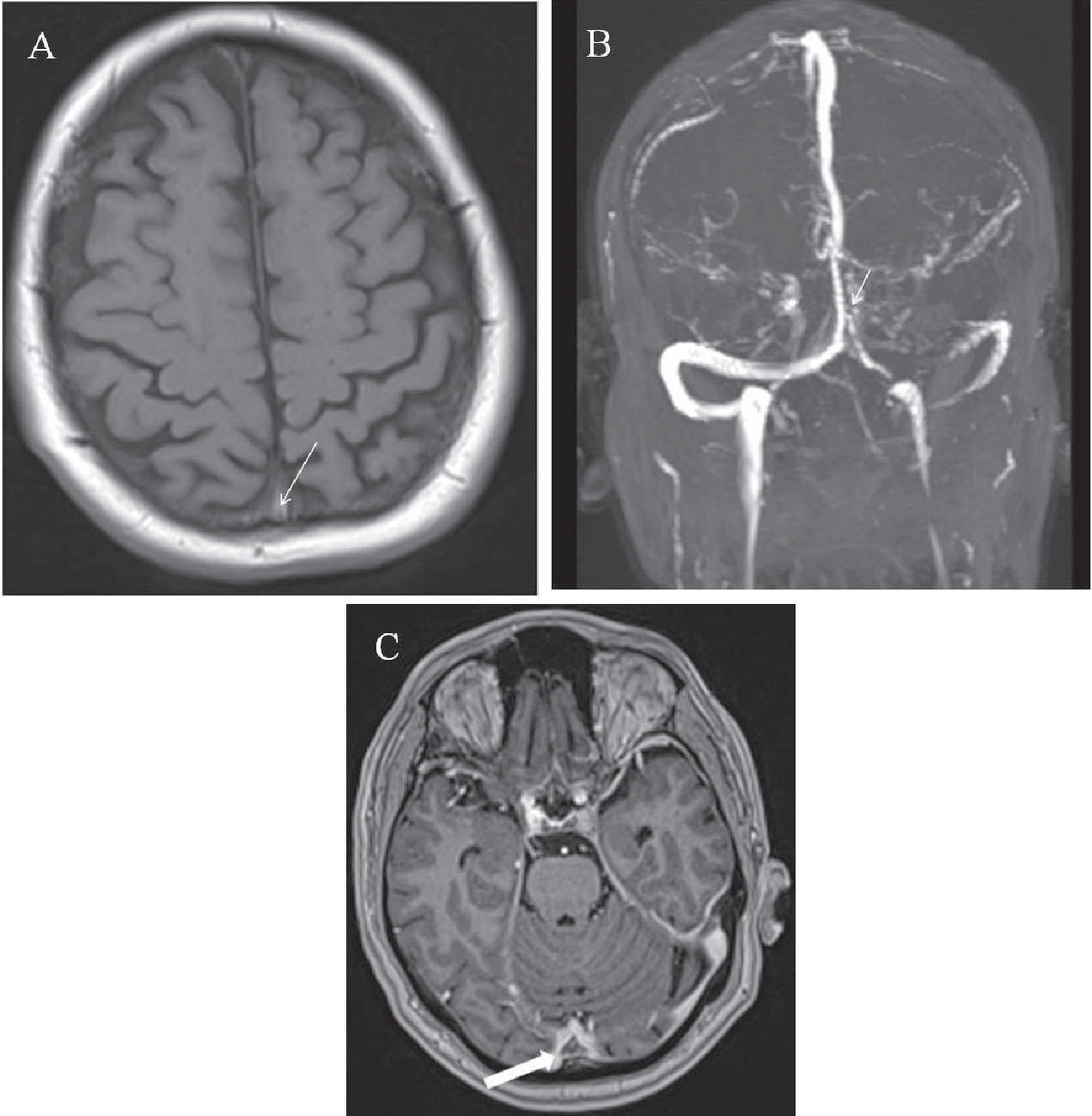
The patient’s ECG, urinalysis, complete blood counts, biochemical, and coagulation profiles were within normal limits. MRI examination of the brain revealed evidence of hyperintense signal intensity on T1-weighted sequences in the region of the torcula herophilli and the superior sagittal sinus (
Figure 1A). MR venography examination revealed a filling defect in the torcula herophilli extending into the superior sagittal sinus (
Figure 1B), and contrast-enhanced, magnetization-prepared rapid acquisition gradient echo imaging confirmed the presence of a thrombus (
Figure 1C). The results of a diffusion-weighted MRI examination were normal. A final diagnosis of TGA caused by a cerebral venous thrombosis was made.
The patient was placed on anticoagulation therapy with low molecular weight heparin. He regained his premorbid neurological state within 6 hours (by approximately 3.00 p.m.) but was unable to recall the events occurring between 9.00 a.m. and 3.00 p.m. He was kept under observation for the next 72 hours and was discharged.
Discussion
TGA is characterized by transient, sudden loss of memory and incapacity to acquire new information that lasts a few hours,
1 with patients showing complete recovery in less than 24 hours.
2 An individual in a state of TGA exhibits no other signs of impaired cognitive functioning and has no focal deficits.
1 Most cases occur in the sixth and seventh decade (age range, 21–92 years).
3 There are no apparent long-term sequelae, and recurrence is uncommon
4; however, some studies have reported an annual recurrence rate of 2.5% to just over 5.0%.
1,5The etiology of TGA has been debated for years. The clinical symptoms suggest that the site of neurologic involvement would be the medial temporal lobe and hippocampus, as this area is involved in the formation and retrieval of new episodic memories.
6 Epilepsy,
1 migraine,
2 and ischemia
3 have been proposed as potential causative mechanisms for TGA. Because of the longer duration of TGA episodes and a lower atherosclerotic risk burden,
2,5 arterial ischemia appears less likely to be an etiological factor for TGA. The Valsalva maneuver has been found to be frequently associated with TGA,
7 and hypertension has been observed as a predominant vascular risk factor.
8 However, because of the lack of a definitive evidence for a single causative mechanism, multifactorial etiology seems a more likely causative mechanism for TGA.
2It has been hypothesized that venous congestion attributable to retrograde flow in the internal jugular veins occurs more frequently during the Valsalva maneuver, causing ischemia or spreading depression in the medial temporal lobes, suggesting that dysfunctional venous circulation is part of the pathogenesis.
7 Many cases of TGA seem to be associated with factors of increased risk of cerebral venous thrombosis, including polycythemia, anti-phospholipid antibodies, venous hypertension, female sex, and more. It has been suggested that most cases of TGA may be due to small thrombi in the deep cerebral venous system.
9 In our patient, TGA appears to be related to venous congestion secondary to cerebral venous thrombosis. The thrombus in the venous sinus must have impeded venous return by the superior vena cava, thus allowing transient retrograde transmission of elevated venous pressure to the cerebral venous system, which in turn might have resulted in venous ischemia of the mesial temporal lobes leading to TGA. A PubMed search revealed only one case report of TGA associated with the cerebral venous system.
10The present case report highlights cerebral venous thrombosis as one of the possible etiological mechanisms behind TGA and demonstrates the need for its investigation. Furthermore, small venous thrombi may be difficult to visualize, even when using modern imaging technology; therefore, detailed imaging of cerebral venous sinuses with thin section, contrast-enhanced MR venography or CT venography could be considered in all patients of TGA. In addition, there may be a role for markers of thrombosis (e.g., D-dimer) in such cases; however, this role needs further evaluation.


