Full access
Departments
Published Online: 29 July 2015

Anti-Ma2 Antibody Encephalitis Manifesting as Cognitive Impairment and Psychosis

Publication: The Journal of Neuropsychiatry and Clinical Neurosciences
To the Editor: Anti-Ma2 antibody encephalitis is a rare autoimmune disease and is a paraneoplastic syndrome related with systemic cancers.14 The literature includes a few case reports in which anti-Ma2 antibody encephalitis involved the brainstem and limbic system; these patients showed neuropsychiatric manifestations, including neuro-ophthalmologic abnormalities, dizziness, headache, ataxia, seizure, and drowsiness.14 Given the rarity of the condition and the need to test levels of specific antibodies, diagnosing anti-Ma2 antibody encephalitis is a very difficult and challenging process. Therefore, a physician’s knowledge of and experience with autoimmune encephalitis are important for proper diagnosis and treatment. This report describes a patient with anti-Ma2 antibody encephalitis and a hidden malignant neoplasm who initially manifested acute cognitive impairment and psychosis.

Case Report

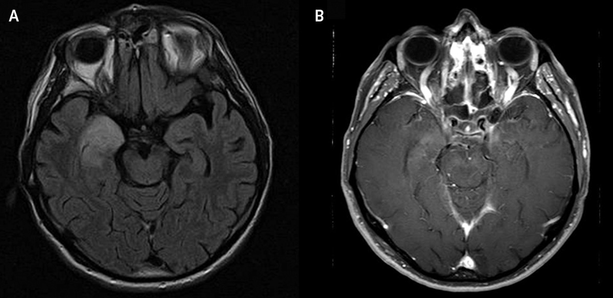
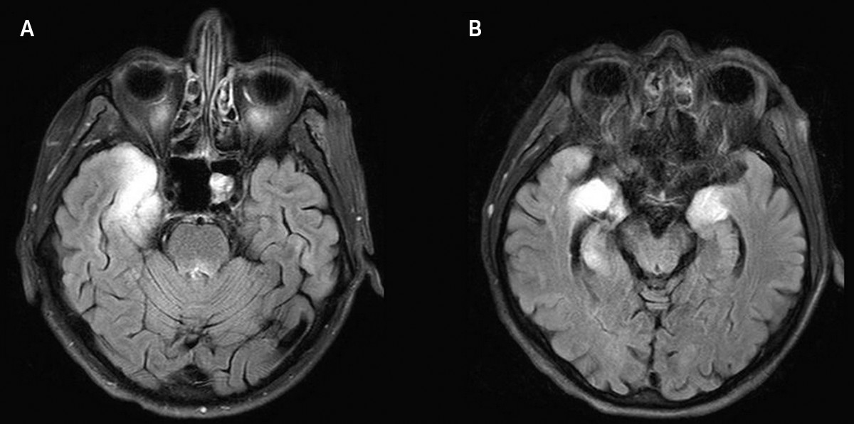
A 64-year-old man was seen at our hospital with a 2-month history of memory impairment, decreased speech, depressed mood, anxiety, disorganized language, aggressiveness, and excessive sleepiness. Results on neurologic examination revealed lethargy and cognitive impairment, but there was no evidence of cranial nerve abnormalities, motor weakness, or pathological reflexes. A brain MRI scan showed an infiltrative mass lesion in the right medial temporal lobe, suggestive of a brain tumor (Figure 1). Before surgery to remove the mass, the patient’s neuropsychological symptoms rapidly progressed and he exhibited decreased mentality, agitation, and aggressiveness. A follow-up brain MRI scan revealed further aggravation of the right medial temporal lobe lesion and new lesions involving the left temporal lobe (Figure 2). Because these findings were inconsistent with a diagnosis of brain tumor, evaluations for herpes simplex viral encephalitis and limbic encephalitis were performed. A CSF analysis showed elevated levels of WBCs (13/mm3) and protein (78 mg/dL). We performed a CT scan of the chest, neck, abdomen, and pelvis to exclude the possibility of hidden malignant neoplasms. A chest CT scan and [18F]-fluorodeoxyglucose positron emission tomography showed an ovoid nodule in the right apex with multiple lymph nodes in the hilum and mediastinum, indicating lung cancer (Figure 3). We also measured serum levels of anti-Hu, anti-Yo, anti-CV2/CRMP5, anti-Ri, anti-Ma2, and anti-amphiphysin antibodies associated with well-known paraneoplastic limbic encephalitis, as well as antibodies targeting neuronal cell surface antigens, ion channels (voltage-gated potassium channels), and ligand-gated ion channels (N-methyl-d-aspartic acid, AMPA, and GABA-B receptor channels). Only anti-Ma2 antibodies were detected. On the basis of these findings, the patient was diagnosed with anti-Ma2 antibody encephalitis and lung cancer, and he underwent a right upper lung lobectomy.
FIGURE 1. Brain MRI Scan Using (A) Fluid-Attenuated Inversion Recovery and (B) Gadolinium-Enhanced T1-Weighted Imaging Revealing a High-Signal Mass Lesion With Mild Enhancement in the Right Medial Temporal Lobe
FIGURE 2. Follow-Up Brain MRI Scan Using Fluid-Attenuated Inversion Recovery Revealing a Much-Aggravated Lesion in the Right Medial Temporal Lobe and Newly Developed Lesions Involving the Left Temporal Lobesa
a These findings were inconsistent with a diagnosis of brain tumor.
FIGURE 3. (A) Chest CT Scan and (B) [18F]Fluorodeoxyglucose Positron Emission Tomography Scan Showing an Ovoid Nodule in the Right Apex With Multiple Lymph Nodes at Both the Hilum and Mediastinum, Indicating Lung Cancer (White Arrow)

Discussion

Paraneoplastic limbic encephalitis is an autoimmune-mediated disease involving damage to the limbic system owing to antibodies generated in response to a remote malignant neoplasm.57 Anti-Ma2 antibody encephalitis is very rare and is characterized by neuro-ophthalmologic abnormalities, ataxia, seizure, dysarthria, and decreased mentality.14 However, unlike previous reports, our patient showed dominant psychiatric symptoms of memory impairment, decreased speech, depressed mood, anxiety, disorganized language, and aggressiveness, but there was no clear evidence of seizure, ataxia, neuro-ophthalmologic abnormalities, or systemic tumor. Therefore, although the patient manifested unusual symptoms, he did not visit the hospital for 2 months because his family assumed that he was experiencing psychiatric problems (e.g., depression, stress, and anxiety). His inflammatory brain lesion might have slowly progressed; his early psychiatric symptoms were consistent with the initial MRI finding of damage to the right medial temporal lobe and hippocampus. As his encephalopathy advanced, he experienced drowsiness and cognitive impairment. A second MRI scan revealed abnormalities in the limbic system, including the insular and temporal cortices and thalamus. When imaging findings and neuropsychiatric symptoms are anatomically unmatched, subsequent MRI scan and further evaluations are warranted to investigate the possibility of autoimmune encephalitis.
There have been reports of psychiatric manifestations of anti-Ma2 antibody encephalitis; however, this is a rare disease that is likely to be misdiagnosed as psychosis, brain tumor, or infection. This unusual case course serves to remind clinicians that patients who experience a first episode of rapidly progressing cognitive impairment and psychosis of an uncertain origin should undergo brain MRI scan, and the possibility of anti-Ma2 antibody encephalitis and hidden malignant neoplasms should be considered.

References

1.
Dalmau J, Graus F, Villarejo A, et al: Clinical analysis of anti-Ma2-associated encephalitis. Brain 2004; 127:1831–1844
2.
Garcia-Reitboeck P, Thompson G, Johns P, et al: Upbeat nystagmus in anti-Ma2 encephalitis. Pract Neurol 2014; 14:36–38
3.
Abdulkader MM, Yousef MM, Abdelhadi MK, et al: Microscopic dysgerminoma associated with anti-Ma2 paraneoplastic encephalitis in a patient with gonadal dysgenesis. Int J Gynecol Pathol 2013; 32:277–282
4.
Sahashi K, Sakai K, Mano K, et al: Anti-Ma2 antibody related paraneoplastic limbic/brain stem encephalitis associated with breast cancer expressing Ma1, Ma2, and Ma3 mRNAs. J Neurol Neurosurg Psychiatry 2003; 74:1332–1335
5.
Darnell RB, Posner JB: Paraneoplastic syndromes involving the nervous system. N Engl J Med 2003; 349:1543–1554
6.
Bennett JL, Galetta SL, Frohman LP, et al: Neuro-ophthalmologic manifestations of a paraneoplastic syndrome and testicular carcinoma. Neurology 1999; 52:864–867
7.
Graus F, Delattre JY, Antoine JC, et al: Recommended diagnostic criteria for paraneoplastic neurological syndromes. J Neurol Neurosurg Psychiatry 2004; 75:1135–1140

Information & Authors

Information

Published In

Go to The Journal of Neuropsychiatry and Clinical Neurosciences
The Journal of Neuropsychiatry and Clinical Neurosciences
Pages: e221 - e222
PubMed: 26222977

History

Published in print: Summer 2015
Published online: 29 July 2015

Authors

Affiliations

Moo-Seok Park, M.D.
From the Department of Neurology, Chung-Ang University Hospital, Chung-Ang University College of Medicine, Seoul, Republic of Korea.
Jae-Han Bae, M.D.
From the Department of Neurology, Chung-Ang University Hospital, Chung-Ang University College of Medicine, Seoul, Republic of Korea.
Hae-Bong Jeong, M.D.
From the Department of Neurology, Chung-Ang University Hospital, Chung-Ang University College of Medicine, Seoul, Republic of Korea.
Jeong-Min Kim, M.D., Ph.D.
From the Department of Neurology, Chung-Ang University Hospital, Chung-Ang University College of Medicine, Seoul, Republic of Korea.
Oh-Sang Kwon, M.D., Ph.D.
From the Department of Neurology, Chung-Ang University Hospital, Chung-Ang University College of Medicine, Seoul, Republic of Korea.
Young Chul Youn, M.D., Ph.D.
From the Department of Neurology, Chung-Ang University Hospital, Chung-Ang University College of Medicine, Seoul, Republic of Korea.
Suk-Won Ahn, M.D., Ph.D.
From the Department of Neurology, Chung-Ang University Hospital, Chung-Ang University College of Medicine, Seoul, Republic of Korea.

Notes

Send correspondence to Suk-Won Ahn, M.D., Ph.D.; e-mail: [email protected]

Funding Information

This research was supported by grant NRF-2014R1A1A1005484 from the Basic Science Research Program through the National Research Foundation of Korea, funded by the Ministry of Science, ICT and Future Planning.

Metrics & Citations

Metrics

Citations

Export Citations

If you have the appropriate software installed, you can download article citation data to the citation manager of your choice. Simply select your manager software from the list below and click Download.

For more information or tips please see 'Downloading to a citation manager' in the Help menu.

Format
Citation style
Style
Copy to clipboard

There are no citations for this item

View Options

View options

PDF/ePub

View PDF/ePub

Get Access

Login options

Already a subscriber? Access your subscription through your login credentials or your institution for full access to this article.

Personal login Institutional Login Open Athens login
Purchase Options

Purchase this article to access the full text.

PPV Articles - Journal of Neuropsychiatry and Clinical Neurosciences

PPV Articles - Journal of Neuropsychiatry and Clinical Neurosciences

Not a subscriber?

Subscribe Now / Learn More

PsychiatryOnline subscription options offer access to the DSM-5-TR® library, books, journals, CME, and patient resources. This all-in-one virtual library provides psychiatrists and mental health professionals with key resources for diagnosis, treatment, research, and professional development.

Need more help? PsychiatryOnline Customer Service may be reached by emailing [email protected] or by calling 800-368-5777 (in the U.S.) or 703-907-7322 (outside the U.S.).
Media

Figures

Other

Tables

Share

Share

Share article link

Share