Full access
Windows to the Brain
Published Online: 27 January 2016

Working Memory Models: Insights From Neuroimaging

Publication: The Journal of Neuropsychiatry and Clinical Neurosciences
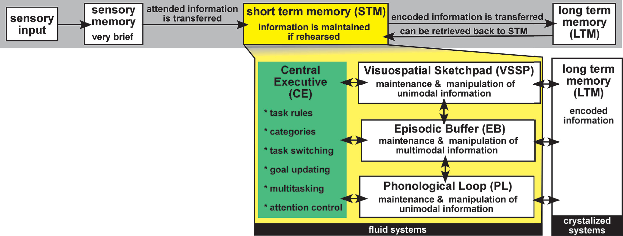
Memory, the ability to maintain access to information in the absence of sensory input, is fundamental to all goal-directed behavior. Early models of memory were grounded in the logic models of the rising world of computer science and information theory.11 One popular model (Atkinson and Shiffrin Model) developed at that time proposed three stages of declarative memory: sensory memory (sense-specific registers), short term memory (STM), and long term memory (LTM). Information was lost if not successfully passed to the next stage (Figure 1).1 Although flawed, this model continues to provide a simple and easy approach to understanding memory. One assumption that proved to be inconsistent with research was that there is a single STM system. The low level of interference when concurrent tasks utilized different senses indicated that STM was not a unitary store. The multicomponent model of working memory (WM) essentially reconceptualized STM by expanding it into more than one component and adding a set of active processes (Figure 1).2 This model of WM continues to be the most widely used conceptual map.1217 The original publication has been cited nearly 10,000 times in more than 40 years since its initial presentation.16 Due to the importance of intact WM for daily functioning, foundational understanding of WM theory, tests, and recent neuroimaging is of value for both researchers and clinicians working in neuroscience-related fields.
FIGURE 1. The Atkinson and Shiffrin Model of memory is diagramed in the gray panel. Sensory information is perceived and suppressed via selective attention processes, then briefly held in the limited-capacity modality-specific sensory register. Information enhanced by selective attention enters into short term memory (STM). The process of rehearsal (maintenance) can sustain information in STM. Information in STM is automatically encoded (stored, consolidated) into long term memory (LTM).1 Although flawed, this model continues to provide a simple and easy approach to understanding memory. Assumptions that proved to be inconsistent with research included that any information in STM will transfer to LTM and that STM is required for access to LTM. Additionally, the low level of interference when concurrent tasks utilized different senses indicated that STM was not a unitary store. The multicomponent model of working memory (WM) essentially reconceptualized STM by expanding it into more than one component (yellow) and adding a set of active processes.2 Three buffers support short term processing of visual-spatial (VSSP), auditory-verbal (PL), and multimodal (EB) information, under the control of the central executive (CE, green). The CE directs all other aspects of memory processes and attention. CE processes overlap with tasks commonly referred to as executive functions.

Cognitive Psychology

The concepts of STM and WM are related, as both represent processes that provide temporary storage of information (i.e. maintenance of information for a short period of time in the absence of perceptual input). A key difference is that the concept of WM adds processes that support manipulation of the information, providing a basis for goal-directed behaviors.2,12,13,16,17 The multicomponent model of WM continues to evolve, but currently includes: the phonological loop (PL), the visuospatial sketchpad (VSSP), the episodic buffer (EB), and the central executive (CE) (Figure 1). The PL and VSSP provide temporary maintenance and manipulation of domain-specific information under the attentional control of the CE. The PL provides access to auditory-verbal information, and is perhaps the most researched aspect of this model. The VSSP provides access to visual information. The need for two components was based on research indicating far less interference between different types of domain-specific information during WM tasks than would be expected if there were a single component. Other domains of information (e.g. tactile, smell, taste) are much less studied. The EB component of WM was added to the model much later to provide the capacity to integrate information across domains and temporarily maintain and manipulate multidimensional representations.2 The EB is also thought to be responsible for the process underlying the cognitive strategy of collapsing related data together (“chunking” information) to increase attentional capacity. The CE component provides higher-order control processing. It has been dissociated into multiple independent but related processes, such as attentional control, multitasking, retrieval strategy switching, selective inhibition, and mental manipulation.2 There are two major views of the CE. The first conceptualizes the CE from a model of general intellectual ability; the second views the CE from the perspective of information manipulation. It is important to emphasize that the multicomponent model of WM is a cognitive model that does not propose biological substrates. Delineation of the neural substrates of the WM components is now an area of active investigation.

Neuropsychology

The concept of intelligence provides a context to understand other cognitive abilities. Intelligence is generally defined as general mental capacity. There are numerous theories of intelligence, with many including WM as a component. The Cattell-Horn-Carroll (CHC) model of intelligence, a comprehensive and widely accepted theory, is hierarchical.18,19 General intellectual ability (g) encompasses the comprehensive representation of global intelligence. Within g are various broad abilities (e.g. fluid intelligence [mental efficiency] and crystallized intelligence [acquired knowledge]), each of which can be further segmented into numerous narrow abilities.20 Although the model encompasses a STM component, WM is not directly incorporated. However, abilities such as general sequential reasoning (under the fluid intelligence factor) and memory span (under the general memory and learning factor) require use of WM. Thus, WM is represented in (but not actually part of) the model. Numerous studies have found moderate to high correlations between WM and g.21 Two mainstream intelligence tests (Stanford-Binet and the Wechsler Intelligence Scales [WIS]) directly incorporate WM into their batteries, comprising a full scale intelligence quotient (IQ score). The relationship between WM and intelligence is partially mediated by how intelligence is measured. Of particular note, the WIS Working Memory Index includes Digit Span, Arithmetic, and Letter-Number Sequencing, three commonly used tests in both clinical and research settings. As the WIS are some of the most popular tests of intellectual ability in current use, many IQ estimates will inevitably be greatly influenced by WM ability.
Although conceptualized as an active STM process, WM tasks are commonly classified as attentional in neuropsychology. One major text introduces WM within the more global memory paradigm but describes tests of WM as the combination of storage and manipulation of information.22 WM tests are included among tests of attention, not with tests of memory. Commonly used WM tests do not directly correlate to the multicomponent WM model. WM tests are classified generally, or at most, divided by modality (visual versus verbal). In general, WM tests can be grossly divided into two categories: complex span and complex tracking. Additionally, there is a division between those tests commonly used in research and those commonly used clinically. One study assessed whether research WM tests (operation span, listening span, and N-back) were consistent with those used clinically (WIS and Wechsler Memory Scale tests).23 The three research tests and WAIS-III Letter-Number Sequencing were the most strongly correlated. The model containing those four measures provided the best fit to the latent WM variable. Thus, WIS Letter-Number Sequencing (a complex span test) may be a common metric for translation across the settings.
Two commonly used tests of complex tracking are Paced Auditory Serial Addition Test (PASAT) and N-back. During the PASAT, single-digit numbers are presented at various time intervals and the respondent must add each number to the immediately preceding number, though there are a number of versions of the test available.24 The PASAT is frequently included in fixed multiple sclerosis batteries due to its sensitivity to impairments common to that disorder.25,26 Psychometrically, the task seems to load heavily on processing speed, attention, and executive functions.27 Although the test is difficult and many find it aversive, it was reported to be the third most popular test of attention among neuropsychologists according to one survey.28 The N-back paradigm is a category of complex tracking tests that is frequently used in neuroimaging studies. A series of stimuli (verbal or nonverbal) are presented, requiring the respondent to signal when the new stimulus matches the one presented “n” items before. Thus, this test requires both WM and vigilance. Increasing the span (or “n”) increases the complexity of the test. The stimuli can be numbers, letters, shapes, or location of a stimulus in space.
In contrast, complex span tests focus more on attentional capacity. A commonly used test is Digits Backward. Strings of digits are presented with the respondent reciting the digits in reverse order. On subsequent trials the length of the string (digit span) is increased. The test is heavily researched, easy to administer, and a regular subtest in the WIS. Current versions combine Digits Backward with the simpler Digits Forward and the Digit Sequencing tests. However, factor analyses indicate that simple span and complex span tasks are related but not equivalent.29 Digits Backward is an ideal test for use as part of a bedside exam due to its ease and speed of administration. A Digits Forward span should be completed first. The typical forward span for adults is seven digits; the typical backward span is four or five digits correct.22,30,31 Numerous other complex span tasks are in use (e.g. reversed spelling, mental arithmetic, WIS Letter-Number Sequencing). The Corsi block tapping test, has been computerized, making it useful for neuroimaging studies.32 The stimulus is an array of 3D blocks arranged in space. The examiner taps the blocks in a particular order, with the respondent then tapping the blocks in the same (or reverse) order. On subsequent trials, the number of taps increases (similar to digit span). A series of dual procedure and suppression experiments using the Corsi forward and backward paradigms found that the Corsi test employs VSSP and CE but not PL, suggesting that the Corsi test fits well with the multicomponent model of WM.33 The importance of this finding is that most WM tests employ PL and CE, with fewer tests available to elicit VSSP.

Neurobiology

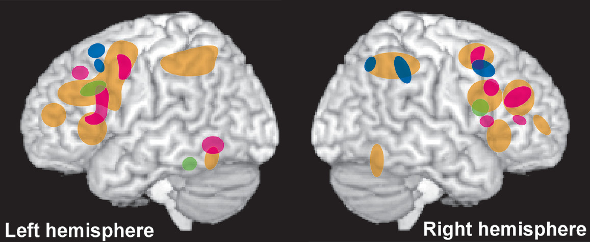
The classic approach for identifying the neural architecture supporting particular aspects of function is the study of patients with focal brain lesions.2,13 Studies that establish correlations between morphological and performance variables (i.e. lesion-deficit analyses) provide a basis for identifying areas of the brain that support specific abilities. As intelligence and WM are strongly interrelated, the most informative studies include a sufficiently broad range of performance measures to support assessment of both. A recent series of studies utilized voxel-based lesion-symptom mapping (each voxel designated as intact or lesioned) to delineate the neural architecture underlying the major types of intelligence (general, fluid) and major components of WM (domain-specific information maintenance, information monitoring, information manipulation, cognitive flexibility).3,4,6 Both general and fluid intelligence were associated with widely distributed networks of cortical areas (ventrolateral and dorsolateral prefrontal [PFC], primary motor and somatosensory, inferior and superior parietal, and superior temporal cortices) and associated major white matter tracts. General intelligence was primarily left lateralized, whereas fluid intelligence was primarily right lateralized (Figure 2).3,6 The areas associated with general intelligence were similar in locations but greater in extent than reported in a previous study.34 Domain specific WM lateralized by domain (Figure 2), with verbal/ numeric primarily associated with areas in the left hemisphere (dorsolateral PFC, superior parietal and superior temporal cortices, angular gyrus) and spatial primarily associated with areas in the right hemisphere (dorsolateral PFC and superior parietal cortices).6 Both overlapped with areas associated with intelligence in dorsolateral PFC and superior parietal cortices.3,6 Cognitive flexibility and the information manipulation aspect of WM were both associated with primarily left hemisphere networks of cortical areas (Figure 2). Cognitive flexibility overlapped areas associated with general intelligence throughout that network.3,4 Some of the left hemisphere areas associated with information manipulation overlapped with areas associated with general intelligence (superior parietal and superior temporal cortices), whereas others (angular gyrus) did not.3,6 In addition, some areas in the right hemisphere associated with fluid intelligence were also associated with information manipulation (dorsolateral PFC). The information monitoring aspect of WM was associated with a primarily right hemisphere network of cortical areas (ventrolateral PFC, inferior parietal and middle temporal cortices) that overlapped the fluid intelligence network only in inferior parietal cortex.6
FIGURE 2. A recent series of studies utilized voxel-based lesion-symptom mapping (each voxel designated as intact or lesioned) to delineate the neural architecture underlying the major types of intelligence (general, fluid) and major components of WM (domain-specific information maintenance, information monitoring, information manipulation, cognitive flexibility).36 Visually summarized are the cortical areas most associated with fluid intelligence and several aspects of WM in the final model.6 Fluid intelligence (red) was associated with a primarily right lateralized network of frontal and parietal areas. Domain specific WM lateralized by domain, with verbal/numeric WM (green) primarily associated with areas in the left hemisphere and spatial WM (blue) primarily associated with areas in the right hemisphere. Areas associated with information manipulation aspects of WM (yellow) partially overlapped with both general intelligence (not illustrated) and verbal/numeric WM in the left hemisphere and fluid intelligence in the right hemisphere. The information monitoring aspect of WM (purple) was associated with a primarily right hemisphere network of cortical areas, but overlapped the fluid intelligence network only in inferior parietal cortex.
Studies in healthy individuals are also informative. A meta-analysis of the association between PFC volume and thickness and tests of executive functioning found a moderate effect size for volume and a weak effect size for thickness.35 In the regional analysis, both lateral and medial PFC volumes were significantly correlated with executive functioning.35 Similarly, larger gray matter volume was associated with better executive performance both at the global (whole brain) and regional (PFC, insular and cerebellar clusters) levels in another study.36 A study quantified several measures of cortical morphology (gray matter volume, cortical surface area, cortical thickness) to identify areas supporting aspects of intelligence (fluid, crystalized, spatial) and basic cognitive processing (WM capacity, executive updating, attention, processing speed).37 Statistical parametric mapping of gray matter volume indicated that all three intelligence factors were associated with partially overlapping clusters in right dorsolateral PFC. Spatial intelligence was also associated with clusters in right inferior temporal cortex (fusiform gyrus) and caudate. WM capacity was associated with a cluster in right dorsolateral PFC that overlapped fluid and crystalized intelligence. WM was also associated with clusters in right primary somatosensory cortex and left frontal and occipital cortices. Attention was associated with clusters within frontal, temporal and occipital cortices.37 A study that estimated size of visual cortex using both structural (landmark based parcellation) and functional (retinotopic mapping) measures found that the size of V1 (first stage of visual processing) correlated strongly with visual WM capacity.38 As noted by the authors, these findings suggest that individual differences in the size of domain-specific cortices may contribute to individual differences in domain-specific WM performance.
Functional imaging provides a range of methods for probing the neural architecture supporting particular aspects of brain function. Task-activated functional imaging studies are most commonly used to identify areas of the brain that are more active during performance of a particular task. Most functional imaging studies utilize functional magnetic resonance imaging (fMRI), which provides an indirect measure of neuronal activity based on local changes in blood flow. The majority of studies compare signal intensity of relatively large groups of voxels (cluster or region) across conditions (univariate analyses). Multivariate pattern analytic (MVPA) techniques have recently been introduced that utilize machine learning techniques to identify patterns of activated voxels that encode task-related information. Comparisons across types and phases of tasks have the potential to provide insight into the brain areas supporting different components of WM. One example is comparing tasks that require similar manipulations of information but utilize different information domains. Processes related to maintaining domain-specific information (e.g. visual, verbal) might be expected to localize differently, whereas domain-general processes (e.g. remembering rules or goals, ignoring distractors) might be expected to localize similarly.
Several recent meta-analyses of functional imaging studies reported bilateral activation of a network of frontal, parietal, temporal, cerebellar, and thalamic areas across WM tasks (e.g. N-back, Sternberg, delayed matching to sample, prospective memory) and stimuli types (Figure 3).710 The meta-analysis that compared by gender also found a few differences, such as greater activation of limbic areas in females and parietal areas in males.8 Increasing WM load (increasing task difficulty or adding the presence of distractors) was associated with increased activations in almost all areas (exceptions were rostral lateral PFC and thalamus), suggesting a network supporting domain-general executive control processing (Figure 3).7 A study utilizing MVPA reported successful cross-modal (visual WM task, verbal WM task) prediction of WM-load-sensitive areas, further supporting a common domain-general neural substrate.39 A study utilizing graph-based modularity analysis found that WM tasks activated multiple areas within the executive control (lateral PFC, superior parietal lobule) and salience (anterior insular and dorsal cingulate cortices) networks.40 Connectivity within the executive control network and between the executive control network and salience network increased with increasing WM load.40
COVER and FIGURE 3. Several recent meta-analyses of functional imaging studies reported bilateral activation of a network of frontal, parietal, temporal, cerebellar, and thalamic areas across WM tasks and stimuli types.710 Illustrated here are data from one study.7 Main effect of WM (gold) was associated with the expected wide spread network of areas (see COVER for insula and thalamus). Increasing WM load (pink) was associated with increased activations in almost all the same areas, suggesting a network supporting domain-general executive control processes. Areas most associated with object identity (green) were in a similar region of prefrontal cortex but ventral to areas most associated with object location (blue), supporting the proposed dorsal/ventral gradient in prefrontal functions.7
Some areas, most commonly within lateral PFC and parietal cortex, display sustained activation during the delay period in WM tasks.12,13,17,41,42 One interpretation has been that this represents domain specific information being maintained in an active state. However, fMRI studies utilizing MVPA have reported that patterns of activity in lateral PFC contain higher order information required for task performance (e.g. task rules, stimulus-response mapping, categories based on multiple domains of information) rather than domain specific information.12,13,41,42 These findings support the alternative view that sustained activity in lateral PFC and parietal cortex indicates allocation of cognitive resources (e.g. attention) to maintain domain specific information elsewhere (top-down modulation). Congruent with this view, MVPA studies identified patterns of activity in sensorimotor cortices during delay periods consistent with population encoding of domain specific information even in the absence of elevated activity.12,13,41,42 Patterns were predictive across memory types (WM, LTM), suggesting a common neural substrate for WM and LTM. An fMRI study using MVPA reported dynamic shifts in patterns of activity when a retrocue (cue presented when the stimulus is no longer available) signaled a change in the stimulus dimension required for task performance.43 This suggests that what is encoded is not a static representation of all information contained in the stimulus, but rather specifically favors task-relevant aspects.
The multicomponent model of WM is a conceptual model, informed by and developed based on cognitive psychology research. Advances in identifying neural correlates of processes that contribute to WM have provided insights into the underlying neural architecture. For example, in the multicomponent model WM and LTM are functionally distinct systems. There is growing evidence from neuroimaging research, particularly MVPA studies, that WM and LTM are neuroanatomically colocated. This apparent disparity has been reconciled in state-based models of WM, which postulate that these types of memory differ by activation state rather than location in the brain.12,13,15,17,41 In these models, attention is the key factor determining the accessibility of memory content. Terminology and number of activation states varies, but the basic postulates are similar. WM contains information that is the focus of attention (region of direct access). Other information ranges from less accessible (latent WM, activated LTM) to not directly accessible (LTM). The hierarchical process model also incorporates timescale and information complexity to distinguish the nature of memory processing in domain-specific areas from that occurring in higher order areas.15

Conclusions

WM is a critical cognitive ability that has emerged as an important domain in comprehensive neuropsychological evaluation. WM can be conceptualized as a set of active processes in declarative memory systems that occur between the sensory register and stable, theoretically permanent memory stores (LTM). The multicomponent model of WM was developed to address aspects of the passive model of STM that did not match research findings. Conceived of as a component of general intellectual ability, it is also classified as an attentional skill. Thus, WM has become a complicated and multifaceted construct of mental ability. Tests of WM typically involve either complex span or complex tracking. Many tests do not translate well to the bedside exam and tests used in research often differ from those commonly used clinically. WM tests utilizing mental arithmetic and reverse span are the most practical to employ in bedside or less-formal evaluation. Imaging studies frequently place WM abilities in dorsolateral PFC and in frontoparietal networks. There is a relatively consistent lateralized effect between the verbal (PL) and visual-spatial (VSSP) aspects of WM. As imaging studies continue to inform WM conceptualization, cognitive models will likely continue to evolve.

References

1.
Shiffrin RM, Atkinson RC: Storage and retrieval processes in long-term memory. Psychol Rev 1969; 76:179–193
2.
Baddeley A: Working memory: theories, models, and controversies. Annu Rev Psychol 2012; 63:1–29
3.
Barbey AK, Colom R, Solomon J, et al: An integrative architecture for general intelligence and executive function revealed by lesion mapping. Brain 2012; 135:1154–1164
4.
Barbey AK, Koenigs M, Grafman J: Dorsolateral prefrontal contributions to human working memory. Cortex 2013; 49:1195–1205
5.
Barbey AK, Colom R, Grafman J: Architecture of cognitive flexibility revealed by lesion mapping. Neuroimage 2013; 82:547–554
6.
Barbey AK, Colom R, Paul EJ, et al: Architecture of fluid intelligence and working memory revealed by lesion mapping. Brain Struct Funct 2014; 219:485–494
7.
Rottschy C, Langner R, Dogan I, et al: Modelling neural correlates of working memory: a coordinate-based meta-analysis. Neuroimage 2012; 60:830–846
8.
Hill AC, Laird AR, Robinson JL: Gender differences in working memory networks: a BrainMap meta-analysis. Biol Psychol 2014; 102:18–29
9.
Chu C, Fan L, Eickhoff CR, et al: Co-activation Probability Estimation (CoPE): An approach for modeling functional co-activation architecture based on neuroimaging coordinates. Neuroimage 2015; 117:397–407
10.
Cona G, Scarpazza C, Sartori G, et al: Neural bases of prospective memory: a meta-analysis and the “Attention to Delayed Intention” (AtoDI) model. Neurosci Biobehav Rev 2015; 52:21–37
11.
Greenwood JD: A conceptual history of psychology. New York, NY, McGraw-Hill, 2009
12.
D’Esposito M, Postle BR: The cognitive neuroscience of working memory. Annu Rev Psychol 2015; 66:115–142
13.
Eriksson J, Vogel EK, Lansner A, et al: Neurocognitive Architecture of Working Memory. Neuron 2015; 88:33–46
14.
Fenesi B, Sana F, Kim JA, et al: Reconceptualizing working memory in educational research. Educ Psychol Rev 2015; 27:333–351
15.
Hasson U, Chen J, Honey CJ: Hierarchical process memory: memory as an integral component of information processing. Trends Cogn Sci 2015; 19:304–313
16.
Logie RH, Cowan N: Perspectives on working memory: introduction to the special issue. Mem Cognit 2015; 43:315–324
17.
Martini M, Furtner MR, Maran T, et al: Information maintenance in working memory: an integrated presentation of cognitive and neural concepts. Front Syst Neurosci 2015; 9:104
18.
McGrew KS: CHC theory and the human cognitive abilities project: Standing on the shoulders of the giants of psychometric intelligence research. Intelligence 2009; 37:1–10
19.
McGrew KS, Wendling BJ: Cattell-Horn-Carroll cognitive-achievement relations: What we have learned from the past 20 years of research. Psychol Sch 2010; 47:651–675
20.
Newton JH, McGrew KS: Introduction to the special issue: Current research in Cattell-Horn-Carroll-Based Assessment. Psychol Sch 2010; 47:621–634
21.
Chooi W-T: Working Memory and Intelligence: A Brief Review. J Educ Develop Psychol 2012; 2
22.
Lezak MD, Howieson DB, Bigler ED, et al: Neuropsychological assessment, 5th ed. New York, NY, Oxford University Press, 2012
23.
Shelton JT, Elliott EM, Hill BD, et al: A Comparison of Laboratory and Clinical Working Memory Tests and Their Prediction of Fluid Intelligence. Intelligence 2009; 37:283
24.
Gronwall DM: Paced auditory serial-addition task: a measure of recovery from concussion. Percept Mot Skills 1977; 44:367–373
25.
Rosti E, Hämäläinen P, Koivisto K, et al: PASAT in detecting cognitive impairment in relapsing-remitting MS. Appl Neuropsychol 2007; 14:101–112
26.
Strober L, Englert J, Munschauer F, et al. Sensitivity of conventional memory tests in multiple sclerosis: comparing the Rao Brief Repeatable Neuropsychological Battery and the Minimal Assessment of Cognitive Function in MS. Multiple sclerosis (Houndmills, Basingstoke, England). 2009;15(9):1077-1084.
27.
Tombaugh TN: A comprehensive review of the Paced Auditory Serial Addition Test (PASAT). Arch Clin Neuropsychol 2006; 21:53–76
28.
Rabin LA, Barr WB, Burton LA: Assessment practices of clinical neuropsychologists in the United States and Canada: a survey of INS, NAN, and APA Division 40 members. Arch Clin Neuropsychol 2005; 20:33–65
29.
Reynolds CR: Forward and backward memory span should not be combined for clinical analysis. Arch Clin Neuropsychol 1997; 12:29–40
30.
Baddeley A: The magical number seven: still magic after all these years? Psychol Rev 1994; 101:353–356
31.
Weinberg J, Diller L, Gerstman L, et al: Digit span in right and left hemiplegics. J Clin Psychol 1972; 28:361
32.
Berch DB, Krikorian R, Huha EM: The Corsi block-tapping task: methodological and theoretical considerations. Brain Cogn 1998; 38:317–338
33.
Vandierendonck A, Kemps E, Fastame MC, et al. Working memory components of the Corsi blocks task. British journal of psychology (London, England: 1953). 2004;95(Pt 1):57-79.
34.
Gläscher J, Rudrauf D, Colom R, et al: Distributed neural system for general intelligence revealed by lesion mapping. Proc Natl Acad Sci USA 2010; 107:4705–4709
35.
Yuan P, Raz N: Prefrontal cortex and executive functions in healthy adults: a meta-analysis of structural neuroimaging studies. Neurosci Biobehav Rev 2014; 42:180–192
36.
Ruscheweyh R, Deppe M, Lohmann H, et al: Executive performance is related to regional gray matter volume in healthy older individuals. Hum Brain Mapp 2013; 34:3333–3346
37.
Colom R, Burgaleta M, Román FJ, et al: Neuroanatomic overlap between intelligence and cognitive factors: morphometry methods provide support for the key role of the frontal lobes. Neuroimage 2013; 72:143–152
38.
Bergmann J, Genç E, Kohler A, et al: Neural Anatomy of Primary Visual Cortex Limits Visual Working Memory. Cereb Cortex 2014; bhu168
39.
Majerus S, Cowan N, Péters F, et al: Cross-Modal Decoding of Neural Patterns Associated with Working Memory: Evidence for Attention-Based Accounts of Working Memory. Cereb Cortex 2014; bhu189 [Epub ahead of print]
40.
Liang X, Zou Q, He Y, et al: Topologically Reorganized Connectivity Architecture of Default-Mode, Executive-Control, and Salience Networks across Working Memory Task Loads. Cereb Cortex 2015; bhu316 [Epub ahead of print]
41.
Larocque JJ, Lewis-Peacock JA, Postle BR: Multiple neural states of representation in short-term memory? It’s a matter of attention. Front Hum Neurosci 2014; 8:5
42.
Sreenivasan KK, Curtis CE, D’Esposito M: Revisiting the role of persistent neural activity during working memory. Trends Cogn Sci 2014; 18:82–89
43.
Lewis-Peacock JA, Drysdale AT, Postle BR: Neural Evidence for the Flexible Control of Mental Representations. Cereb Cortex 2015; 25:3303–3313

Information & Authors

Information

Published In

Go to The Journal of Neuropsychiatry and Clinical Neurosciences
The Journal of Neuropsychiatry and Clinical Neurosciences
Pages: A4 - 5
PubMed: 26844960

History

Published in print: Winter 2016
Published online: 27 January 2016

Authors

Details

Robert D. Shura, Psy.D.
Dr. Shura is affiliated with the Mental Health Service Line and the Veterans Affairs Mid Atlantic Mental Illness Research, Education, and Clinical Center at the W.G. Hefner Veterans Affairs Medical Center in Salisbury, North Carolina and the Department of Psychiatry at Wake Forest School of Medicine in Winston-Salem, North Carolina. Drs. Hurley and Taber are affiliated with the Research and Academic Affairs Service Line and the Veterans Affairs Mid Atlantic Mental Illness Research, Education, and Clinical Center at the W.G. Hefner Veterans Affairs Medical Center in Salisbury, North Carolina. Dr. Hurley is affiliated with the Departments of Psychiatry and Radiology at Wake Forest School of Medicine in Winston-Salem, North Carolina, and the Menninger Department of Psychiatry and Behavioral Sciences at Baylor College of Medicine in Houston, Texas. Dr. Taber is affiliated with the Division of Biomedical Sciences at the Via College of Osteopathic Medicine in Blacksburg, Virginia, and the Department of Physical Medicine and Rehabilitation at Baylor College of Medicine in Houston, Texas
Robin A. Hurley, M.D
Dr. Shura is affiliated with the Mental Health Service Line and the Veterans Affairs Mid Atlantic Mental Illness Research, Education, and Clinical Center at the W.G. Hefner Veterans Affairs Medical Center in Salisbury, North Carolina and the Department of Psychiatry at Wake Forest School of Medicine in Winston-Salem, North Carolina. Drs. Hurley and Taber are affiliated with the Research and Academic Affairs Service Line and the Veterans Affairs Mid Atlantic Mental Illness Research, Education, and Clinical Center at the W.G. Hefner Veterans Affairs Medical Center in Salisbury, North Carolina. Dr. Hurley is affiliated with the Departments of Psychiatry and Radiology at Wake Forest School of Medicine in Winston-Salem, North Carolina, and the Menninger Department of Psychiatry and Behavioral Sciences at Baylor College of Medicine in Houston, Texas. Dr. Taber is affiliated with the Division of Biomedical Sciences at the Via College of Osteopathic Medicine in Blacksburg, Virginia, and the Department of Physical Medicine and Rehabilitation at Baylor College of Medicine in Houston, Texas
Katherine H. Taber, Ph.D.
Dr. Shura is affiliated with the Mental Health Service Line and the Veterans Affairs Mid Atlantic Mental Illness Research, Education, and Clinical Center at the W.G. Hefner Veterans Affairs Medical Center in Salisbury, North Carolina and the Department of Psychiatry at Wake Forest School of Medicine in Winston-Salem, North Carolina. Drs. Hurley and Taber are affiliated with the Research and Academic Affairs Service Line and the Veterans Affairs Mid Atlantic Mental Illness Research, Education, and Clinical Center at the W.G. Hefner Veterans Affairs Medical Center in Salisbury, North Carolina. Dr. Hurley is affiliated with the Departments of Psychiatry and Radiology at Wake Forest School of Medicine in Winston-Salem, North Carolina, and the Menninger Department of Psychiatry and Behavioral Sciences at Baylor College of Medicine in Houston, Texas. Dr. Taber is affiliated with the Division of Biomedical Sciences at the Via College of Osteopathic Medicine in Blacksburg, Virginia, and the Department of Physical Medicine and Rehabilitation at Baylor College of Medicine in Houston, Texas

Notes

Corresponding author: Send correspondence to Dr. Hurley; e-mail: [email protected].

Metrics & Citations

Metrics

Citations

Export Citations

If you have the appropriate software installed, you can download article citation data to the citation manager of your choice. Simply select your manager software from the list below and click Download.

For more information or tips please see 'Downloading to a citation manager' in the Help menu.

Format
Citation style
Style
Copy to clipboard

View Options

View options

PDF/EPUB

View PDF/EPUB

Login options

Already a subscriber? Access your subscription through your login credentials or your institution for full access to this article.

Personal login Institutional Login Open Athens login
Purchase Options

Purchase this article to access the full text.

PPV Articles - Journal of Neuropsychiatry and Clinical Neurosciences

PPV Articles - Journal of Neuropsychiatry and Clinical Neurosciences

Not a subscriber?

Subscribe Now / Learn More

PsychiatryOnline subscription options offer access to the DSM-5-TR® library, books, journals, CME, and patient resources. This all-in-one virtual library provides psychiatrists and mental health professionals with key resources for diagnosis, treatment, research, and professional development.

Need more help? PsychiatryOnline Customer Service may be reached by emailing [email protected] or by calling 800-368-5777 (in the U.S.) or 703-907-7322 (outside the U.S.).

Media

Figures

Other

Tables

Share

Share

Share article link

Share