Full access
Clinical and Research Reports
Published Online: 3 May 2019

Impaired Empathy Versus General Hypoemotionality in Frontotemporal Dementia

Publication: The Journal of Neuropsychiatry and Clinical Neurosciences

Abstract

Objective:

Impaired empathy is a diagnostic feature of behavioral variant frontotemporal dementia (bvFTD), but it is not clear whether it is caused by a primary impairment in empathy or by general emotional blunting.

Methods:

Patients with bvFTD who met criteria for loss of empathy (N=10) and patients with Alzheimer’s disease (N=15) were assessed with a measure for empathy (Socioemotional Dysfunction Scale [SDS]) and a measure for general emotion (Scale for Emotional Blunting [SEB]). All patients underwent neuroimaging. Both patient groups and a healthy control group (N=18) were evaluated by using autonomic responses (skin conductance responses [SCR]) to pictures from the Internal Affective Picture System (IAPS) (presence or absence of empathy stimuli and high versus low emotion).

Results:

All participants reported understanding the content and others’ perspectives on the empathy pictures; however, only patients with bvFTD showed impaired empathic behavior on the SDS, which persisted after adjusting for measures from the emotion scale (SEB). Patients with bvFTD had significantly lower SCR for all the IAPS stimuli, including for empathy pictures, which also persisted after adjusting for emotional content. On MRI analysis, SCR across groups significantly correlated with the volume of the dorsal anterior cingulate gyrus (dACC).

Conclusions:

These results indicate that patients with bvFTD have decreased empathic behavior with or without emotional blunting, and they exhibit decreased psychophysiological responses to empathy stimuli, independent of general emotion. These preliminary findings suggest a specific impairment in emotional empathy, possibly related to impairment of the emotional appraisal role of the dACC.
Behavioral variant frontotemporal dementia (bvFTD) results in emotional blunting and low levels of emotional arousal (1, 2). This disorder commonly affects mesiolimbic frontal, anterior temporal, and anterior insular brain regions involved in emotional processing (37), and hypoemotionality underlies apathy and many of the other socioemotional characteristics of bvFTD (5). Dysfunction of emotional reactivity and responsiveness can significantly contribute to empathic disengagement and impaired interpersonal reciprocity in bvFTD (8, 9).
Loss of empathy is an important clinical diagnostic criterion for bvFTD and could result from an overall emotional blunting (5). Empathy, or the identification (ability to understand and resonate) with the emotional experience of others (10), is a complex construct that includes both cognitive components, such as perspective taking, and affective components, such as affect sharing (1113). Previous studies of bvFTD have found deficits in cognitive empathy (14), emotional empathy (8, 1517), and both (1822). Affect sharing includes emotional contagion, variously defined as a rapid and automatic synchronization of emotional and physiological states with another person (11, 23). Prior research in bvFTD suggests that patients with bvFTD are psychophysiologically hyporesponsive, and hence generally impaired in responding to emotion stimuli (24). This emotional blunting could be primarily responsible for the loss of empathy in bvFTD.
Another possibility, however, given the prominence of empathic behavioral impairments in bvFTD, is that patients with the disorder have specific problems in emotional empathy or emotional contagion not simply due to general emotional hyporeactivity. This is further supported by the locus of neuropathology in bvFTD, with involvement of the ventromedial prefrontal cortex, anterior cingulate cortex (ACC), insula, and right anterior temporal lobe (2528), structures that particularly mediate empathic behavior. In lesions studies, emotional contagion was shown to be impaired by lesions in a predominately right-sided network involving inferior frontal and orbitofrontal regions, as well as the amygdala, temporal pole, anterior insula, and ACC plus involvement of the uncinate fasciculus (15, 17, 29).
The presence of a specific deficit in emotional contagion rather than a generalized emotional hyporeactivity may be distinguishable with psychophysiological measures. Skin conductance responses (SCR) are a measure of autonomic activity that can reflect specific impairment in emotional empathy or general emotional hypoarousal (24). In bvFTD and traumatic brain injury, decreased SCR have corresponded with reduced emotional reactivity to low-level emotional arousal (24, 30) and to complex high-level emotional empathy (31, 32), and elevated SCR have corresponded with affective mentalization (33). Studies using stimuli designed to evoke empathy (empathy stimuli) have tended to focus on emotional aspects rather than a specific empathic nature of the stimulus responses (34, 35). Yet there may be differential psychophysiological responses to empathy stimuli irrespective of general emotional responsiveness. Hence, studies of empathic responsiveness need to assess responsivity to both empathy stimuli and stimuli designed to evoke emotion in general (emotion stimuli).
This study investigates whether impaired empathy in bvFTD is a specific behavioral deficit that is not just due to general hypoemotionality or impaired emotional arousal. Because patients with bvFTD have decreased insight and self-awareness, we used alternate measures of empathy. Informant-based behavior scale and SCR measures of empathy are compared while controlling for behavior scale and SCR measures of general emotional blunting among patients with bvFTD compared with patients with early-onset Alzheimer’s disease. The purpose of the age-matched Alzheimer’s disease comparison group was to control for general effects of cognitive impairments on the outcome measures. The SCR psychophysiology additionally requires the inclusion of healthy control (HC) subjects. We hypothesized that there would be a disproportionate impairment in reactivity to empathy stimuli in the bvFTD group compared with the Alzheimer’s disease and HC groups.

Methods

Participants

Participants were community-based patients with bvFTD or Alzheimer’s disease recruited from the UCLA Behavioral Neurology Program. All patients underwent clinical evaluation, and all had mild to moderate impairment as defined by the Mini-Mental State Examination (MMSE) and Montreal Cognitive Assessment (MoCA). Inclusion criteria were age at onset before 65 years and ability to understand English. Exclusion criteria were 1) being unable to respond to basic neuropsychological testing, 2) presenting with evidence of cortical infarction or other cortical or significant subcortical lesions on brain MRI (i.e., all subcortical lesions except mild white matter capping of the lateral ventricles, based on Fazekas scale lesion stages 1–3) (36), and 3) receiving beta-blocker drugs. This study was approved by the UCLA Institutional Review Board, and patients and caregivers provided written informed consent.
Patients with bvFTD (N=10) presented with progressive behavioral changes, such as decline in empathic interpersonal conduct, impairment in regulation of personal conduct, emotional blunting, and loss of insight into their disease, and they met clinical criteria for probable bvFTD on the basis of the International Consensus Criteria for bvFTD (5). In addition, on caregiver report and initial presentation, all the bvFTD participants met criterion C1, “early loss of sympathy or empathy,” characterized by diminished response to other people’s needs and feelings (5). The clinical diagnosis of probable bvFTD was confirmed by the presence of predominant frontal and anterior temporal involvement on structural or functional neuroimaging. None of the patients in the bvFTD group had a family history of bvFTD or related disorders, nor did they have neurogenetic testing.
The comparison group consisted of 15 patients with early-onset Alzheimer’s disease (age at onset <65 years). On clinical evaluation, these patients met the National Institute on Aging-Alzheimer Association guidelines (37). The Alzheimer’s disease group was matched according to demographic factors relevant to dementia course, including age, gender, years of education, and ethnicity. The groups were not significantly different on clinical characteristics, including disease duration or gross cognition on the MMSE and MoCA. None of the patients in the Alzheimer’s disease group, compared with patients in the bvFTD group, had loss of sympathy or empathy, per caregiver report. For both patient groups, we recruited caregiver informants to provide data related to behavioral measures for each patient. Both bvFTD and Alzheimer’s patients were receiving standard care medication for dementia, with no psychoactive medications other than sertraline or citalopram.
Eighteen HCs volunteered as an additional comparison group solely for the psychophysiology part of the study. They were matched on age (within 3 years), sex, and education (within 3 years) to the two patient groups. None of the control subjects had a history of neurologic or psychiatric disease or were using medications that could interfere with the psychophysiological testing.

Behavioral Scales

Caregivers for the two dementia groups filled out the following two behavioral questionnaires to assess empathic dysfunction and emotional blunting.
1.
Socioemotional Dysfunction Scale (SDS) used to assess empathy (38): The SDS is a 40-item informant-based rating scale that is completed by a spouse, family member, caregiver, or other informant living with the patient. It is a modification of the Social Competency Questionnaire (39), which is a self-report personality measure composed primarily of items reflecting empathy (Table 1), with additional measures reflecting the International Consensus Criteria for bvFTD, particularly “loss of sympathy or empathy” (5). Although the SDS is heavily weighted to empathy, it does clearly distinguish its cognitive and emotional components. Written instructions are, “For each item, base ratings on a comparison with typical behavior before disease symptoms emerged.” Informants rate items regarding the participant’s empathic behavior on a 5-point Likert scale (ranging from 1=very inaccurate to 5=very accurate). The 25 items are summed, yielding a total raw score, with higher scores suggestive of greater empathic dysfunction. Potential scores on the measure range from 40 to 200, and prior age-matched unpublished norms for 12 control subjects are 86.0 (SD=32.83) (38). This scale has demonstrated the ability to discriminate bvFTD from Alzheimer’s disease and an internal consistency of α=0.977 (38).
2.
Scale for Emotional Blunting (SEB) used to assess general emotionality (40): The SEB was completed by caregivers for present-day functioning (after disease onset). It was administered aurally to the caregiver by a research assistant, and the patient was not present during data collection. The SEB was initially developed to characterize negative symptoms in schizophrenia (40, 41), but has proven to be an effective instrument in assessing the presence of overall hypoemotionality in bvFTD (1, 42). The SEB includes 16 affect, behavior, and thought items (Table 1), with a factor analysis of items revealing two factors: lack of emotional expression and avolition (41). Each item is scored on a three-point scale where 0=condition absent, 1=slightly present or doubtful, and 2=clearly present. Potential scores on the measure range from 0 to 3, and prior age-matched norms are reported as 1.25 (SD=1.28) (1). In the initial validation sample, interrater reliability was 0.83, with a reliability coefficient of 0.77 (Kendall’s coefficient of concordance) (p<0.01) (40).
TABLE 1. Items from the Social Dysfunction Scale (SDS) and the Scale for Emotional Blunting (SEB)a
SDSSEB
Does not anticipate people’s reactions to his/her behavior.Absent, shallow, incongruous mood.
Does not understand how people react to him/her.Constricted affect (narrow range).
Does not establish rapport with small talk.Unvarying affect (lacks modulation).
Has decreased self-consciousness or embarrassability.Expressionless face.
Does not pay attention to “common courtesies” (e.g., opening doors for others).Unvarying monotonous voice.
Is not interested in and concerned about others.Difficult to excite emotions/unresponsive.
Does not go out of his/her way to help others.Lacks spontaneity.
Does not express concern or provide comfort when others experience sadness or loss.Indifferent to surroundings.
Does not reciprocate expressions of warmth after receiving the consideration of others.Indifferent/unconcern for own present situation.
Does not give positive feedback to others.Indifference/unconcern for future (lacks plans, ambition, desires, drive).
a
For further details on the SEB and SDS, see Mendez et al. (1) and Barsuglia et al. (38).

Psychophysiological Assessment

Stimuli presentation.

The participants were instructed to view pictures displayed on a 32-inch 60Hz LCD HDTV monitor, presented at level with their field of vision and located two feet in front of the participant. The procedure was done at approximately the same time of day (10:30 a.m.) for all participants, who had refrained from caffeine 2 hours before testing.
Digital images were chosen from the standardized International Affective Picture System (IAPS) and each presented for a six-second period. From the larger dataset, an initial 100 stimuli were initially chosen by the investigators as reflecting either the presence or absence of an empathy-eliciting situation of either positive or negative valence (e.g., empathy: burned child, dad hugging baby, elderly couple’s loving self-gaze, injured person, an assault; nonempathy: people in neutral situations, fire, cockroach on food, dirty toilet, pollution). The pictures were then sorted based on presence or absence of emotional arousal and, specifically, whether of high or low emotion (regardless of positive or negative valence). The mean IAPS arousal scores for the empathy (5.69 [SD=2.60]) and nonempathy (4.99 [SD=2.51]) pictures were not significantly different (t=1.37, df=98, p=0.174), but the mean IAPS arousal scores for high-emotion (6.08 [SD=2.61]) and low-emotion (4.60 [SD=2.43]) pictures were statistically significant (t=2.93, df=98, p<0.01). Among these stimuli, the investigators chose 10 representative stimuli for each of four categories: empathy-high emotion, empathy-low emotion, nonempathy-high emotion, and nonempathy-low emotion. There were approximately equal numbers of positive and negative valence pictures in all the four categories of stimuli (valence was not a primary variable of interest). For further validation, blinded members of the research team independently assessed these final 40 IAPS stimuli for agreement on classification into the four categories, and there were no disagreements on classification of any of the 40 IAPS.
Superlab Pro (Cedrus Corporation, San Pedro, Calif.) software was used to display pictures in a randomized order. Two blocks of passive picture viewing were conducted with an intersession interval of 10 minutes. In each block, the participants passively viewed five pictures for six seconds each across all categories (20 pictures). Interstimulus interval between two pictures was 22 seconds, during which a blank screen was displayed on the monitor. The participants were monitored using a video camera during the sessions. The participants were debriefed at the end of the second session for their comprehension of the content of the pictures, and they were assessed for any discomfort or adverse reactions.

Psychophysiological recording and data reduction.

Skin conductance (SC) was also continuously recorded using BIOPAC hardware (MP150 System) with the skin conductance module (GSR 100 C). Electrodermal electrodes were placed on the palmar surface of the index and middle fingers of the participant’s right hand. Acquisition parameters were set at 5 µS/V, low pass filter 1 Hz with no high pass filter. Sampling rate was 31.2 5Hz. SC results were processed for values at each second. The average baseline value was determined for each individual from resting SC levels collected during the 2.5-minute baseline period preceding the stimuli. SCR values (phasic responses from baseline to peak amplitude) were collected on presentation of each stimulus picture and analyzed for each participant. Individual baseline means prior to each stimulus presentation were subtracted from the initial peak SC values. SC measures assumed a latency period of 0.8–1 second before the onset of rise times with peak SCR in the 3- to 4-second range.

Post-IAPS viewing debriefing.

The participants were debriefed with a standard script: “This concludes this portion of the research.” The formal debriefing included the following script:
Thank you again for participating in this experiment. This process was designed to test your physiological responses to different types of stimuli. The still pictures you looked at come from the International Affective Picture System and have been used before in many different studies.
In order to assure that the participants understood the pictures, including the depicted experience of people pictured in the IAPS, they were then asked to describe their experience and their understanding of the pictures (including emotional or positive or negative), as well as to report any sense of unpleasantness or discomfort. This was particularly noted for the empathy pictures, in which understanding content included the ability to report another person’s perspective.

Neuroimaging

The bvFTD and Alzheimer’s disease patients underwent MRI with a standardized protocol on a 1.5-T high-resolution scanner. Images were acquired in the coronal plane using an MPRAGE sequence with the following acquisition parameters: TR=2,000 ms, TE=2.49 ms, TI=900 ms, flip angle=8°, slice thickness=1 mm, field of view=25.6 cm, voxel size=1.0×1.0×1.0 mm3. Two cortical labels per hemisphere were extracted from the Desikan-Killiany Atlas from the native space of each subject’s T1-weighted structural MRI scan using FreeSurfer version 5.3 (http://surfer.nmr.mgh.harvard.edu). For best results, the FreeSurfer cortical delineations were manually adjusted as needed using control points in the white and gray matter structures. From the total atlas regions of interest (ROIs) of 68, this study examined mean volumes in 40 cortical areas, including all of the frontal-temporal-parietal ROIs, excluding the occipital cortex and subcortical regions that are not primarily implicated in the literature on empathy and emotion (11). Subcortical regions included limbic structures involved with emotion regulation and functioning.

Statistical Analysis

A one-way analysis of variance (ANOVA) was conducted to examine group differences in demographic variables such as age and education; a chi-square test was conducted to analyze categorical variables such as ethnicity and sex. A one-way ANOVA was conducted to analyze group differences observed on the SDS and SEB scores. SCR was tested for normality by using Shapiro-Wilks analysis and found to lack normality. Accordingly, as recommended for obtaining normal distributions of SCR (23), the log-transformation of the SCR was calculated and compared between groups (bvFTD or Alzheimer’s versus HC) using a one-way ANOVA for each picture category. Independent sample t tests were used to compare the 40 cortical gray matter thickness ROIs between bvFTD and Alzheimer’s. Across the sample, Pearson correlations (r) were computed to examine the relations between SCR scores and the ROI volumes. For further analysis of multiple correlations, we also performed the Benjamini-Hochberg procedure to control the false discovery rate at 0.1 and then determined which corrected values met the Benjamini-Hochberg corrected significance levels (q*) to <0.01 (43, 44).

Results

No significant differences were observed on age at examination, sex, ethnicity, and years of education across the three study groups. All of the patients in the bvFTD group had a history of decreased empathic behavior compared with none of the participants in the Alzheimer’s and HC groups. As expected, both dementia groups had lower MMSE scores compared with the HC group, and the bvFTD group had significantly higher empathic function (SDS) and emotional blunting (SEB) scores compared with the Alzheimer’s disease group (Table 2). The worse SDS scores for the bvFTD patients remained even after adjusting for SEB scores (F=15.04, df=2, 23, p<0.001). In an additional comparison with historical data from age-matched normal control subjects, bvFTD patients performed significantly worse on the SDS (t=4.2, p=0.0004) but not Alzheimer’s patients, and both patient groups performed significantly worse on the SEB (p<0.001) (t=58.24 and t=61.95, respectively).
TABLE 2. Clinical characteristics of patients with behavioral variant frontotemporal dementia (bvFTD) compared with patients with Alzheimer’s disease and healthy control (HC) subjectsa
CharacteristicbvFTD group (N=10)AD group (N=15)HC group (N=18)p
 N%N%N% 
Male gender440.0640.0739.0n.s.
Caucasian10100.01493.01583.0n.s.
 MeanSDMeanSDMeanSD 
Age (years)60.89.457.34.255.08.4n.s.
Education (years)15.92.915.92.115.91.7n.s.
Duration of illness in years3.923.373.762.01n.s.
Montreal Cognitive Assessment17.336.4916.935.78n.s.
Mini-Mental State Examinationb25.93.223.24.629.50.8<0.0001
Socioemotional Dysfunction Scalec1463473.825.2<0.0001
Scale for Emotional Bluntingd23.55.96.716.49<0.0001
a
Raw scores for the Montreal Cognitive Assessment and Mini-Mental State Examination were not further adjusted for age and education, given the lack of group differences and variability and high education level of the study participants. n.s.=nonsignificant.
b
The bvFTD group and Alzheimer’s disease group performed worse than the HC group (F=16.48, df=2, 40, Tukey’s b post hoc p<0.05).
c
t=5.106, df=23.
d
t=6.564, df=23.

Psychophysiological Results

The mean logSCR differed significantly between the bvFTD (0.324 [SD=0.205]), Alzheimer’s (0.542 [SD=0.174]), and HC(0.405 [SD=0.207]) groups across all the IAPS picture stimuli (F=607.85, df=2, 15, p<0.001), with post hoc (least significant difference, Tukey’s b) of the bvFTD group compared with the Alzheimer’s disease group (p<0.001), the bvFTD group compared with the HC group (p<0.01), and the Alzheimer’s disease group compared with the HC group (p<0.001). For all conditions, the mean logSCR was lowest for the bvFTD patients compared with Alzheimer’s patients and HCs (Alzheimer’s patients showed a higher mean logSCR compared with the other two groups). There were no significant group differences based on the four types of stimuli. When emotion (high emotion, low emotion) was added as a covariate, the group differences remained unchanged (F=405.18, df=2, 15, p<0.001), with the same post hoc differences for empathy pictures (i.e., lowest scores for the bvFTD group).

Neuroimaging

On analysis of FreeSurfer extracted ROIs, there were significant group differences between the two dementia groups when using bilateral averages of the left and right ROIs (total of 20 comparisons). The bvFTD group had significantly decreased mean volumes in four ROI measures referable to the frontal lobes (overall frontal volume, pars orbitalis, caudal anterior cingulate, and lateral orbitofrontal) compared with the Alzheimer’s group. In contrast, the Alzheimer’s group had decreased volumes in parietal (inferior, superior, supramarginal, and precuneus) and inferior temporal ROIs compared with the bvFTD group.
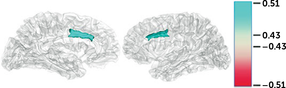
Additional correlations were performed between the logSCR scores for each of the four stimulus categories and the ROI volume measures (Figure 1). There were no significant correlations within groups except when examined across both dementia groups using a significance level of p and q*<0.01. For the logSCR results, there were significant correlations between both empathy and high emotion and the gray volume of the left >right dorsal ACC (0.517 and 0.512, respectively) (Figure 1).
FIGURE 1. Psychophysiological correlations across the behavioral variant frontotemporal dementia and Alzheimer’s disease groupsa
a Pearson correlations are shown. Blue regions represent significant positive correlations of skin conductance responses with the dorsal anterior cingulate gyrus.

Discussion

Among patients with bvFTD, this study showed decreased emotional empathy beyond the extent of hypoemotionality characteristic of bvFTD. The bvFTD patients had decreased empathy behavioral scale scores, regardless of performance on an emotional blunting scale, and the bvFTD patients had decreased SC changes to empathy stimuli that persisted after adjusting for responses to emotion stimuli. These findings indicate that patients with bvFTD are disproportionately impaired in empathy compared with general emotion stimuli. Moreover, across groups, the correlation of SCR to empathy and emotion stimuli with the volume of the dACC suggests a role for this area in emotional empathy.
BvFTD is an example of disease that targets the empathic behavioral centers of the brain (45). Patients with this disorder present with alterations in empathic behavior ranging from disengagement and lack of consideration for others to sociopathic acts with a loss of the emotional aspects of morality (5, 46, 47). From a neuroanatomical point of view, bvFTD is clearly centered in areas critical to the “empathic brain,” such as the ventromedial prefrontal cortex, ACC, anterior insula, amygdale, and right anterior temporal lobe (48). These areas are part of neural networks dedicated to empathic functioning and self-awareness, particularly the salience network centered in the ACC and frontoinsular regions (49).
A major consideration is whether loss of emotional empathy in bvFTD results for a generalized autonomic hyporesponsiveness to emotion or whether there is a specific impairment in the emotional contagion aspect of empathy. In addition to behavioral scales, psychophysiological measures are particularly useful for differentiating the effects of empathy from general emotional reactivity. In this study, the SCR among the bvFTD patients remained low to empathy stimuli, regardless of responses to emotion stimuli. An incidental finding in this psychophysiological study is that the Alzheimer’s patients had higher SCR consistent with reported high baseline anxiety among early-onset patients with Alzheimer’s disease and neurodegeneration of temporal lobe structures involved in emotion detection and inhibition (23, 50, 51).
Some investigators have reported an increased overall threshold for psychophysiological responsiveness among patients with bvFTD (52), implying decreased resting sympathetic tone, and a higher threshold for emotional reactivity in general (53). The bvFTD patients did have emotional blunting and an impaired reactivity to emotion stimuli. The results of this study, however, suggest the presence of decreased emotional empathy beyond the presence of generalized emotional blunting, as control for emotion on the behavioral scales and psychophysiology did not eliminate measures of decreased empathy, and the psychophysiological responses to empathy and to emotion were also dissociable.
On neuroimaging, the ACC is implicated in empathizing or vicariously processing another’s rewards (54, 55); it may be abnormal in psychopathy and autism (5660). In this study, the SCR to both empathic and emotion stimuli were strongly correlated with the neuroimaging volume in the dACC, suggesting that this area contributes to emotional empathy through emotional appraisal. Traditionally, investigators have divided the ACC into a cognitive region in the dACC involved in error detection and executive tasks and an emotion-related region in the rostral-ventral ACC involved in assessing and regulating emotion (61). However, recent literature has indicated that the dACC, particularly in its anterior section, evaluates whether stimuli are sufficiently motivationally significant to induce an emotional reaction (62). This general emotional appraisal role relates to emotional empathy when the ACC distinguishes whether the stimuli affect others rather than oneself (6366). Empathy involves a vicarious reward prediction that depends on the ability to predict when others are likely to receive rewards (56). Studies indicate that a subregion of the ACC in the gyrus (ACCg) processes and signals anticipated rewards for other people, as well as for oneself (6771). The ACCg is significantly positively associated with trait emotion contagion and trait empathy (56).
This preliminary study had several important limitations. First, the number of participants was relatively small. This particularly affected the strength of the neuroimaging findings; the only significant findings evident on across-groups correlations used a liberal significance level. The participant numbers, however, were sufficient to reveal significant and insightful differences between the groups on the behavioral scales and psychophysiology measures. Second, this study corroborates reports of a high degree of anxiety and heightened emotional contagion in early stages of Alzheimer’s disease (23, 50, 51). However, this does not alter the main finding of this study, that worse empathy among the bvFTD patients is not explained by emotional blunting. Third, there are limitations in the informant behavior ratings, as the SDS does not clearly distinguish cognitive and emotional aspects of empathy and the SEB has some overlap with emotional empathy. Fourth, the behavioral scales and neuroimaging analysis did not include HC, where there is normative data. Finally, the study was limited by focusing on empathic versus emotion stimuli without further analysis of valence (positivity/negativity of the emotion) or of other specific emotions, such as disgust sensitivity.
In conclusion, the present study highlights decreased responsiveness to empathy stimuli among patients with bvFTD, a disorder that starts and focuses in frontotemporal areas of the empathic brain. A comparison with patients with Alzheimer’s disease suggests that there is disproportionate impairment in reactivity to empathy stimuli among those with bvFTD not due to their general autonomic hyporesponsiveness. This report is a first, preliminary exploration, and future studies are planned in order to expand and further evaluate these observations in a greater number of patients with bvFTD.

References

1.
Mendez MF, McMurtray A, Licht E, et al: The Scale for Emotional Blunting in patients with frontotemporal dementia. Neurocase 2006; 12:242–246
2.
Joshi A, Barsuglia JP, Mather MJ, et al: Evaluation of emotional blunting in behavioral variant frontotemporal dementia compared to Alzheimer’s disease. Dement Geriatr Cogn Disord 2014; 38:79–88
3.
Neary D, Snowden JS, Gustafson L, et al: Frontotemporal lobar degeneration: a consensus on clinical diagnostic criteria. Neurology 1998; 51:1546–1554
4.
Piguet O, Hornberger M, Mioshi E, et al: Behavioural-variant frontotemporal dementia: diagnosis, clinical staging, and management. Lancet Neurol 2011; 10:162–172
5.
Rascovsky K, Hodges JR, Knopman D, et al: Sensitivity of revised diagnostic criteria for the behavioural variant of frontotemporal dementia. Brain 2011; 134:2456–2477
6.
Kersaitis C, Halliday GM, Kril JJ: Regional and cellular pathology in frontotemporal dementia: relationship to stage of disease in cases with and without Pick bodies. Acta Neuropathol 2004; 108:515–523
7.
Kril JJ, Macdonald V, Patel S, et al: Distribution of brain atrophy in behavioral variant frontotemporal dementia. J Neurol Sci 2005; 232:83–90
8.
Baez S, Manes F, Huepe D, et al: Primary empathy deficits in frontotemporal dementia. Front Aging Neurosci 2014; 6:262
9.
Shany-Ur T, Rankin KP: Personality and social cognition in neurodegenerative disease. Curr Opin Neurol 2011; 24:550–555
10.
Singer T, Lamm C: The social neuroscience of empathy. Ann N Y Acad Sci 2009; 1156:81–96
11.
Decety J, Jackson PL: The functional architecture of human empathy. Behav Cogn Neurosci Rev 2004; 3:71–100
12.
Shamay-Tsoory SG, Aharon-Peretz J, Perry D: Two systems for empathy: a double dissociation between emotional and cognitive empathy in inferior frontal gyrus versus ventromedial prefrontal lesions. Brain 2009; 132:617–627
13.
Cox CL, Uddin LQ, Di Martino A, et al: The balance between feeling and knowing: affective and cognitive empathy are reflected in the brain’s intrinsic functional dynamics. Soc Cogn Affect Neurosci 2012; 7:727–737
14.
Rankin KP, Kramer JH, Miller BL. Patterns of cognitive and emotional empathy in frontotemporal lobar degeneration. Cogn Behav Neurol 2005;18:28–36
15.
Hillis AE: Inability to empathize: brain lesions that disrupt sharing and understanding another’s emotions. Brain 2014; 137:981–997
16.
Shamay-Tsoory SG: The neural bases for empathy. Neuroscientist 2011; 17:18–24
17.
Leigh R, Oishi K, Hsu J, et al: Acute lesions that impair affective empathy. Brain 2013; 136:2539–2549
18.
Cerami C, Dodich A, Canessa N, et al: Neural correlates of empathic impairment in the behavioral variant of frontotemporal dementia. Alzheimers Dement 2014; 10:827–834
19.
Dermody N, Wong S, Ahmed R, et al: Uncovering the neural bases of cognitive and affective empathy deficits in Alzheimer’s disease and the behavioral-variant of frontotemporal dementia. J Alzheimers Dis 2016; 53:801–816
20.
Oliver LD, Mitchell DG, Dziobek I, et al: Parsing cognitive and emotional empathy deficits for negative and positive stimuli in frontotemporal dementia. Neuropsychologia 2015; 67:14–26
21.
Rankin KP, Gorno-Tempini ML, Allison SC, et al: Structural anatomy of empathy in neurodegenerative disease. Brain 2006; 129:2945–2956
22.
Eslinger PJ, Moore P, Anderson C, et al: Social cognition, executive functioning, and neuroimaging correlates of empathic deficits in frontotemporal dementia. J Neuropsychiatry Clin Neurosci 2011; 23:74–82
23.
Sturm VE, Yokoyama JS, Seeley WW, et al: Heightened emotional contagion in mild cognitive impairment and Alzheimer’s disease is associated with temporal lobe degeneration. Proc Natl Acad Sci USA 2013; 110:9944–9949
24.
Joshi A, Mendez MF, Kaiser N, et al: Skin conductance levels may reflect emotional blunting in behavioral variant frontotemporal dementia. J Neuropsychiatry Clin Neurosci 2014; 26:227–232
25.
Irish M, Hodges JR, Piguet O: Right anterior temporal lobe dysfunction underlies theory of mind impairments in semantic dementia. Brain 2014; 137:1241–1253
26.
Sturm VE, Sollberger M, Seeley WW, et al: Role of right pregenual anterior cingulate cortex in self-conscious emotional reactivity. Soc Cogn Affect Neurosci 2012; 8:468–474
27.
Bickart KC, Brickhouse M, Negreira A, et al: Atrophy in distinct corticolimbic networks in frontotemporal dementia relates to social impairments measured using the Social Impairment Rating Scale. J Neurol Neurosurg Psychiatry 2014; 85:438–448
28.
Bertoux M, Funkiewiez A, O’Callaghan C, et al: Sensitivity and specificity of ventromedial prefrontal cortex tests in behavioral variant frontotemporal dementia. Alzheimers Dement 2013; 9(Suppl):S84–S94
29.
Oishi K, Faria AV, Hsu J, et al: Critical role of the right uncinate fasciculus in emotional empathy. Ann Neurol 2015; 77:68–74
30.
Rushby JA, Fisher AC, McDonald S, et al: Autonomic and neural correlates of dysregulated arousal in severe traumatic brain injury. Int J Psychophysiol 2013; 89:460–465
31.
Rushby JA, McDonald S, Fisher AC, et al: Brain volume loss contributes to arousal and empathy dysregulation following severe traumatic brain injury. Neuroimage Clin 2016; 12:607–614
32.
de Sousa A, McDonald S, Rushby J: Changes in emotional empathy, affective responsivity, and behavior following severe traumatic brain injury. J Clin Exp Neuropsychol 2012; 34:606–623
33.
Kalbe E, Grabenhorst F, Brand M, et al: Elevated emotional reactivity in affective but not cognitive components of theory of mind: a psychophysiological study. J Neuropsychol 2007; 1:27–38
34.
Derouesné C, Lacomblez L, Fiori N, et al: L’apathie dans la démence frontemporale et la maladie d’Alzheimer : existe-t-il des profils distincts? Geriatr Psychol Neuropsychiatr Vieil 2012; 10:107–115
35.
Drago V, Foster PS, Chanei L, et al: Emotional indifference in Alzheimer’s disease. J Neuropsychiatry Clin Neurosci 2010; 22:236–242
36.
Fazekas F, Chawluk JB, Alavi A, et al: MR signal abnormalities at 1.5 T in Alzheimer’s dementia and normal aging. AJR Am J Roentgenol 1987; 149:351–356
37.
McKhann GM, Knopman DS, Chertkow H, et al: The diagnosis of dementia due to Alzheimer’s disease: recommendations from the National Institute on Aging-Alzheimer’s Association workgroups on diagnostic guidelines for Alzheimer’s disease. Alzheimers Dement 2011; 7:263–269
38.
Barsuglia JP, Kaiser NC, Wilkins SS, et al: A scale of socioemotional dysfunction in frontotemporal dementia. Arch Clin Neuropsychol 2014; 29:793–805
39.
Schneider RJ, Ackerman PL, Kanfer R: To “act wisely in human relations”: exploring the dimensions of social competence. Pers Individ Dif 1996; 21:469–481
40.
Abrams R, Taylor MA: A rating scale for emotional blunting. Am J Psychiatry 1978; 135:226–229
41.
Berenbaum SA, Abrams R, Rosenberg S, et al: The nature of emotional blunting: a factor-analytic study. Psychiatry Res 1987; 20:57–67
42.
Boone KB, Miller BL, Swartz R, et al: Relationship between positive and negative symptoms and neuropsychological scores in frontotemporal dementia and Alzheimer’s disease. J Int Neuropsychol Soc 2003; 9:698–709
43.
Benjamini Y, Yekutieli D: Quantitative trait loci analysis using the false discovery rate. Genetics 2005; 171:783–790
44.
Hochberg Y, Benjamini Y: More powerful procedures for multiple significance testing. Stat Med 1990; 9:811–818
45.
Mendez MF, McMurtray A, Chen AK, et al: Functional neuroimaging and presenting psychiatric features in frontotemporal dementia. J Neurol Neurosurg Psychiatry 2006; 77:4–7
46.
Mendez MF: The unique predisposition to criminal violations in frontotemporal dementia. J Am Acad Psychiatry Law 2010; 38:318–323
47.
Fong SS, Navarrete CD, Perfecto SE, et al: Behavioral and autonomic reactivity to moral dilemmas in frontotemporal dementia versus Alzheimer’s disease. Soc Neurosci 2017; 12:409–418
48.
Seeley WW, Zhou J, Kim EJ: Frontotemporal dementia: what can the behavioral variant teach us about human brain organization? Neuroscientist 2012; 18:373–385
49.
Seeley WW: Anterior insula degeneration in frontotemporal dementia. Brain Struct Funct 2010; 214:465–475
50.
Joshi A, Jimenez E, Mendez MF: Initial heart rate reactivity to socioemotional pictures in early-onset Alzheimer’s disease. J Alzheimers Dis 2017; 60:1325–1332
51.
Kaiser NC, Liang LJ, Melrose RJ, et al: Differences in anxiety among patients with early- versus late-onset Alzheimer’s disease. J Neuropsychiatry Clin Neurosci 2014; 26:73–80
52.
Hoefer M, Allison SC, Schauer GF, et al: Fear conditioning in frontotemporal lobar degeneration and Alzheimer’s disease. Brain 2008; 131:1646–1657
53.
Dreissen YE, Bakker MJ, Koelman JH, et al: Exaggerated startle reactions. Clin Neurophysiol 2012; 123:34–44
54.
Lamm C, Decety J, Singer T: Meta-analytic evidence for common and distinct neural networks associated with directly experienced pain and empathy for pain. Neuroimage 2011; 54:2492–2502
55.
Engen HG, Singer T: Empathy circuits. Curr Opin Neurobiol 2013; 23:275–282
56.
Lockwood PL, Apps MA, Roiser JP, et al: Encoding of vicarious reward prediction in anterior cingulate cortex and relationship with trait empathy. J Neurosci 2015; 35:13720–13727
57.
Lockwood PL, Sebastian CL, McCrory EJ, et al: Association of callous traits with reduced neural response to others’ pain in children with conduct problems. Curr Biol 2013; 23:901–905
58.
Simms ML, Kemper TL, Timbie CM, et al: The anterior cingulate cortex in autism: heterogeneity of qualitative and quantitative cytoarchitectonic features suggests possible subgroups. Acta Neuropathol 2009; 118:673–684
59.
Brazil IA, Mars RB, Bulten BH, et al: A neurophysiological dissociation between monitoring one’s own and others’ actions in psychopathy. Biol Psychiatry 2011; 69:693–699
60.
Anderson NE, Kiehl KA: The psychopath magnetized: insights from brain imaging. Trends Cogn Sci 2012; 16:52–60
61.
Bush G, Luu P, Posner MI: Cognitive and emotional influences in anterior cingulate cortex. Trends Cogn Sci 2000; 4:215–222
62.
Etkin A, Egner T, Kalisch R: Emotional processing in anterior cingulate and medial prefrontal cortex. Trends Cogn Sci 2011; 15:85–93
63.
Oliveira Silva P, Maia L, Coutinho J, et al: Empathy by default: correlates in the brain at rest. Psicothema 2018; 30:97–103
64.
Decety J, Svetlova M: Putting together phylogenetic and ontogenetic perspectives on empathy. Dev Cogn Neurosci 2012; 2:1–24
65.
Ochsner KN, Gross JJ: The cognitive control of emotion. Trends Cogn Sci 2005; 9:242–249
66.
Ruby P, Decety J: What you believe versus what you think they believe: a neuroimaging study of conceptual perspective-taking. Eur J Neurosci 2003; 17:2475–2480
67.
Apps MA, Ramnani N: The anterior cingulate gyrus signals the net value of others’ rewards. J Neurosci 2014; 34:6190–6200
68.
Rogers RD, Ramnani N, Mackay C, et al: Distinct portions of anterior cingulate cortex and medial prefrontal cortex are activated by reward processing in separable phases of decision-making cognition. Biol Psychiatry 2004; 55:594–602
69.
Sallet J, Quilodran R, Rothé M, et al: Expectations, gains, and losses in the anterior cingulate cortex. Cogn Affect Behav Neurosci 2007; 7:327–336
70.
Vogt BA, Nimchinsky EA, Vogt LJ, et al: Human cingulate cortex: surface features, flat maps, and cytoarchitecture. J Comp Neurol 1995; 359:490–506
71.
Apps MA, Lockwood PL, Balsters JH: The role of the midcingulate cortex in monitoring others’ decisions. Front Neurosci 2013; 7:251

Information & Authors

Information

Published In

Go to The Journal of Neuropsychiatry and Clinical Neurosciences
The Journal of Neuropsychiatry and Clinical Neurosciences
Pages: 378 - 385
PubMed: 31046591

History

Received: 3 September 2018
Revision received: 30 December 2018
Accepted: 11 January 2019
Published online: 3 May 2019
Published in print: Fall 2019

Keywords

  1. Empathy
  2. Frontotemporal Dementia
  3. Alzheimer’s Disease
  4. Skin Conductance

Authors

Affiliations

Mario F. Mendez, M.D., Ph.D. [email protected]
The Departments of Neurology, Psychiatry and Biobehavioral Sciences, and Medicine, Statistic Core, David Geffen School of Medicine, University of California at Los Angeles (Mendez, Jimenez); the VA Greater Los Angeles Healthcare System (Mendez, Carr, Jimenez); the Imaging Genetics Center, Mark and Mary Stevens Neuroimaging and Informatics Institute, University of Southern California, Los Angeles (Riedel, Thompson); and the Departments of Engineering, Neurology, Ophthalmology, Pediatrics, Psychiatry, and Radiology, University of Southern California, Los Angeles (Thompson).
Andrew R. Carr
The Departments of Neurology, Psychiatry and Biobehavioral Sciences, and Medicine, Statistic Core, David Geffen School of Medicine, University of California at Los Angeles (Mendez, Jimenez); the VA Greater Los Angeles Healthcare System (Mendez, Carr, Jimenez); the Imaging Genetics Center, Mark and Mary Stevens Neuroimaging and Informatics Institute, University of Southern California, Los Angeles (Riedel, Thompson); and the Departments of Engineering, Neurology, Ophthalmology, Pediatrics, Psychiatry, and Radiology, University of Southern California, Los Angeles (Thompson).
Elvira E. Jimenez, M.P.H.
The Departments of Neurology, Psychiatry and Biobehavioral Sciences, and Medicine, Statistic Core, David Geffen School of Medicine, University of California at Los Angeles (Mendez, Jimenez); the VA Greater Los Angeles Healthcare System (Mendez, Carr, Jimenez); the Imaging Genetics Center, Mark and Mary Stevens Neuroimaging and Informatics Institute, University of Southern California, Los Angeles (Riedel, Thompson); and the Departments of Engineering, Neurology, Ophthalmology, Pediatrics, Psychiatry, and Radiology, University of Southern California, Los Angeles (Thompson).
Brandalyn C. Riedel, M.S.G.
The Departments of Neurology, Psychiatry and Biobehavioral Sciences, and Medicine, Statistic Core, David Geffen School of Medicine, University of California at Los Angeles (Mendez, Jimenez); the VA Greater Los Angeles Healthcare System (Mendez, Carr, Jimenez); the Imaging Genetics Center, Mark and Mary Stevens Neuroimaging and Informatics Institute, University of Southern California, Los Angeles (Riedel, Thompson); and the Departments of Engineering, Neurology, Ophthalmology, Pediatrics, Psychiatry, and Radiology, University of Southern California, Los Angeles (Thompson).
Paul M. Thompson, Ph.D.
The Departments of Neurology, Psychiatry and Biobehavioral Sciences, and Medicine, Statistic Core, David Geffen School of Medicine, University of California at Los Angeles (Mendez, Jimenez); the VA Greater Los Angeles Healthcare System (Mendez, Carr, Jimenez); the Imaging Genetics Center, Mark and Mary Stevens Neuroimaging and Informatics Institute, University of Southern California, Los Angeles (Riedel, Thompson); and the Departments of Engineering, Neurology, Ophthalmology, Pediatrics, Psychiatry, and Radiology, University of Southern California, Los Angeles (Thompson).

Notes

Send correspondence to Dr. Mendez ([email protected]).

Competing Interests

The authors report no financial relationships with commercial interests.

Funding Information

National Institutes of HealthNational Institutes of Health: R01AG034499; RF1 AG041915; RF1AG051710
Supported by the National Institute on Aging (grant R01AG034499), and supported in part by NIH (grants U54 EB020403, RF1 AG041915, and RF1AG051710 to Drs. Riedel and Thompson).

Metrics & Citations

Metrics

Citations

Export Citations

If you have the appropriate software installed, you can download article citation data to the citation manager of your choice. Simply select your manager software from the list below and click Download.

For more information or tips please see 'Downloading to a citation manager' in the Help menu.

Format
Citation style
Style
Copy to clipboard

There are no citations for this item

View Options

View options

PDF/ePub

View PDF/ePub

Get Access

Login options

Already a subscriber? Access your subscription through your login credentials or your institution for full access to this article.

Personal login Institutional Login Open Athens login
Purchase Options

Purchase this article to access the full text.

PPV Articles - Journal of Neuropsychiatry and Clinical Neurosciences

PPV Articles - Journal of Neuropsychiatry and Clinical Neurosciences

Not a subscriber?

Subscribe Now / Learn More

PsychiatryOnline subscription options offer access to the DSM-5-TR® library, books, journals, CME, and patient resources. This all-in-one virtual library provides psychiatrists and mental health professionals with key resources for diagnosis, treatment, research, and professional development.

Need more help? PsychiatryOnline Customer Service may be reached by emailing [email protected] or by calling 800-368-5777 (in the U.S.) or 703-907-7322 (outside the U.S.).

Media

Figures

Other

Tables

Share

Share

Share article link

Share