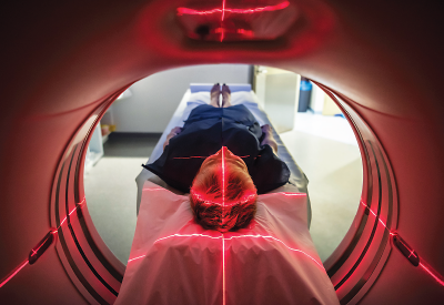
Brain imaging is revealing differences between individuals at high risk for psychosis that will help clinicians distinguish those who are likely to progress to acute psychosis, those whose symptoms will remain subsyndromal, and those who may recover.
Among patients with existing psychosis, these advances may also help distinguish those who will respond to antipsychotic treatment from those who will not, said Phillip McGuire, M.D., a professor of psychiatry and cognitive neuroscience at Kings College, London. He presented a plenary lecture at the International Congress of Schizophrenia Research in San Diego in March.
Specifically, McGuire said imaging advances point to important differences among patients in a triad of brain regions—the hippocampus, mid-brain region, and striatal cingulate region—and neurochemical differences in dopamine and glutamate activation in those regions. The hope is that imaging results, along with other relevant predictive data (demographics, family history, etc.) could be analyzed in a handheld device allowing a clinician to make determinations about risk stratification at the patient’s presentation.
The findings can also help facilitate the development of alternative treatments for those who do not respond to existing treatments, he said.
McGuire noted that advances are being made in predicting outcome using other strategies—including peripheral biomarkers and clinical/demographic predictor tools—but he focused his remarks on neuroimaging.
“Neuroimaging has been quite successful at a group level in differentiating individuals who are clinically identical but are likely to have different outcomes,” he said. “These are not trivial differences, but clinically significant differences, in outcome. One of the major research efforts now is to translate these findings into tools that can be used in clinical practice. That is the goal over the next five or 10 years. Once these tools have been developed, we would like to be able to develop alternative treatments for different stratified groups.”
The neuroimaging findings are important because they can help resolve a critical problem in the identification and treatment of at-risk individuals: How can clinicians better predict who, among those deemed at clinical high risk, will actually convert to acute psychosis?
The last decade and a half has seen an enormous focus on early identification of patients at clinical high risk, resulting in criteria that were included in Section 3 of DSM-5. Those criteria encompass such factors as family history, social withdrawal, deficits in function, and attenuated psychotic symptoms. The criteria can reliably predict progression to psychosis approximately 30 percent of the time, but a sizable portion of at-risk individuals will have persistent subsyndromal symptoms without developing psychosis, or may even spontaneously recover.
But which patients will fall into which category, and how can clinicians avoid the problem of false-positives? Moreover, with the exception of clozapine, existing antipsychotic treatments have relied for decades on dopamine blockade even though a significant number of patients with psychosis do not respond to D2 antipsychotic antagonists.
“This is not an academic issue but is grounded in real clinical practice,” McGuire said. “A key problem in the management of clinical high-risk psychosis is that it is very difficult to predict clinical outcome on the basis of clinical presentation alone. If we had predictive biomarkers, we could intervene more selectively—perhaps more assertively in patients we were confident would have a psychotic disorder and with a lighter touch in patients who may not convert to psychosis or who might even spontaneously recover.
“In patients with established psychosis, we know that antipsychotics will work in two-thirds of patients, but for up to a third the response will be disappointing.”
McGuire provided an overview of neuronal imaging as it pertains to risk stratification over the last 15 years, beginning with MRI findings showing that the subset of CHR patients who develop psychosis have a smaller hippocampus and higher levels of resting-state activity in the mid-brain, hippocampus, and basal ganglia. Imaging has also shown elevated glutamate in the hippocampus among those who develop psychosis.
“The important concept here is that at the clinical level, these individuals are indistinguishable, but at the imaging level, there are important differences,” he said.
Neurochemical abnormalities regarding treatment response that have been found using imaging have been especially revealing, particularly with regard to dopamine activity, long regarded as a common factor in schizophrenia. McGuire said patients who respond to treatment show classically elevated levels of striatal dopamine function; however, in those who don’t respond, glutamate is elevated while dopamine function is comparable to that of controls.
“Conceptually this is important because it is the first evidence that the psychotic population is heterogenous in terms of neurochemistry,” McGuire said, and it points to the need for alternative forms of antipsychotic treatment.
Ultimately, McGuire said the goal is to translate these findings into usable tools in the clinical setting. “A key consideration is that all of these findings are at the group level revealing mean differences between one group and another,” he said. “In clinical practice, you have to make a decision about the patient sitting in front of you. The challenge is to develop a test powerful enough to work with data from a single individual.
“The real deliverable goal is not a scientific paper describing mean differences but a tool that clinicians can use in practice—such as an iPad device that would allow a clinician to enter data from an individual patient and get a readout regarding the patient’s likely outcome.” ■


