Noninvasive Brain Stimulation Boosts Phobia Therapies
Noninvasive brain stimulation may enhance the effectiveness of exposure therapy for people with specific phobias, suggests a study in the Journal of Anxiety Disorders.
Researchers at the University of Texas and colleagues recruited 49 people with fears of contamination, snakes, or spiders. The participants were randomized to receive 20 minutes of transcranial direct current stimulation (tDCS) to the prefrontal cortex or a sham stimulation prior to a 30-minute session of exposure therapy (for example, confronting the phobia stimulus in a safe setting). The participants came back one month later for a follow-up assessment, which included a 30-second exposure to patients’ phobia.
Participants in both groups reported reduced emotional distress about their phobias (assessed on a scale of 1 to 100) at the follow-up visit, but the differences were more pronounced in participants who received active tDCS; participants who received tDCS scored an average of 11 points lower on the distress scale than those who received sham stimulation. Participants who received tDCS also demonstrated fewer avoidance behaviors (for example, how close they came to the fearful stimuli) than those in the sham group.
“[A]ctive tDCS was found to especially benefit individuals with more severe phobic symptoms and elevated fearful reactivity to anxiety, as well as individuals who exhibited persistent emotional distress,” the investigators wrote. “This investigation provides novel preliminary support for the use of tDCS to enhance in vivo exposure.”
Some Patients With Depression May Benefit From Add-On Minocycline
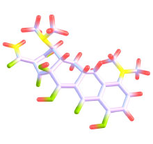
People experiencing depression and elevated inflammation may experience symptom improvements if given the antibiotic minocycline in combination with antidepressants, according to a study published in Neuropsychopharmacology. Minocycline has broad anti-inflammatory properties in addition to its antibiotic properties.
Researchers at Kings College London and colleagues enrolled 39 adults aged 25 to 60 with depression who were not responding to their current medication. For inclusion in the trial, study participants had to have at least what the authors considered to be low-grade inflammation: C-reactive protein (CRP) concentrations of at least 1 mg/L.
The participants were randomly assigned to receive add-on minocycline (200 mg/day) or placebo for four weeks. They were assessed using the Hamilton Depression Rating Scale (HAM-D) at the start of the study and at follow-up four weeks later.
There were no differences in changes in HAM-D scores between the minocycline and placebo groups after four weeks. However, when the participants were further divided according to inflammation levels (low: CRP 1 to 3 mg/L; high: CRP 3+ mg/L), differences between the groups emerged.
HAM-D scores in the participants with high inflammation fell by 12 points after four weeks in those taking minocycline, compared with 3.5 points among patients with high inflammation taking placebo. In the low-inflammation group, HAM-D scores fell by 2.4 points and 2.1 points for the minocycline and placebo groups, respectively. Five of the six patients with high inflammation receiving add-on minocycline achieved at least a partial response (25% or greater reduction in depression symptoms).
“Although replications in larger samples are needed, we believe our study has a potentially important clinical impact, as we moved a step toward the identification of personalized treatments for depression,” the researchers wrote.
Variable Gait May Signal Alzheimer’s Disease
Neurodegenerative disorders such as Alzheimer’s impair not only cognition and memory, but also movement. Researchers have now identified a set of abnormalities in gait that may be able to distinguish patients with Alzheimer’s from those with other neurodegenerative conditions. The findings were published in Alzheimer’s and Dementia.
Investigators at the Lawson Health Research Institute in London, Ontario, and colleagues conducted comprehensive gait assessments of 500 adults aged 60 years and older. The participants included adults with normal cognition, mild cognitive impairment, Alzheimer’s, Lewy body dementia, Parkinson’s disease dementia, or frontotemporal dementia. The investigators used videos and pressure sensors to assess four central components of gait: rhythm, pace, posture, and variability (the consistency across multiple steps).
The analysis showed that gait rhythm, posture, and variability were all significantly worse in individuals with mild cognitive impairment or dementia relative to people with no impairment. However, only deficits in gait variability correlated with individuals’ cognitive scores, as measured using the Montreal Cognitive Assessment, with lower scores indicating with more gait variability. The association was strongest among people with Alzheimer’s.
The researchers found that a prediction tool using gait variability data could accurately differentiate those with Alzheimer’s from those with other neurodegenerative disorders with about 77% accuracy.
“Increased gait variability may reflect the progression of cognitive impairment in neurodegenerative diseases and potentially with specificity for Alzheimer’s disease dementia, which is the archetypal cortical cognitive disorder,” the researchers wrote.
Schizophrenia Risk Variants in Placental Genes Tied to Slower Cognitive Development
Genetic variants of placental genes associated with schizophrenia risk are also associated with the risk of early developmental problems, reports a study in PNAS. Compared with infants without such placenta-associated risk factors, those with high genetic risk were more likely to have reduced brain volume at birth and slower cognitive development.
“Understanding the trajectories leading to neurodevelopmental disorders is a big challenge but a necessary one to design strategies aimed at prevention,” wrote the researchers from the Lieber Institute for Brain Development in Baltimore and colleagues. Previous work by this group showed that early life complications (such as preeclampsia) can alter gene expression in the placenta and increase the risk of schizophrenia.
For the current study, the researchers collected blood samples from 242 infants who experienced complications in utero and calculated the placental genetic risk (PlacGRS) for a range of disorders, including attention-deficit/hyperactivity disorder, autism, depression, and schizophrenia. All the infants were given cognitive assessments at one and two years of age.
The researchers found a strong association between higher PlacGRS score for schizophrenia and lower brain volume at birth. Higher PlacGRS scores were also associated with worse cognitive performance when the children were one and two years. The association was weaker at age two, which may reflect the greater role of parenting and the environment as children age.
The researchers did not find any associations between PlacGRS scores and cognitive problems for other disorders.
Equine Therapy Associated With Changes in Brain Structure and Connectivity
There is evidence that some people with posttraumatic stress disorder (PTSD) benefit from equine-assisted therapy. A study in Human Brain Mapping now provides evidence that this therapeutic approach can lead to functional and structural changes in the brain.
Researchers at Columbia University and colleagues conducted MRI scans on 19 veterans with PTSD who were part of a larger clinical trial exploring the benefits of equine-assisted therapy. Equine-assisted therapy involved eight weekly 90-minute group sessions. The MRI scans were taken at the start of the study and after eight weeks. PTSD and depression symptoms of the participants were assessed before and after the eight-week period, as well as at a three-month follow-up.
The MRI scans showed that participants had greater connectivity in the caudate nucleus after eight weeks of equine therapy. (The caudate nucleus processes memories and is also part of the brain’s reward network.) The changes in connectivity in this region correlated with the degree of symptom improvement, which was measured with the Clinician Administered PTSD Scale. Further, the researchers found that participants with higher caudate nucleus connectivity at the start of the study reported greater symptom improvements.
The MRI scans also showed that eight weeks of equine-assisted therapy was associated with reduced gray matter in both the caudate nucleus and thalamus, a brain region regulating fear and anxiety.
“The findings suggest that [equine-assisted therapy] may target reward circuitry responsiveness and produce a caudate pruning effect from pre- to post-treatment,” the authors concluded. ■


