Full access
Letters
Published Online: 1 October 2011

Reversible Carbamazapine Encephalopathy With Frontal Intermittent Rhythmic Delta Activity

Publication: The Journal of Neuropsychiatry and Clinical Neurosciences
To the Editor: In this report, we describe the clinical and EEG features of a patient with focal epilepsy who developed toxic encephalopathy at the beginning of low-dose treatment with carbamazepine. It is noteworthy that the pattern of EEG showed typical bursts of frontal intermittent rhythmic delta activities (FIRDA), which to our knowledge, have never been described during adult CBZ intoxication.

Case Report

A 70-year-old housewife with a past medical history of hypertension, was admitted to our hospital after three episodes of simple partial motor seizures characterized by clonic movements involving the head and the right arm (mean duration: 90 seconds). On evaluation, the patient was alert, and no focal neurological signs were observed. Blood sodium levels were in the normal range (139 mEq/lter; VN 135–145 mEq/lter). Potassium and calcium levels were normal as well. EEG nonictal recording was normal. Gadolinium-enhancement brain MRI displayed small lacunae that mostly involved the basal ganglia, suggesting a vascular nature for the seizures. Antiepileptic treatment with 200 mg carbamazepine (CBZ) was started, with rapid and complete seizure control. Three days later, while on a CBZ dose of 300 mg/day, she developed confusion, hypersomnia, memory impairment, and psychomotor slowness. EEG recordings demonstrated a mild, generalized slowing, mostly in theta range, associated with bursts of frontal intermittent rhythmic delta activity (FIRDA; Figure 1). no epileptiform activity was present. Extensive blood tests, including neoplastic markers, ammonia concentration, and CSF analysis, did not reveal abnormalities. The CBZ level (22.6 μg/ml) was above the high-normal level (normal range: 6–12 μg/ml). CBZ was discontinued and replaced by valproic acid (VPA), starting at 200 mg/day, then slowly increased up to 1,000 mg/day, administered twice daily. The patient dramatically improved within a few days and, on EEG, diffuse slowing and FIRDA disappeared, with full recovery of physiological activities. At 8-month follow-up, FIRDA was not observed on a routine EEG recording. The patient was seizure-free, and VPA, which had been maintained at the dosage of 1,000 mg/day (serum level: 54 μg/ml; therapeutic range: 50–100 μg/ml) was well tolerated without any side effects.
FIGURE 1. EEG Recordings Showing a Mild, Generalized Slowing, Mostly in the Theta Range, Associated With Bursts of Frontal Intermittent Rhythmic Delta Activity (FIRDA)

Discussion

Although usually well tolerated, carbamazepine is known to induce many neurological adverse effects, including tremor, somnolence, ataxia, nystagmus, and worsening of preexisting epileptic syndromes.1 These reactions may occur with toxic drug levels and even within the normal therapeutic range. The underlying pathophysiology of these phenomena is still unclear, and an idiosyncratic reaction should also be considered. In the majority of cases, the encephalopathy is acute and reversible, and CBZ withdrawal leads to a normalization of the clinical and EEG picture. In our patient, there was a clear temporal relationship between drug administration, drug serum levels (above normal range), and the development of the encephalopathy. The causative effect of CBZ is confirmed by rapid recovery, after drug discontinuation, of both clinical and EEG abnormalities. CBZ occasionally induces EEG changes even in seizure-free patients. The predominant, associated EEG findings are a diffuse slowing of background activity, the development of focal slow-wave activity, and intensification of secondary bilateral synchrony. Burst suppression pattern, pseudo-periodic sharp-wave activity, and alpha-coma pattern have also been observed during CBZ intoxication.
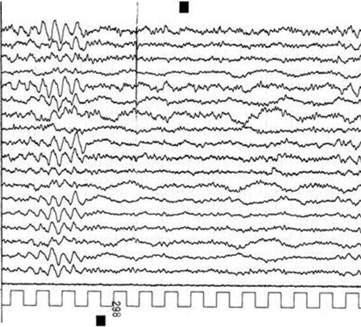
2 In our case, EEG findings consisted predominantly of FIRDA occurring on a diffuse, slowing background. To the best of our knowledge, a similar pattern has never been described during adult CBZ encephalopathy. “There is a single report of 16-year-old girl with a large, toxic overdose of carbamazepine, in which EEG was dominated by occipital delta activity (OIRDA), because of which authors hypothesized brainstem disorders.3 FIRDA was originally described by Cobb4 in 1945. It is a rhythmic slow-wave discharge localized in the frontal regions, with a frequency of 1.5 to 4.0 c/sec. Although the pathophysiological significance of FIRDA is unknown, it may reflect many pathological conditions, such as deep midline lesions and posterior fossa tumors.5 However, it has also been reported in association with subcortical lesions, hydrocephalus, cerebral edema, increased intracranial pressure, and acute basilar migraine.6
FIRDA is often observed in metabolic encephalopathy, especially in patients with diffuse or focal brain lesions.7 For example, FIRDA often occur in dialytic encephalopathy in subjects with chronic renal failure. Moreover, the association between FIRDA and metabolic dysfunction is still unclear. More recently, several reports have correlated FIRDA with long-term medication, especially antipsychotic agents.8 In such cases, EEG abnormalities occur in a dose-dependent manner and are predominantly observed in schizophrenic patients receiving high daily dosages of antipsychotic drugs. On the other hand, FIRDA are quite common in elderly patients with partial epilepsy and with cerebrovascular disorders. In a study by Fariello, 44% of patients with FIRDA had focal structural lesions; 34%, diffuse structural; and 22% had nonstructural disease.9 Brain MRI revealed small lacunae, mostly involving the basal ganglia, whereas our patient's EEG recording was normal before CBZ treatment; thus, a clear relationship between a toxic level of CBZ and FIRDA is seen in the case of our patient. Our evidence suggests that a toxic CBZ dose can trigger EEG abnormalities in a patient with structural brain lesions, even if mild. We conclude that patients with chronic, mostly ischemic brain lesions may be predisposed to develop FIRDA during acute CBZ encephalopathy, as well as during acute metabolic derangement.

References

1.
Horvath J, Coeytaux A, Jallon P, et al.: Carbamazepine encephalopathy masquerading as Creutzfeldt-Jakob disease. Neurology 2005; 65:650–651
2.
Bertram M, Fabian CW, Schwarz S, et al.: Massive carbamazepine overdose: clinical and neurophysiological findings. J Neurol 1998; 245:745–747
3.
Howard RS, Trend PS, Townsend HR: EEG appearances in acute carbamazepine toxicity. Hum Exp Toxicol 1990; 9:313–315
4.
Cobb WA: Rhythmic slow discharges in the electroencephalogram. J Neurol Neurosurg Psychiatry 1945; 8:65–78
5.
Daly D, Whelan JL, Bickford RG, et al.: The electroencephalogram in cases of tumors of the posterior fossa and third ventricle. Electroencephalogr Clin Neurophysiol 1953; Suppl 5:203–216
6.
Pietrini V, Terzano MG, D'Andrea G, et al.: Acute confusional migraine: clinical and electroencephalographic aspects. Cephalalgia 1987; 7:29–37
7.
Watemberg N, Alehan F, Dabby R, et al.: Clinical and radiologic correlates of frontal intermittent rhythmic delta activity. J Clin Neurophysiol 2002; 19:535–539
8.
Koshino Y, Murata I, Murata T, et al.: Frontal intermittent delta activity in schizophrenic patients receiving antipsychotic drugs. Clin Electroencephalogr 1993; 24:13–18
9.
Fariello RG, Orreson W, Blanco G, et al.: Neuroradiological correlates of frontally predominant intermittent rhythmic delta activity. Electroenceph Clin Neurophysiol 1982; 54:194–202

Information & Authors

Information

Published In

Go to The Journal of Neuropsychiatry and Clinical Neurosciences
The Journal of Neuropsychiatry and Clinical Neurosciences
Pages: E21 - E22
PubMed: 22231332

History

Published online: 1 October 2011
Published in print: Fall 2011

Authors

Affiliations

Antonio Milia, M.D.
UO Neurologia Riabilitiva Ospedale Santissima Trinita Cagliari, Italy
Giuseppina Pilia, M.D.
UO Neurologia Riabilitiva Ospedale Santissima Trinita Cagliari, Italy
Maria Giuseppina Mascia, M.D.
UO Neurologia Riabilitiva Ospedale Santissima Trinita Cagliari, Italy
Stefano Firinu, M.D.
UO Neurologia Riabilitiva Ospedale Santissima Trinita Cagliari, Italy

Notes

Correspondence: [email protected]

Metrics & Citations

Metrics

Citations

Export Citations

If you have the appropriate software installed, you can download article citation data to the citation manager of your choice. Simply select your manager software from the list below and click Download.

For more information or tips please see 'Downloading to a citation manager' in the Help menu.

Format
Citation style
Style
Copy to clipboard

There are no citations for this item

View Options

View options

PDF/ePub

View PDF/ePub

Get Access

Login options

Already a subscriber? Access your subscription through your login credentials or your institution for full access to this article.

Personal login Institutional Login Open Athens login
Purchase Options

Purchase this article to access the full text.

PPV Articles - Journal of Neuropsychiatry and Clinical Neurosciences

PPV Articles - Journal of Neuropsychiatry and Clinical Neurosciences

Not a subscriber?

Subscribe Now / Learn More

PsychiatryOnline subscription options offer access to the DSM-5-TR® library, books, journals, CME, and patient resources. This all-in-one virtual library provides psychiatrists and mental health professionals with key resources for diagnosis, treatment, research, and professional development.

Need more help? PsychiatryOnline Customer Service may be reached by emailing [email protected] or by calling 800-368-5777 (in the U.S.) or 703-907-7322 (outside the U.S.).

Media

Figures

Other

Tables

Share

Share

Share article link

Share