New high-resolution, three-dimensional maps of the brains of children with attention-deficit/hyperactivity disorder (ADHD) indicate significant and specific anatomical differences within areas of the brain thought to control attentional and inhibitory control systems, compared with brain scans of children without ADHD.
The images are thought to be the most advanced to date to reveal the anatomical basis of the disorder. Elizabeth Sowell, M.D., an assistant professor of neurology at the David Geffen School of Medicine at the University of California at Los Angeles, and her colleagues used high-resolution magnetic resonance imaging (MRI) and sophisticated computer analysis to pinpoint more accurately the specific areas of the brain contributing to the symptoms of ADHD.
“Our morphometric procedures allow more precise localization of group differences than do the methods used in previous studies,” Sowell said in a press release. “Our results therefore suggest that the disturbances in prefrontal cortices are localized to more inferior aspects of prefrontal regions than was previously appreciated. Our findings also indicate that prefrontal abnormalities are represented bilaterally, by contrast to the predominantly right-sided findings that were emphasized in other reports.”
Sowell teamed with Bradley Peterson, M.D., the Suzanne Crosby Murphy Associate Professor of Psychiatry at Columbia University and the New York State Psychiatric Institute, to image 27 children (11 girls and 16 boys) with ADHD and compare them with 46 children without ADHD who were matched for age and sex. The results of the study, funded by several NIH grants as well as the Suzanne Crosby Murphy Endowment at Columbia, appear in the November 22, 2003, issue of Lancet.
Localizing Differences
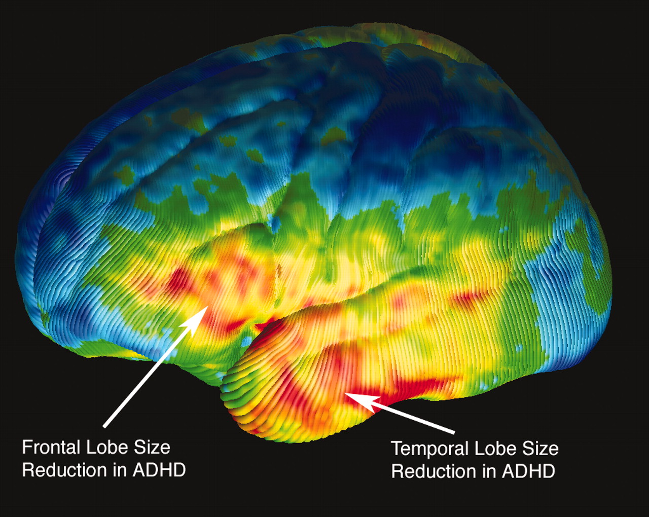
A three-dimensional, high-resolution MRI image of the brain of a patient with ADHD shows reductions (in yellow and red) in the size of specific areas within the frontal and temporal lobes. (UCLA Laboratory of Neuro Imaging)Sowell and Peterson observed significant differences in brain structure in the frontal cortices of both sides of the brains of the children with ADHD, with reduced regional brain size mainly confined to small areas of the dorsal prefrontal cortices (see images). Children with ADHD also were observed to have reduced brain size in anterior temporal areas, again on both sides of their brains.
In addition, substantial increases were noted in the volume of gray matter in large areas of the posterior temporal and inferior parietal cortices of children with ADHD, compared with children in the control group. These regions have previously been identified as areas of the brain controlling attention and impulse control.
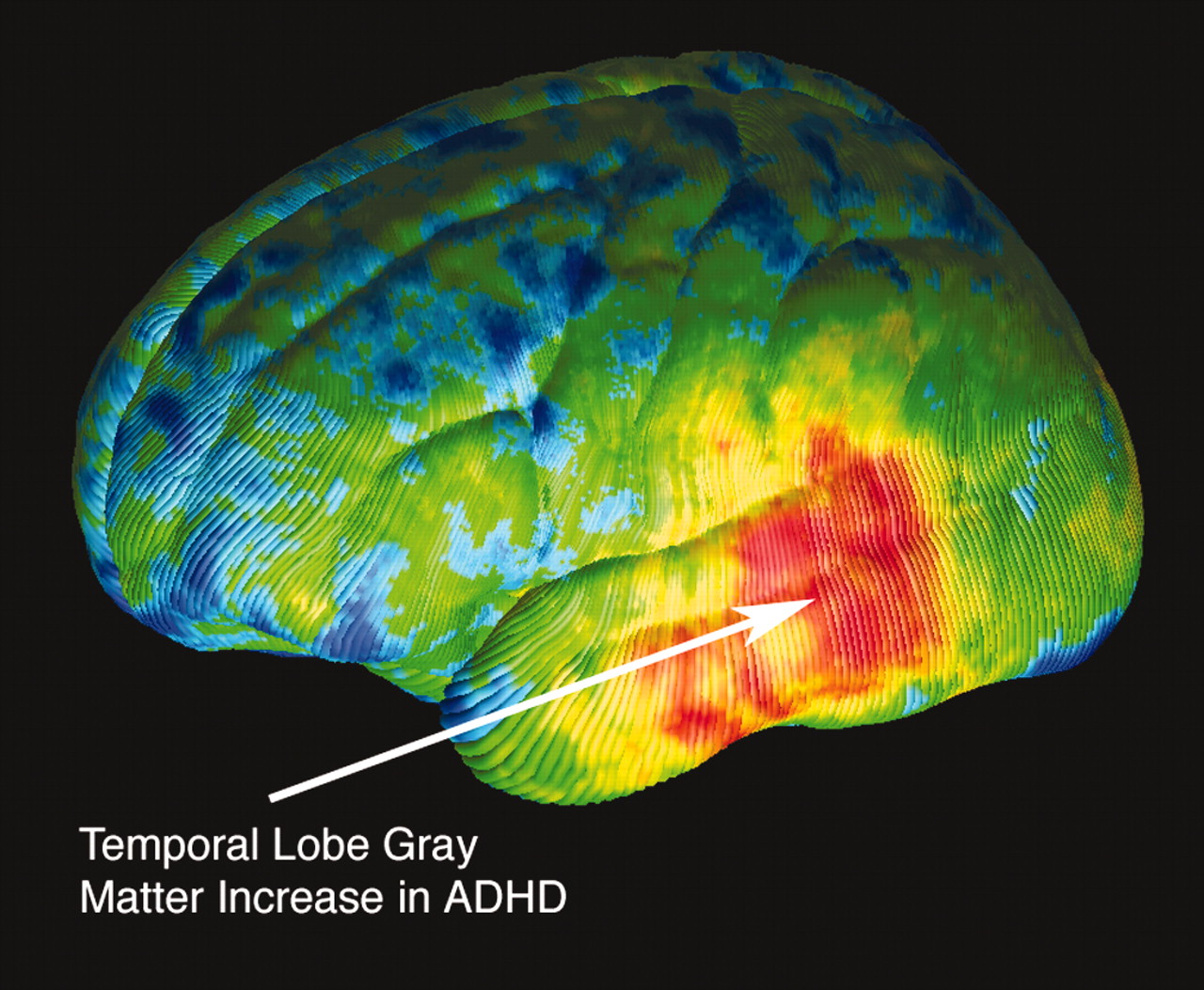
A three-dimensional, high-resolution MRI image of the brain of a patient with ADHD shows regional increases in the density of gray matter. Areas in yellow and red average between 10 percent and 24 percent more gray matter than those of the average control subject. (UCLA Laboratory of Neuro Imaging)“Disordered impulse control is often the most clinically debilitating symptom in children with ADHD,” noted Peterson in the press release. “These findings may help us to understand the sites of action of the medications used to treat ADHD, particularly stimulant medications. In conjunction with other imaging techniques, these findings may also help us develop new therapeutic agents, given our knowledge of the cellular and neurochemical makeup of the brain regions where we detected the greatest abnormalities.”
Building Complex Images
All of the children were imaged using the same MRI scanner at Yale University. The basic images were then processed at the UCLA Laboratory of Neuro Imaging using complex computer systems to build three-dimensional surface maps of each subject’s brain. The researchers then painstakingly identified on each brain image a series of 80 standardized anatomical landmarks, which were used to create a composite average for both the ADHD group and the control group. In this way, each child’s brain could be matched spatially, preserving differences in brain size and shape.
The resulting three-dimensional map of each child’s brain was indeed high resolution, representing more than 65,000 individual points on the surface of the cortex. The points on each subject’s brain were then compared with the average composite image created for both the ADHD group and the control group.
The researchers assessed differences in boys and girls, individually as well as combined, since recent studies have suggested that abnormalities in brain activity and structure may differ between boys and girls with ADHD. However, no significant differences were found. The researchers cautioned, however, that because their sample size was fairly small, differences may not have been appreciable.
The majority of the children with ADHD imaged in this study were taking stimulant medications at the time of imaging or had taken stimulant medications in the past. Sowell notes that it is not possible to determine with any significant certainty whether the anatomical differences they observed are due, in part or whole, to medication. However, other research has noted no anatomical changes associated with intermediate-term use of stimulant medications by children and adolescents with ADHD.
The reductions in size of prefrontal regions observed by Sowell and her colleagues are consistent with other reports of reduced frontal lobe volumes in children with ADHD. The more advanced imaging methods and analysis used in the current study, however, suggest that those reductions are localized to more inferior aspects of the prefrontal regions than was previously realized. Taken together, the evidence base continues to build, supporting smaller and hypo-functional lateral prefrontal cortices in children with ADHD.
Sowell and her coauthors also noted that “while we measured gray-matter density at the cortical surface, arguably a reduction of white matter in the same region could result in an apparent abundance of gray matter.” The authors emphasized that, in fact, total white matter volume was reduced in the children with ADHD, but only at a level of significance suggesting a trend.
Lastly, the team attempted to correlate the severity of symptoms in the children with ADHD with the anatomical abnormalities they discovered. They found that children with lesser volumes of gray matter generally were more inattentive, whereas children with significantly larger frontal lobes had higher levels of hyperactivity.
“ADHD,” Sowell and Peterson concluded in the study, “is almost certainly a disorder of heterogeneous etiologies that have correspondingly heterogeneous neuro-anatomical underpinnings.”
They noted that their sample size was too small to permit a complete ADHD subgroup analysis; however, they called for further studies to confirm differing anatomical and functional disturbances in different areas of the brain’s action-attentional network.
An abstract of “Cortical Abnormalities in Children and Adolescents With Attention-Deficit Hyperactivity Disorder” is posted online at www.thelancet.com/journal/vol362/iss9397/abs/llan.362.9397.original_research.27787.1. ▪
If you have the appropriate software installed, you can download article citation data to the citation manager of your choice. Simply select your manager software from the list below and click Download.
For more information or tips please see 'Downloading to a citation manager' in the Help menu.
View Options
View options
Get Access
Login options
Already a subscriber? Access your subscription through your login credentials or your institution for full access to this article.
PsychiatryOnline subscription options offer access to the DSM-5-TR® library, books, journals, CME, and patient resources. This all-in-one virtual library provides psychiatrists and mental health professionals with key resources for diagnosis, treatment, research, and professional development.
Need more help? PsychiatryOnline Customer Service may be reached by emailing [email protected] or by calling 800-368-5777 (in the U.S.) or 703-907-7322 (outside the U.S.).
If the address matches an existing account you will receive an email with instructions to retrieve your username
Create a new account
Change Password
Password Changed Successfully
Your password has been changed
Login
Reset password
Can't sign in? Forgot your password?
Enter your email address below and we will send you the reset instructions
If the address matches an existing account you will receive an email with instructions to reset your password.
Change Password
Congrats!
Your Phone has been verified
×
As described within the American Psychiatric Association (APA)'s Privacy Policy and Terms of Use, this website utilizes cookies, including for the purpose of offering an optimal online experience and services tailored to your preferences. Please read the entire Privacy Policy and Terms of Use. By closing this message, browsing this website, continuing the navigation, or otherwise continuing to use the APA's websites, you confirm that you understand and accept the terms of the Privacy Policy and Terms of Use, including the utilization of cookies.