Full access
Images in Neuroscience
Published Online: 1 July 2007

Human White Matter Atlas
Publication: American Journal of Psychiatry
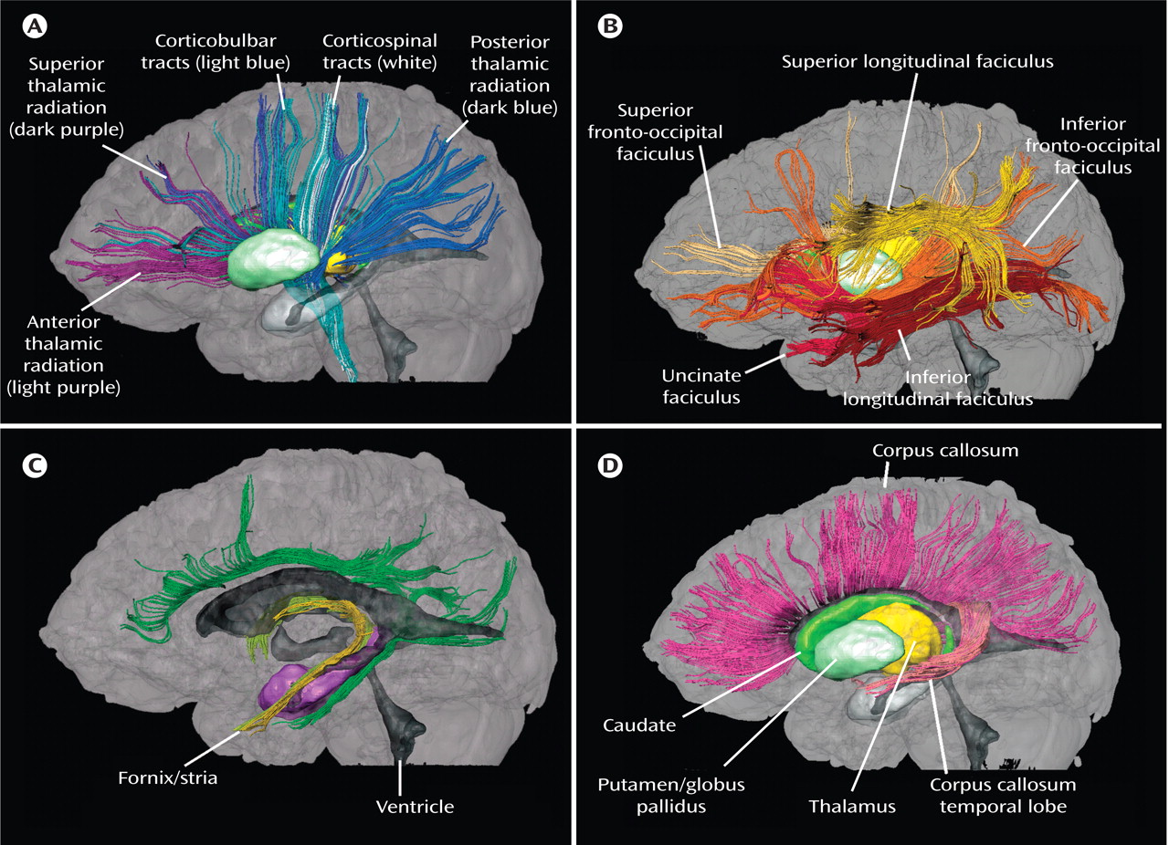
The axonal membrane and the myelin sheets around axons restrict water diffusion perpendicular to the fiber orientation in white matter but not along the axon’s main axis, which leads to anisotropic diffusion of water. Over the last decade, the quantitative measurement of this anisotropy by using what has been called diffusion tensor imaging (DTI) has become well established in research applications, revealing the macroscopic three-dimensional architecture of white matter and the constituent white matter tracts. The depiction of white matter architecture in orientation-based color coding is a visualization approach in which the image brightness depicts diffusion anisotropy and a red/green/blue color scheme indicates tract orientation. Using a multiple region-of-interest (ROI) approach that exploits existing anatomic knowledge of tract trajectories, white matter bundles of interest can be reconstructed non-invasively in vivo . We have created a human white matter brain atlas, consisting of projection, association, and commissural fibers as well as brainstem fibers, examples of which are depicted here.
Figure 1. These figures, taken from a human white matter brain atlas (Mori S, Wakana S, Nagae-Poetscher LM, van Zijl PCM: MRI Atlas of Human White Matter. Amsterdam, Elsevier, 2005), are left lateral views from three-dimensional depictions of fiber pathways, which were developed from normal DTI images. The thalamus, caudate, and putamen and globus pallidus are depicted for anatomic orientation. In box A (Projection and Thalamic Fibers), the anterior, superior, and posterior thalamic radiation derive from the thalamus. The corticobulbar and corticospinal tracts form the projection system. In box B (Association Fibers), the superior and inferior longitudinal faciculus and the superior and inferior fronto-occipital faciculus are organized around the basal ganglia. In box C (Limbic System Fibers), the cingulum, fornix, and stria terminalis fibers are depicted; the ventricular system around which limbic fibers are organized is dark gray. In box D (Callosal Fibers), corticocortical connections pass through the corpus callosum. The subset of these tracts that project to the temporal lobe are pink.

Footnote

Address reprint requests to Dr. Tamminga, UT Southwestern Medical Center, Department of Psychiatry, 5323 Harry Hines Blvd., #NE5.110, Dallas, TX 75390-9070; [email protected] (e-mail).

Information & Authors

Information

Published In

Go to American Journal of Psychiatry
American Journal of Psychiatry
Pages: 1005
PubMed: 17606649

History

Published online: 1 July 2007
Published in print: July, 2007

Authors

Details

Peter van Zijl, PH.D.

Metrics & Citations

Metrics

Citations

Export Citations

If you have the appropriate software installed, you can download article citation data to the citation manager of your choice. Simply select your manager software from the list below and click Download.

For more information or tips please see 'Downloading to a citation manager' in the Help menu.

Format
Citation style
Style
Copy to clipboard

View Options

View options

PDF/EPUB

View PDF/EPUB

Get Access

Login options

Already a subscriber? Access your subscription through your login credentials or your institution for full access to this article.

Personal login Institutional Login Open Athens login
Purchase Options

Purchase this article to access the full text.

PPV Articles - American Journal of Psychiatry

PPV Articles - American Journal of Psychiatry

Not a subscriber?

Subscribe Now / Learn More

PsychiatryOnline subscription options offer access to the DSM-5-TR® library, books, journals, CME, and patient resources. This all-in-one virtual library provides psychiatrists and mental health professionals with key resources for diagnosis, treatment, research, and professional development.

Need more help? PsychiatryOnline Customer Service may be reached by emailing [email protected] or by calling 800-368-5777 (in the U.S.) or 703-907-7322 (outside the U.S.).
Media

Figures

Other

Tables

Share

Share

Share article link

Share