Full access
Letters to the Editor
Published Online: 1 October 2007

Temporal Stability of Individual Differences in Amygdala Reactivity
To The Editor: Functional magnetic resonance imaging (fMRI) is used extensively to assess relative activation of the amygdala to emotionally provocative stimuli. Heightened amygdala reactivity is associated with trait negative affect (1), symptom severity in depression (2), and polymorphisms of disease liability genes (3) . The magnitude of these associations is constrained by the reliability of study measurements, and, for dimensions of individual differences, by their reproducibility over time. However, little research has addressed the temporal stability of individual differences in amygdala reactivity. Although moderate reliability of interindividual variability in amygdala activation has been demonstrated on short-term retesting (4), we are unaware of evidence of trait-like stability over lengthier intervals. We recently evaluated the stability of amygdala reactivity in 13 volunteers who completed similar (albeit nonidentical) fMRI protocols separated by at least 1 year.
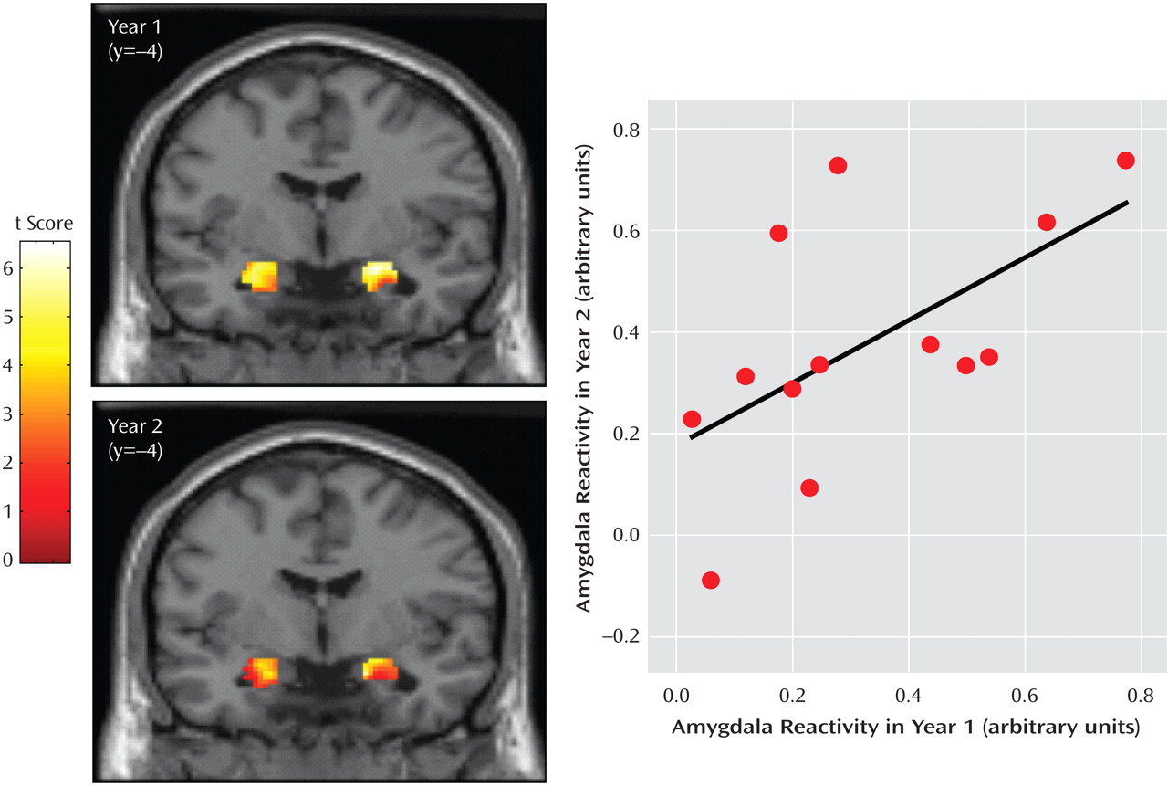
Thirteen subjects (10 men; mean age=45.9, range=35–55) who were in good health and without DSM-IV axis I psychopathology were scanned on the same 3T platform, with a median test-retest interval of 21 months (range=13–22 months). Amygdala reactivity to threat-related stimuli (angry and fearful faces at year 1; angry faces only at year 2) was determined using an archival paradigm known to reliably engage the amygdala (5) . Across subjects, significant amygdala activation was observed during both sessions ( Figure 1 ).
Figure 1. Amygdala Reactivity a
a The scans show mean bilateral amygdala reactivity to threat-related facial expressions for both sessions. Year 1: Right: 24, –4, –12, 154 voxels; t=4.59, p<0.001; Left: –20, –11, –16, 155 voxels; t=3.74, p<0.001. Year 2: Right: 22, –3, –12, 142 voxels; t=7.12, p<0.001; Left: –22, –8, –13, 150 voxels; t=6.63, p<0.001). Amygdala reactivity was calculated using random-effects analyses in SPM2 correcting for multiple comparisons (p<0.05) over the volume of the amygdala (for complete details of the fMRI challenge paradigm, blood-oxygen-level-dependent acquisition parameters, and statistical analyses, see [3]). The scatter plot of the bivariate correlation (r=0.59, p<0.035) shows mean right amygdala reactivity from the clusters for years 1 and 2.
Reliability was calculated as the intraclass correlation coefficient (ICC) for activation values in years 1 and 2. For the right amygdala, there was a 91% overlap in suprathreshold voxels across sessions; for activation magnitude (mean maximum voxel-level percent blood oxygen-level dependent signal change), the ICC was 0.59 (p<0.013), denoting moderate stability ( Figure 1 ). Notably, year 2 values for all except one participant ranked within four positions of their year 1 rank (median rank change=2; r s =0.71, p<0.007). There was also significant session overlap in left amygdala voxels (95%), but retest reliability of activation magnitude was not significant (ICC=–0.08).
In summary, we found significant long-term reproducibility of individual differences in fMRI-assessed right amygdala reactivity, suggesting that such reactivity comprises an enduring dimension of individual differences (4) . We speculate that our observed right laterality bias reflects the visuospatial processing demands of our paradigm, which preferentially engages right hemisphere circuits, and not underlying differences in the inherent biological properties of the right and left amygdala. This is underscored by the finding of left but not right temporal stability in an earlier study (4), in which paradigm characteristics may have contributed to the observed opposite laterality bias. It is also possible that our data underestimate the stability of amygdala reactivity, given the many methodological factors that might compromise reliability of measurements (e.g., variation in protocol or stimulus attributes, as well as between-session shifts in scanner characteristics, such as signal-to-noise ratio and tissue inhomogeneity-related artifacts). Our current observation confirms an important property (i.e., temporal stability) of a neural phenotype that figures increasingly in research on personality and temperament, psychopathology, and genetically driven variability in behavior. Further longitudinal investigation will be useful in evaluating the dispositional nature of amygdala reactivity and its role in liability to mood and anxiety disorders.

Footnotes

The authors report no competing interests.
Supported by NIH grants PO1 HL040962 (Dr. Manuck); KO1 MH072837 (Dr. Hariri); KO MH074769 (Dr. Forbes); and NARSAD Young Investigator Awards (Drs. Hariri and Forbes).
This letter (doi: 10.1176/appi.ajp.2007.07040609) was accepted for publication in July 2007.

References

1.
Stein MB, Simmons AN, Feinstein JS, Paulus MP: Increased amygdala and insula activation during emotion processing in anxiety-prone subjects. Am J Psychiatry 2007; 164:318–327
2.
Abercrombie HC, Schaefer SM, Larson CL, Oakes TR, Lindgren KA, Holden JE, Perlman SB, Turski PA, Krahn DD, Benca RM, Davidson RJ: Metabolic rate in the right amygdala predicts negative affect in depressed patients. Neuroreport 1998; 9:3301–3307
3.
Brown SM, Peet E, Manuck SB, Williamson DE, Dahl RE, Ferrell RE, Hariri AR: A regulatory variant of the human tryptophan hydroxylase-2 gene biases amygdala reactivity. Mol Psychiatry 2005; 10:884–888
4.
Johnstone T, Somerville LH, Alexander AL, Oakes TR, Davidson RJ, Kalin NH, Whalen PJ: Stability of amygdala BOLD response to fearful faces over multiple scan sessions. Neuroimage 2005; 25:1112–1123
5.
Hariri AR, Drabant EM, Weinberger DR: Imaging genetics: perspectives from studies of genetically driven variation in serotonin function and corticolimbic affective processing. Biol Psychiatry 2006; 59:888–897

Information & Authors

Information

Published In

Go to American Journal of Psychiatry
American Journal of Psychiatry
Pages: 1613 - 1614
PubMed: 17898358

History

Published online: 1 October 2007
Published in print: October, 2007

Authors

Affiliations

STEPHEN B. MANUCK, Ph.D.
ERIKA E. FORBES, Ph.D.
AHMAD R. HARIRI, Ph.D.

Metrics & Citations

Metrics

Citations

Export Citations

If you have the appropriate software installed, you can download article citation data to the citation manager of your choice. Simply select your manager software from the list below and click Download.

For more information or tips please see 'Downloading to a citation manager' in the Help menu.

Format
Citation style
Style
Copy to clipboard

There are no citations for this item

View Options

View options

PDF/ePub

View PDF/ePub

Get Access

Login options

Already a subscriber? Access your subscription through your login credentials or your institution for full access to this article.

Personal login Institutional Login Open Athens login
Purchase Options

Purchase this article to access the full text.

PPV Articles - American Journal of Psychiatry

PPV Articles - American Journal of Psychiatry

Not a subscriber?

Subscribe Now / Learn More

PsychiatryOnline subscription options offer access to the DSM-5-TR® library, books, journals, CME, and patient resources. This all-in-one virtual library provides psychiatrists and mental health professionals with key resources for diagnosis, treatment, research, and professional development.

Need more help? PsychiatryOnline Customer Service may be reached by emailing [email protected] or by calling 800-368-5777 (in the U.S.) or 703-907-7322 (outside the U.S.).

Media

Figures

Other

Tables

Share

Share

Share article link

Share