Abstract

Objective:

Major depressive disorder is associated with an increased risk of mortality and aging-related diseases. The authors examined whether major depression is associated with higher epigenetic aging in blood as measured by DNA methylation (DNAm) patterns, whether clinical characteristics of major depression have a further impact on these patterns, and whether the findings replicate in brain tissue.

Method:

DNAm age was estimated using all methylation sites in blood of 811 depressed patients and 319 control subjects with no lifetime psychiatric disorders and low depressive symptoms from the Netherlands Study of Depression and Anxiety. The residuals of the DNAm age estimates regressed on chronological age were calculated to indicate epigenetic aging. Major depression diagnosis and clinical characteristics were assessed with questionnaires and psychiatric interviews. Analyses were adjusted for sociodemographic characteristics, lifestyle, and health status. Postmortem brain samples of 74 depressed patients and 64 control subjects were used for replication. Pathway enrichment analysis was conducted using ConsensusPathDB to gain insight into the biological processes underlying epigenetic aging in blood and brain.

Results:

Significantly higher epigenetic aging was observed in patients with major depression compared with control subjects (Cohen’s d=0.18), with a significant dose effect with increasing symptom severity in the overall sample. In the depression group, epigenetic aging was positively and significantly associated with childhood trauma score. The case-control difference was replicated in an independent data set of postmortem brain samples. The top significantly enriched Gene Ontology terms included neuronal processes.

Conclusions:

As compared with control subjects, patients with major depression exhibited higher epigenetic aging in blood and brain tissue, suggesting that they are biologically older than their corresponding chronological age. This effect was even more profound in the presence of childhood trauma.
A growing body of literature suggests that major depressive disorder is associated with increased risks of mortality and aging-related phenotypes and diseases, including cardiovascular disease, diabetes, obesity (1), cancer (2), cognitive impairment (3), and frailty (4). Given the associated negative impact on quality of life and health care costs (5), it is of interest to investigate whether patients with major depression are prone to accelerated aging.
The literature provides evidence for advanced biological aging in major depressive disorder, as indicated by shorter telomere length (6, 7) and advanced brain aging (8). Recently, alternative markers of biological age derived from DNA methylation (also known as “epigenetic clocks”) have been developed. Chronological age can be accurately predicted from methylation data and yields estimates of DNA methylation age (DNAm age) (9, 10). Furthermore, DNAm age can be studied as either “decelerated” or “accelerated” by regressing it on chronological age to obtain a measure of epigenetic aging. Thus, DNAm age is a promising candidate for reliably investigating accelerated or premature aging in major depression.
Previous studies have shown epigenetic aging in individuals with Down’s syndrome (11), HIV-positive patients (12), and individuals with obesity (13). In addition, epigenetic aging has been associated with poorer physical and cognitive fitness (14), increased smoking and alcohol use (15), cancer (16), Alzheimer’s disease (17), cardiovascular disease (16), and an increased risk of mortality (18). A few studies have investigated DNAm age in relation to schizophrenia (19, 20), life stress (21), and posttraumatic stress disorder (22, 23), with mixed findings. However, studies examining epigenetic aging in relation to major depressive disorder are currently lacking.
In this study, we examined whether major depression is associated with higher epigenetic aging in blood, using a large, clinically well phenotyped sample to further explore associations with clinical characteristics, and we sought to replicate the findings in postmortem brain tissue. We used a sequencing-based approach (24, 25) that yields almost complete coverage of the CpG methylome, which allowed us to obtain the most accurate DNAm age estimates for our sample and to better explore the biological processes underlying epigenetic aging.

Method

The Netherlands Study of Depression and Anxiety

Study participants were from the Netherlands Study of Depression and Anxiety (NESDA), an ongoing longitudinal multicenter cohort study designed to investigate the long-term course and consequences of depressive and anxiety disorders (26). NESDA’s 2,981 participants (ages 18–65) include patients with a current or lifetime diagnosis of depression and/or an anxiety disorder and control subjects with no lifetime depressive disorder or anxiety disorders. Participants were recruited from the general population, general practices, and mental health organizations in order to reflect various settings and the entire range of psychopathology. Presence of major depression was ascertained with the DSM-IV-based Composite International Diagnostic Interview, version 2.1 (CIDI) (27), administered by trained research staff. Exclusion criteria were a clinically overt primary diagnosis of other psychiatric conditions (e.g., psychotic disorders, obsessive-compulsive disorder, bipolar disorder, and severe substance use disorders) and not being fluent in Dutch. The study was approved by the ethical committees of all participating centers, and participants provided written informed consent.
The total sample of 1,130 participants was divided into a control group (no lifetime psychiatric disorders and low depressive symptoms, as indicated by a score <14 on the Inventory of Depressive Symptomatology [28]) (N=319) and a group with current major depressive disorder (a score ≥14 on the Inventory of Depressive Symptomatology within the past 6 months) (N=811). Individuals who did not meet criteria for either of the two groups were excluded. The sample selection was further based on good-quality genome-wide association study genotype information available from a previous investigation (29).

Assessed Phenotypes

Data on sex, education level (in years), and body mass index (BMI) were collected during interviews. Alcohol use was recorded as mean number of drinks per week. Smoking behavior was measured by plasma cotinine levels, an adequate marker for calculating recent tobacco exposure (30). Physical activity was assessed using the International Physical Activity Questionnaire and indicated by total metabolic equivalent (MET) minutes per week. Health status was assessed as number of chronic diseases for which participants received medical treatment.
In all participants, depression severity was measured with the self-report version of the 30-item Inventory of Depressive Symptomatology (28). Childhood trauma was assessed using the childhood trauma interview from the Netherlands Mental Health Survey and Incidence Study, with personal history questions that included a structured inventory of trauma exposure during childhood. Finally, use of antidepressants was assessed through container inspection and categorized using the World Health Organization’s Anatomical Therapeutic Chemical Classification System: tricyclic antidepressants, selective serotonin reuptake inhibitors, and other antidepressants.
In participants with major depressive disorder, the duration of depression was measured by the Life Chart Interview, utilizing a calendar method to assess the percentage of time in which symptoms were present during the past 4 years (31). Current comorbid anxiety (panic disorder, generalized anxiety disorder, agoraphobia, social phobia) and alcohol disorder, as well as age at onset of depression were assessed with the CIDI. A more detailed description of all phenotypes is provided in the data supplement that accompanies the online edition of this article.

DNA Methylation Measurements

To assay the methylation status of the approximately 28 million common CpG sites in the human genome, we used an optimized protocol for MBD-seq (25). With this approach, genomic DNA is fragmented and the methylated fragments are then bound to the MBD2 protein, which has a high affinity for methylated DNA. The nonmethylated fraction is washed away, and only the methylation-enriched fraction is sequenced (for more details, see the online data supplement). This optimized protocol assesses about 94% of the CpGs in the methylome (25). The sequenced reads were aligned to the reference genome (build hg19/GRCh37) with Bowtie 2 (32) using local and gapped alignment. Aligned reads were further processed using the RaMWAS Bioconductor package (33) to perform quality control and calculate methylation scores for each CpG.

DNAm Age Estimation

While existing algorithms (9, 10) have gone through demonstrations of utility and reliability, estimating DNAm age with those prediction models would have been suboptimal for this study. These algorithms were derived using methylation data from a different platform in study populations with different characteristics (e.g., age distribution). Commonly used methods for assaying DNA methylation depend on the Illumina arrays, platforms that generate variables representing percentage methylated (ranging from 0 to 1). We used MBD-seq, generating methylation data that is semiquantitative (scores may range from 0–20) (24). Because the weights assigned to individual CpGs when making age predictions directly depend on the platform and study population, they will not optimally capture the effects of CpGs on age in the present study. Therefore, we “recalibrated” the DNAm age estimate in a way that is optimal for this study. It is important to emphasize that we aimed to obtain the best possible DNAm age estimates for MBD-seq data in our sample. We have not developed a new clock to be generalized to data from platforms other than MBD-seq. Furthermore, because we already collected MBD-seq data, we did not attempt to reduce the predictor set to the smallest number of CpGs, as pruning sites may reduce the precision of the DNAm age estimates.
Our approach for estimating DNAm age is similar to the one taken by Horvath (10). Specifically, we used elastic nets, a variable selection method that is particularly useful when the number of predictors is much larger than the number of observations (34). Parameter alpha was set to zero (i.e., ridge regression, retaining all sites in the model) where chronological age was used as outcome and methylation sites as predictors. To estimate predictive power and obtain DNAm age estimates for each subject, k-fold cross-validation was used, with k=10. Thus, the sample was randomly partitioned into 10 equally sized subsamples. Of the 10 subsamples, nine were used as training data and the remaining subsample as validation data. This ensured that in samples with the same properties and platform, our results would “replicate” and provide unbiased estimates of DNAm age. In the RaMWAS implementation, a cycle of methylome-wide association studies (MWAS), marker selection, and estimation via ridge regression was repeated in each training data set, with the resulting model applied to the test data to obtain unbiased estimates of DNAm age for each of the k=10 iterations.

Validation of the Use of MBD-seq Data to Estimate DNAm Age

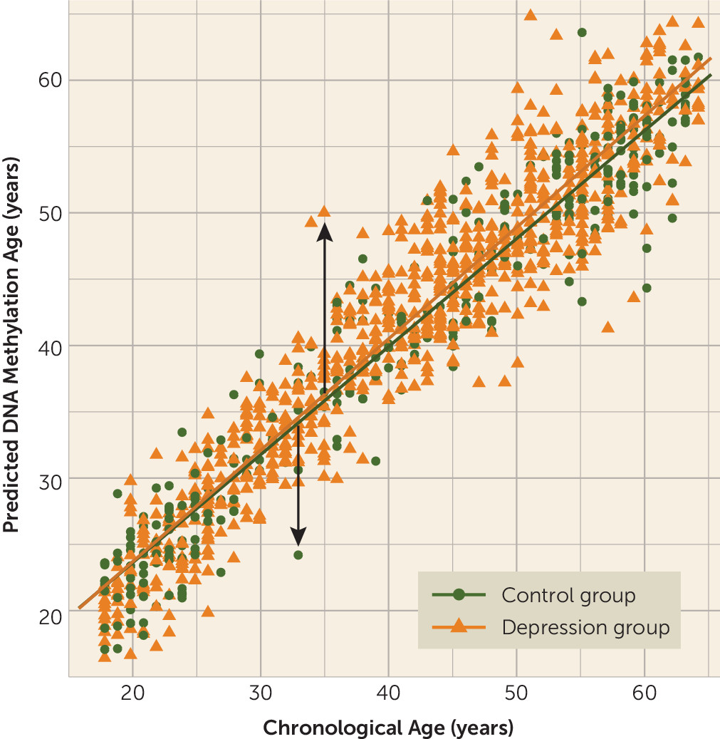
Several analyses were performed to validate the model. First, the model used to estimate DNAm age contained 80,000 CpGs (see Table S1 in the data supplement). Tenfold cross-validation showed that chronological age could be predicted very well, with a correlation of 0.95 (p<0.001) (Figure 1). Second, when analyzing assessed phenotypes in NESDA with DNAm age, we confirmed some similar determinants of DNAm age found in previous studies validating our outcome measure (see Table S3 in the data supplement). Male sex (35) and higher BMI (13, 36, 37) were associated with higher epigenetic aging. Third, to validate calculation of DNAm age, we used both ridge regression and the lasso method (used by Horvath). The additional elastic net model, with parameter alpha set to 0.5, resulted in a comparable correlation of 0.93 between chronological age and predicted DNAm age in our data set (compared with 0.95 with ridge regression and alpha=0), indicating that parameter set point did not have a large impact on our outcome measure. Finally, to ensure that no systematic bias was introduced by training the model on both case subjects and control subjects, we also trained the prediction model in control subjects only. This resulted in a slightly lower correlation between DNAm age estimates and chronological age (r=0.93), since the control subjects represented only a third of our total sample (i.e., lower statistical power). However, the correlation between the DNAm age estimates obtained in the full sample and those obtained using only control subjects was high (r=0.98, p<0.001), indicating that psychiatric status did not have an impact on the estimation of DNAm age.
FIGURE 1. DNA Methylation Age Prediction Using Methyl-CpG Binding Domain Protein-Enriched Genome Sequencing (MBD-seq) in the Netherlands Study of Depression and Anxietya
a The plot shows the prediction of DNA methylation (DNAm) age using MBD-seq across groups in blood. Each circle or triangle represents an individual subject (N=1,130), and the lines indicate regression lines (control group [N=319]: r=0.94, p<0.001; major depression group [N=811]: r=0.96, p<0.001). The arrows indicate the outcome variable epigenetic aging, representing higher epigenetic aging if the individual’s estimated DNAm age exceeds chronological age (upward arrow), whereas negative epigenetic aging indicates lower epigenetic aging (downward arrow).

Postmortem Brain Samples

We pooled data from five brain sample collections from four different brain banks (the Victorian Brain Bank Network, the Harvard Brain Bank, the Netherlands Brain Bank, and the Stanley Medical Research Institute), including a total of 141 brain samples from the Brodmann’s areas 10 and 25 regions. Presence of major depressive disorder (N=74) was determined by at least one psychiatrist by using information obtained from a family member who was well acquainted with the deceased. Control subjects (N=67) had no history of psychiatric disorders. Postmortem interval and pH were recorded in the brain collections, with the exception of the Harvard Brain Bank.
To further test the reliability and validity of our methods, we used the same approach to predict DNAm age in these samples with MBD-seq methylation data generated from the SOLiD 5500 W platform (Life Technologies, Carlsbad, Calif.). The model used to predict DNAm age contained 100,000 CpGs (see Table S2 in the data supplement) and obtained a correlation of 0.69 between predicted DNAm age and chronological age. The lower correlation with age is likely because the methylation data were generated with an older platform with lower quality (e.g., lower alignment of reads) from a more heterogeneous data set. More details about the samples and methods are provided in the data supplement.

Statistical Analyses for Discovery

To investigate case-control differences in epigenetic aging, we conducted linear regression models with epigenetic aging as the outcome and all covariates as predictors. To correct for the relative abundance of cell types that may be differentially associated with major depression, an additional model included cell type proportions as covariates (38). Other linear regression models were used to examine the relationship between epigenetic aging and Inventory of Depressive Symptomatology score across groups and clinical characteristics within patients with major depression. All analyses were corrected for all sociodemographic, lifestyle, and health covariates and used two-tailed tests with a significance threshold of 0.05.

Statistical Analyses for Replication

Within postmortem brain samples, we constructed a linear mixed model in R using the nlme package to account for the heterogeneity of epigenetic aging across brain collections. Thus, brain collection was entered as random effect and sex as fixed effect. The p value was derived by a likelihood ratio test, a hypothesis-driven one-sided test, with p<0.05 considered significant.

Bioinformatics Analyses

To perform enrichment tests of top MWAS findings in brain and blood, we used the shiftR R package with 1 million permutations for each test and used three thresholds (0.5%, 1%, and 5%) to define “top findings.” To account for this “multiple testing,” shiftR uses the same thresholds in the permutations where the test statistic distribution under the null hypothesis is generated from the most significant thresholds or combination of thresholds. A more in-depth description is provided in the data supplement. To gain insight into the overlapping biological pathways affecting epigenetic aging in blood and brain, we used ConsensusPathDB (39) to test whether genes harboring epigenetic aging–associated CpGs were enriched for level 5 Gene Ontology (GO) terms. Methylation sites with p<1×10⁻⁵ were selected and had to be within gene boundaries. At least four genes had to be present in the GO term to be considered. Finally, we also evaluated the overlap between chronological age–associated and epigenetic aging–associated CpG sites to examine whether similar biological processes were involved in chronological and biological aging.

Results

Higher Epigenetic Aging and Major Depression in NESDA

The mean age of the NESDA sample was 41.5 years (SD=13.0, range=18–64), and 64.5% of the sample were female (Table 1). The depression and control groups did not differ significantly in age, but the depression group had a higher proportion of females (p=0.02) and had fewer years of education on average (p<0.001). As anticipated, patients in the depression group reported higher levels of depression severity and use of antidepressants (all p values, <0.001). The mean childhood trauma score was also higher in the depressed group (p<0.001).
TABLE 1. Characteristics of Participants With and Without Depression in the Netherlands Study of Depression and Anxiety in an Analysis of Epigenetic Aging in Major Depressive Disordera
CharacteristicControl Group (N=319)Depression Group (N=811)
Sociodemographic characteristics    
 N%N%
 Female18858.954166.7
MeanSDMeanSD
 Age (years)41.614.6341.512.26
 Education level (years)13.13.1511.53.20
 MeanSDMeanSD
Lifestyle and health characteristics    
 Body mass index25.24.5025.95.31
 Cotinine levels (ng/mL)70.9200.8103.3183.9
 Alcohol intake (mean number of drinks per week)7.107.116.329.12
 Physical activity (1,000 MET minutes per week)3.912.863.493.17
 Number of chronic diseases0.460.740.690.92
 MeanSDMeanSD
Clinical characteristics    
 Inventory of Depressive Symptomatology score5.023.5433.810.9
 Childhood trauma score0.300.691.231.24
 Age at depression onset (years)  27.012.5
 Symptom duration (% of time in past 4 years)  0.390.30
 N%N%
 Comorbid anxiety disorder  53866.4
 Comorbid alcohol disorder  27033.3
 Antidepressant use    
  Tricyclic antidepressants00.0384.7
  Selective serotonin reuptake inhibitors10.324330.0
  Other antidepressants00.09011.2
 MeanSDMeanSD
Epigenetic aging–0.453.370.183.65
a
MET=metabolic equivalents. Epigenetic aging is calculated from unstandardized residuals of DNA methylation age regressed on chronological age.
Epigenetic aging showed (by design) a mean of zero (SD=3.58 years), ranging from −13.26 to 15.00. Patients in the depression group had significantly higher epigenetic aging compared with the control group (b=0.64, t=2.65, p=0.008; effect size, Cohen’s d=0.18), indicating that the patient was estimated to be 0.64 years (or 7.68 months) older on average than the control group after full adjustment for covariates (Table 2). Additional analyses correcting for cell type proportions did not change results and produced a Cohen’s d of 0.14 (see the data supplement). Consistent with a dose-response effect, a fully adjusted linear regression showed that greater epigenetic aging was significantly associated with higher Inventory of Depressive Symptomatology score in the overall sample (β=0.10, p=0.001). As expected from the high correlation between the DNAm age estimates generated by both models (r=0.98), the above-mentioned results remained unchanged when the same analyses were performed with the DNAm age estimates from the control-subjects-only model (see the data supplement).
TABLE 2. Estimated Marginal Means of Epigenetic Aging, by Major Depressive Disorder Status and Association With Depression Severity in the Overall Sample, in Basic and Fully Adjusted Analyses
 Control Group (N=319)Depression Group (N=811)Depression Versus Control GroupInventory of Depressive Symptomatology Score
ModelMeanSE95% CIMeanSE95% CIpCohen’s dβp
Basic adjusteda–0.510.20–0.90, –0.110.200.13–0.05, 0.450.0040.200.12<0.001
Fully adjustedb–0.460.20–0.86, –0.060.180.13–0.07, 0.430.0080.180.100.001
a
Adjusted for sex and education level.
b
Adjusted for sex, education level, body mass index, cotinine levels, alcohol use, physical activity, and number of chronic diseases.

Exploratory Analyses of Epigenetic Aging and Clinical Characteristics

Within the depression groups, we found epigenetic aging to be positively associated with childhood trauma score (β=0.09, p=0.02, see Table 3). The association between epigenetic aging and Inventory of Depressive Symptomatology score in the overall sample did not remain significant when analyzed only within the depression group (β=0.05, p=0.21), likely because of lower variation in symptom severity. No further significant associations with clinical characteristics were found.
TABLE 3. Associations Between Epigenetic Aging and Clinical Characteristics in Patients With Major Depressive Disorder (N=811)a
Variableβpb
Inventory of Depressive Symptomatology score0.050.21
Duration of illness–0.020.58
Age at onset0.030.42
Comorbid anxiety disorder–0.020.53
Comorbid alcohol dependence disorder0.050.21
Childhood trauma score0.090.02
Antidepressant use  
 Tricyclic antidepressants0.020.67
 Selective serotonin reuptake inhibitors–0.040.31
 Other antidepressants–0.040.29
a
Epigenetic aging is calculated from unstandardized residuals of DNA methylation age regressed on chronological age.
b
Analyses are adjusted for sex, education level, body mass index, cotinine levels, alcohol use, physical activity, and number of chronic diseases.
Further analyses revealed that patients in the depression group who had childhood trauma showed the highest epigenetic aging compared with control subjects without childhood trauma (p=0.001, Cohen’s d=0.29), highlighting the fact that this major depression and childhood trauma subgroup is associated with the highest epigenetic aging (see Figure S1 in the data supplement). Notably, greater severity of symptoms of chronic major depression was correlated with childhood trauma (r=0.39, p<0.001), making it difficult to discern which of these two factors drives increased epigenetic aging. Linear regression indeed showed that both childhood trauma (β=0.08, p=0.01) and depression severity score (β=0.07, p=0.03) were significant predictors of epigenetic aging when analyzed in the same model.

Replication in Postmortem Brain Samples

The mean age of the postmortem brain samples was 55.2 years (SD=19.3, range=20–100), and 45.4% of the sample were female. Groups were matched on age and sex. The mean postmortem interval was 35.1 hours (SD=21.1), and the mean pH was 6.51 (SD=0.25) across samples. Epigenetic aging was uncorrelated with pH or postmortem interval. Table 4 summarizes the descriptive characteristics by brain collection. Only the control (N=67) and depression (N=74) samples from the same brain collection were included in analyses (see the data supplement for more detail).
TABLE 4. Descriptive Characteristics of the Postmortem Brain Samples in an Analysis of Epigenetic Aging in Major Depressive Disordera
Brain Collection and CharacteristicControl Group (N=67)Depression Group (N=74)
Brain collection 1bN=30 N=30 
MeanSDMeanSD
 Age (years)51.6312.9451.9318.61
 Postmortem interval (hours)47.0414.6341.7415.74
 pH6.320.216.500.28
N%N%
 Female1756.71860.0
Brain collection 2N=4 N=3 
 MeanSDMeanSD
 Age (years)77.0012.2583.0013.00
 Postmortem interval (hours)NANANANA
 pHNANANANA
N%N%
 Female125.0133.3
Brain collection 3N=9 N=9 
 MeanSDMeanSD
 Age (years)85.678.5786.448.37
 Postmortem interval (hours)5.280.786.591.93
 pH6.560.156.490.23
N%N%
 Female666.7266.7
Brain collection 4N=11 N=22 
 MeanSDMeanSD
 Age (years)48.0011.9542.3211.15
 Postmortem interval (hours)26.729.7930.1412.84
 pH6.640.196.650.13
N%N%
 Female436.41045.5
Brain collection 5N=13 N=10 
 MeanSDMeanSD
 Age (years)51.158.3548.808.35
 Postmortem interval (hours)24.0015.0049.0049.50
 pH6.690.186.640.23
N%N%
 Female00.0110.0
a
Ns refer to numbers of samples left after quality control. NA=not available.
b
Brain collection 1 contains tissue dissected from Brodmann’s area 25; all other collections contain tissue from Brodmann’s area 10.
Our replication findings in independent brain samples supported our findings in NESDA and again showed that the epigenetic aging was higher in the depression group than in the control group (b=1.11, χ2=3.41, p=0.03). The beta indicates that the depression group was estimated to be on average 1.11 years older than the control group. The phenotype information available from the postmortem samples was limited, and therefore we were unable to attempt any replication of the exploratory clinical associations observed, such as childhood trauma, in NESDA.

Enrichment Testing and Gene Ontology Analyses

When evaluating the overlap between both epigenetic aging indicators, we found that after correcting for multiple testing, the top 1% findings from the epigenetic aging MWAS in blood were significantly enriched for CpGs in the top 0.5% of the epigenetic aging MWAS from brain (odds ratio=1.19, p<0.001). To examine possible processes underlying epigenetic aging in both tissues, we performed pathway analyses on the 1,084 overlapping CpGs associated with epigenetic aging, leading to 330 genes (90.7%) that were present in at least one GO category. Subsequently, this resulted in 53 significantly enriched GO terms (see Table S4 in the data supplement). The top GO terms included neurogenesis (p=9.79×10⁻⁹), neuron differentiation (p=5.34×10⁻⁸), and regulation of neuron death (p=4.67×10⁻⁵), indicating that several depression-relevant pathways were enriched in the cross-tissue epigenetic aging indicators.

Discussion

To the best of our knowledge, this is the first time higher epigenetic aging in patients with major depression compared with control subjects has been demonstrated. Exploratory analyses suggested even more pronounced epigenetic aging in patients with major depression who had higher childhood trauma scores. The case-control difference in blood was replicated in postmortem brain tissue. Finally, analyses showed significantly enriched neuronal pathways associated with the overlap between epigenetic aging–associated CpGs from blood and brain tissue.
Replication of our main finding in postmortem brain tissue bolstered confidence in the observed higher epigenetic aging in major depression. Moreover, the significantly enriched overlap suggests that at least some processes affecting epigenetic aging are at play in both blood and brain. There is some evidence that blood and brain show concordance in methylation (40) and epigenetic aging (10). However, considering the interactions between stress, central and peripheral immune processes, and neurobiology (41), it is plausible and likely that epigenetic aging in major depression is also dictated by many systemic processes. Nonetheless, more work is needed to confirm the higher epigenetic aging findings and to better characterize advanced aging–associated genes and their implications in major depression.
DNAm age is just one of the several available markers of biological aging (35). The present study confirms advanced or premature biological aging in major depression with a novel platform and is consistent with the literature regarding telomere length as biological marker of aging in major depression (7, 42). Also in line with other studies (43, 44), post hoc analyses between telomere length and epigenetic aging showed nonsignificant relationships, suggesting that both measures likely independently track different aspects of biological aging. Similarly, other post hoc analyses showed that telomere length did not alter our findings when accounted for, providing further evidence that epigenetic aging captures significant aging signal different from telomere length (see the data supplement).
We found that childhood trauma was positively associated with higher epigenetic aging in patients with major depression. It seems conceivable that major depression and accumulated stress throughout the lifetime due to childhood trauma may alter the epigenetic landscape and influence genomic regulation and function (45). However, this study did not identify additional relationships between higher epigenetic aging and more cumulative clinical characteristics, such as earlier onset age or longer duration of depression. Rather, our findings suggest that higher epigenetic aging in major depression may be driven largely by severity of illness.
Alternatively, childhood trauma may produce long-lasting epigenetic “scars” that have an impact on major depression and advanced or premature aging processes later in life. Individuals with childhood trauma and depressive disorders have an earlier onset age, higher symptom severity, more comorbidities, increased suicidality, and poorer treatment response than patients without childhood trauma (46, 47). As Teicher and Samson (47) have suggested, presence of childhood trauma is associated with a clinically and neurobiologically distinct subtype of depression.
Strikingly, three out of 10 top GO categories enriched across tissues included neuronal pathways. Epigenetic mechanisms are critical in early brain development, adult neurogenesis, and late-stage brain maturation (48). Given that these are all processes that seem markedly aberrant in major depression (49), the implicated pathways suggest that epigenetic aging in major depression directly contributes to symptomology. Additionally, the degree of overlap between the top 1% findings of epigenetic aging from blood and brain and the top 0.5% findings of chronological age in NESDA (odds ratio=85.31, p<0.001, and odds ratio=1.64, p<0.001) was highly significant, suggesting that biological aging overlaps with the same epigenetic processes that underlie chronological aging.
Although effect sizes such as those we observed here are common in depression research (e.g., oxidative stress, brain-derived neurotrophic factor, and cortisol yield effect sizes ranging from 0.15 to 0.31 [5052]), it is possible that this is an underestimate. The reason is that DNAm age is estimated from the residuals of the regression of methylation data on chronological age. This residual variance comprises two components: true unique variance associated with DNAm age, and measurement errors. However, because the residual variance is very small (the correlation between chronological age and methylation data was 0.95), even small measurement errors can have a large negative effect on the reliability of the DNAm age that is defined as reliability=Var(DNAm age)/(Var[DNAm age]+Var[errors of measurement]). As a less than perfect reliability will attenuate the correlation of DNAm age and depression status, the real effect size may have been underestimated.
Strengths of this study are the replication in postmortem brain tissue and inclusion of a large, clinically well characterized, and representative sample, with data that included many potential confounders that did not explain our findings. Given the full methylome coverage, we were also able to examine which biological pathways seemingly underlie epigenetic aging. However, our findings should also be considered against some limitations. A direct comparison of our MBD-seq-based epigenetic clock and existing Illumina array-based clocks was not possible and was beyond the scope of this study. However, a side-by-side comparison is an interesting endeavor for a future methodological study. Furthermore, with the present cross-sectional data, we were not able to disentangle whether greater epigenetic aging in major depression truly reflects aging acceleration over time or whether some individuals have increased epigenetic aging from birth or before adulthood that continues to be stable thereafter (53). Further studies with longitudinal designs are needed to distinguish the two possibilities.
In conclusion, our findings show that DNAm age from both blood and brain of patients with major depression is higher than their corresponding chronological age, which may contribute to their increased risk for mortality and aging-related diseases. Furthermore, higher childhood trauma scores correlated with higher epigenetic aging in patients with major depression. Taken together, our findings suggest that higher methylation aging in major depression is present in both blood and brain and that higher epigenetic aging largely overlaps with the same underpinnings associated with chronological aging. Further research is needed to investigate the causal relationships between age-associated alterations in DNA methylation and major depression.

Supplementary Material

File (appi.ajp.2018.17060595.ds001.pdf)
File (appi.ajp.2018.17060595.ds002.csv)
File (appi.ajp.2018.17060595.ds002.tables1.csv)
File (appi.ajp.2018.17060595.ds003.csv)
File (appi.ajp.2018.17060595.ds003.tables2.csv)
File (appi.ajp.2018.17060595.ds004.tables4.xlsx)
File (appi.ajp.2018.17060595.ds004.xlsx)

References

1.
Penninx BW: Depression and cardiovascular disease: epidemiological evidence on their linking mechanisms. Neurosci Biobehav Rev 2017; 74(Pt B):277–286
2.
Spiegel D, Giese-Davis J: Depression and cancer: mechanisms and disease progression. Biol Psychiatry 2003; 54:269–282
3.
McDermott LM, Ebmeier KP: A meta-analysis of depression severity and cognitive function. J Affect Disord 2009; 119:1–8
4.
Buigues C, Padilla-Sánchez C, Garrido JF, et al: The relationship between depression and frailty syndrome: a systematic review. Aging Ment Health 2015; 19:762–772
5.
Ben-Avraham D, Muzumdar RH, Atzmon G: Epigenetic genome-wide association methylation in aging and longevity. Epigenomics 2012; 4:503–509
6.
Schutte NS, Malouff JM: The association between depression and leukocyte telomere length: a meta-analysis. Depress Anxiety 2015; 32:229–238
7.
Ridout KK, Ridout SJ, Price LH, et al: Depression and telomere length: a meta-analysis. J Affect Disord 2016; 191:237–247
8.
Koutsouleris N, Davatzikos C, Borgwardt S, et al: Accelerated brain aging in schizophrenia and beyond: a neuroanatomical marker of psychiatric disorders. Schizophr Bull 2014; 40:1140–1153
9.
Hannum G, Guinney J, Zhao L, et al: Genome-wide methylation profiles reveal quantitative views of human aging rates. Mol Cell 2013; 49:359–367
10.
Horvath S: DNA methylation age of human tissues and cell types. Genome Biol 2013; 14:R115
11.
Horvath S, Garagnani P, Bacalini MG, et al: Accelerated epigenetic aging in Down syndrome. Aging Cell 2015; 14:491–495
12.
Levine AJ, Quach A, Moore DJ, et al: Accelerated epigenetic aging in brain is associated with pre-mortem HIV-associated neurocognitive disorders. J Neurovirol 2015; 22:366–375
13.
Horvath S, Erhart W, Brosch M, et al: Obesity accelerates epigenetic aging of human liver. Proc Natl Acad Sci USA 2014; 111:15538–15543
14.
Marioni RE, Shah S, McRae AF, et al: The epigenetic clock is correlated with physical and cognitive fitness in the Lothian Birth Cohort 1936. Int J Epidemiol 2015; 44:1388–1396
15.
Beach SRH, Dogan MV, Lei MK, et al: Methylomic aging as a window onto the influence of lifestyle: tobacco and alcohol use alter the rate of biological aging. J Am Geriatr Soc 2015; 63:2519–2525
16.
Perna L, Zhang Y, Mons U, et al: Epigenetic age acceleration predicts cancer, cardiovascular, and all-cause mortality in a German case cohort. Clin Epigenetics 2016; 8:64
17.
Levine ME, Lu AT, Bennett DA, et al: Epigenetic age of the pre-frontal cortex is associated with neuritic plaques, amyloid load, and Alzheimer’s disease related cognitive functioning. Aging (Albany NY) 2015; 7:1198–1211
18.
Chen BH, Marioni RE, Colicino E, et al: DNA methylation-based measures of biological age: meta-analysis predicting time to death. Aging (Albany NY) 2016; 8:1844–1865
19.
Voisey J, Lawford BR, Morris CP, et al: Epigenetic analysis confirms no accelerated brain aging in schizophrenia. NPJ Schizophr 2017; 3:26
20.
McKinney BC, Lin H, Ding Y, et al: DNA methylation age is not accelerated in brain or blood of subjects with schizophrenia. Schizophr Res (Epub ahead of print, Oct 5, 2017)
21.
Zannas AS, Arloth J, Carrillo-Roa T, et al: Lifetime stress accelerates epigenetic aging in an urban, African American cohort: relevance of glucocorticoid signaling. Genome Biol 2015; 16:266
22.
Wolf EJ, Logue MW, Hayes JP, et al: Accelerated DNA methylation age: associations with PTSD and neural integrity. Psychoneuroendocrinology 2016; 63:155–162
23.
Boks MP, van Mierlo HC, Rutten BPF, et al: Longitudinal changes of telomere length and epigenetic age related to traumatic stress and post-traumatic stress disorder. Psychoneuroendocrinology 2015; 51:506–512
24.
Aberg KA, McClay JL, Nerella S, et al: MBD-seq as a cost-effective approach for methylome-wide association studies: demonstration in 1500 case-control samples. Epigenomics 2012; 4:605–621
25.
Chan RF, Shabalin AA, Xie LY, et al: Enrichment methods provide a feasible approach to comprehensive and adequately powered investigations of the brain methylome. Nucleic Acids Res 2017; 45:e97
26.
Penninx BW, Beekman AT, Smit JH, et al: The Netherlands Study of Depression and Anxiety (NESDA): rationale, objectives, and methods. Int J Methods Psychiatr Res 2008; 17:121–140
27.
Wittchen HU: Reliability and validity studies of the WHO–Composite International Diagnostic Interview (CIDI): a critical review. J Psychiatr Res 1994; 28:57–84
28.
Rush AJ, Gullion CM, Basco MR, et al: The Inventory of Depressive Symptomatology (IDS): psychometric properties. Psychol Med 1996; 26:477–486
29.
Boomsma DI, Willemsen G, Sullivan PF, et al: Genome-wide association of major depression: description of samples for the GAIN Major Depressive Disorder Study: NTR and NESDA biobank projects. Eur J Hum Genet 2008; 16:335–342
30.
Benowitz NL, Bernert JT, Caraballo RS, et al: Optimal serum cotinine levels for distinguishing cigarette smokers and nonsmokers within different racial/ethnic groups in the United States between 1999 and 2004. Am J Epidemiol 2009; 169:236–248
31.
Lyketsos CG, Nestadt G, Cwi J, et al: The Life Chart Interview: a standardized method to describe the course of psychopathology. Int J Methods Psychiatr Res 1994; 4:143–155
32.
Langmead B, Salzberg SL: Fast gapped-read alignment with Bowtie 2. Nat Methods 2012; 9:357–359
33.
Shabalin AA, Hattab MW, Clark SL, et al: RaMWAS: fast methylome-wide association study pipeline for enrichment platforms. Bioinformatics (Epub ahead of print, Feb 12, 2018)
34.
Friedman J, Hastie T, Tibshirani R: Regularization paths for generalized linear models via coordinate descent. J Stat Softw 2010; 33:1–22
35.
Jylhävä J, Pedersen NL, Hägg S: Biological age predictors. EBioMedicine 2017; 21:29–36
36.
Quach A, Levine ME, Tanaka T, et al: Epigenetic clock analysis of diet, exercise, education, and lifestyle factors. Aging (Albany NY) 2017; 9:419–446
37.
Weidner CI, Lin Q, Koch CM, et al: Aging of blood can be tracked by DNA methylation changes at just three CpG sites. Genome Biol 2014; 15:R24
38.
Hattab MW, Shabalin AA, Clark SL, et al: Correcting for cell-type effects in DNA methylation studies: reference-based method outperforms latent variable approaches in empirical studies. Genome Biol 2017; 18:24
39.
Kamburov A, Stelzl U, Lehrach H, et al: The ConsensusPathDB interaction database: 2013 update. Nucleic Acids Res 2013; 41:D793–D800
40.
Tylee DS, Kawaguchi DM, Glatt SJ: On the outside, looking in: a review and evaluation of the comparability of blood and brain “-omes.” Am J Med Genet B Neuropsychiatr Genet 2013; 162:595–603
41.
Wohleb ES, Franklin T, Iwata M, et al: Integrating neuroimmune systems in the neurobiology of depression. Nat Rev Neurosci 2016; 17:497–511
42.
Henje Blom E, Han LK, Connolly CG, et al: Peripheral telomere length and hippocampal volume in adolescents with major depressive disorder. Transl Psychiatry 2015; 5:e676
43.
Breitling LP, Saum K-U, Perna L, et al: Frailty is associated with the epigenetic clock but not with telomere length in a German cohort. Clin Epigenetics 2016; 8:21
44.
Marioni RE, Harris SE, Shah S, et al: The epigenetic clock and telomere length are independently associated with chronological age and mortality. Int J Epidemiol (Epub ahead of print, April 13, 2016)
45.
Gassen NC, Chrousos GP, Binder EB, et al: Life stress, glucocorticoid signaling, and the aging epigenome: implications for aging-related diseases. Neurosci Biobehav Rev 2017; 74(Pt B):356–365
46.
Nanni V, Uher R, Danese A: Childhood maltreatment predicts unfavorable course of illness and treatment outcome in depression: a meta-analysis. Am J Psychiatry 2012; 169:141–151
47.
Teicher MH, Samson JA: Childhood maltreatment and psychopathology: a case for ecophenotypic variants as clinically and neurobiologically distinct subtypes. Am J Psychiatry 2013; 170:1114–1133
48.
Matt S, Roth ED, Roth TL: Role of epigenetics in the brain, in Epigenetics in Psychiatry. Edited by Avramopoulos D, Grayson DR, Peedicayil J. San Diego, Elsevier, 2014, pp 79–99
49.
Balu DT, Lucki I: Adult hippocampal neurogenesis: regulation, functional implications, and contribution to disease pathology. Neurosci Biobehav Rev 2009; 33:232–52
50.
Black CN, Bot M, Scheffer PG, et al: Is depression associated with increased oxidative stress? A systematic review and meta-analysis. Psychoneuroendocrinology 2015; 51:164–175
51.
Vreeburg SA, Hoogendijk WJ, van Pelt J, et al: Major depressive disorder and hypothalamic-pituitary-adrenal axis activity: results from a large cohort study. Arch Gen Psychiatry 2009; 66:617–626
52.
Molendijk ML, Bus BAA, Spinhoven P, et al: Serum levels of brain-derived neurotrophic factor in major depressive disorder: state-trait issues, clinical features, and pharmacological treatment. Mol Psychiatry 2011; 16:1088–1095
53.
Kananen L, Marttila S, Nevalainen T, et al: The trajectory of the blood DNA methylome ageing rate is largely set before adulthood: evidence from two longitudinal studies. Age (Dordr) 2016; 38:65

Information & Authors

Information

Published In

Go to American Journal of Psychiatry
American Journal of Psychiatry
Pages: 774 - 782
PubMed: 29656664

History

Received: 1 June 2017
Revision received: 1 December 2017
Revision received: 7 February 2018
Accepted: 20 February 2018
Published online: 16 April 2018
Published in print: August 01, 2018

Keywords

  1. Mood Disorders-Unipolar
  2. Assay Techniques
  3. Genetics

Authors

Details

Laura K.M. Han, M.Sc. [email protected]
From the Department of Psychiatry, VU University Medical Center, Amsterdam Neuroscience, GGZ inGeest, the Amsterdam Public Health Research Institute, Amsterdam; the Center for Biomarker Research and Precision Medicine, School of Pharmacy, Virginia Commonwealth University, Richmond; the Molecular Psychiatry Laboratory, Florey Department of Neuroscience and Mental Health, Melbourne, Australia; and the Centre for Mental Health, Faculty of Health, Arts, and Design, Swinburne University, Hawthorne, Australia.
Moji Aghajani, Ph.D.
From the Department of Psychiatry, VU University Medical Center, Amsterdam Neuroscience, GGZ inGeest, the Amsterdam Public Health Research Institute, Amsterdam; the Center for Biomarker Research and Precision Medicine, School of Pharmacy, Virginia Commonwealth University, Richmond; the Molecular Psychiatry Laboratory, Florey Department of Neuroscience and Mental Health, Melbourne, Australia; and the Centre for Mental Health, Faculty of Health, Arts, and Design, Swinburne University, Hawthorne, Australia.
Shaunna L. Clark, Ph.D.
From the Department of Psychiatry, VU University Medical Center, Amsterdam Neuroscience, GGZ inGeest, the Amsterdam Public Health Research Institute, Amsterdam; the Center for Biomarker Research and Precision Medicine, School of Pharmacy, Virginia Commonwealth University, Richmond; the Molecular Psychiatry Laboratory, Florey Department of Neuroscience and Mental Health, Melbourne, Australia; and the Centre for Mental Health, Faculty of Health, Arts, and Design, Swinburne University, Hawthorne, Australia.
Robin F. Chan, Ph.D.
From the Department of Psychiatry, VU University Medical Center, Amsterdam Neuroscience, GGZ inGeest, the Amsterdam Public Health Research Institute, Amsterdam; the Center for Biomarker Research and Precision Medicine, School of Pharmacy, Virginia Commonwealth University, Richmond; the Molecular Psychiatry Laboratory, Florey Department of Neuroscience and Mental Health, Melbourne, Australia; and the Centre for Mental Health, Faculty of Health, Arts, and Design, Swinburne University, Hawthorne, Australia.
Mohammad W. Hattab, Ph.D.
From the Department of Psychiatry, VU University Medical Center, Amsterdam Neuroscience, GGZ inGeest, the Amsterdam Public Health Research Institute, Amsterdam; the Center for Biomarker Research and Precision Medicine, School of Pharmacy, Virginia Commonwealth University, Richmond; the Molecular Psychiatry Laboratory, Florey Department of Neuroscience and Mental Health, Melbourne, Australia; and the Centre for Mental Health, Faculty of Health, Arts, and Design, Swinburne University, Hawthorne, Australia.
Andrey A. Shabalin, Ph.D.
From the Department of Psychiatry, VU University Medical Center, Amsterdam Neuroscience, GGZ inGeest, the Amsterdam Public Health Research Institute, Amsterdam; the Center for Biomarker Research and Precision Medicine, School of Pharmacy, Virginia Commonwealth University, Richmond; the Molecular Psychiatry Laboratory, Florey Department of Neuroscience and Mental Health, Melbourne, Australia; and the Centre for Mental Health, Faculty of Health, Arts, and Design, Swinburne University, Hawthorne, Australia.
Min Zhao, D.D.S.
From the Department of Psychiatry, VU University Medical Center, Amsterdam Neuroscience, GGZ inGeest, the Amsterdam Public Health Research Institute, Amsterdam; the Center for Biomarker Research and Precision Medicine, School of Pharmacy, Virginia Commonwealth University, Richmond; the Molecular Psychiatry Laboratory, Florey Department of Neuroscience and Mental Health, Melbourne, Australia; and the Centre for Mental Health, Faculty of Health, Arts, and Design, Swinburne University, Hawthorne, Australia.
Gaurav Kumar, Ph.D.
From the Department of Psychiatry, VU University Medical Center, Amsterdam Neuroscience, GGZ inGeest, the Amsterdam Public Health Research Institute, Amsterdam; the Center for Biomarker Research and Precision Medicine, School of Pharmacy, Virginia Commonwealth University, Richmond; the Molecular Psychiatry Laboratory, Florey Department of Neuroscience and Mental Health, Melbourne, Australia; and the Centre for Mental Health, Faculty of Health, Arts, and Design, Swinburne University, Hawthorne, Australia.
Lin Ying Xie, M.Sc.
From the Department of Psychiatry, VU University Medical Center, Amsterdam Neuroscience, GGZ inGeest, the Amsterdam Public Health Research Institute, Amsterdam; the Center for Biomarker Research and Precision Medicine, School of Pharmacy, Virginia Commonwealth University, Richmond; the Molecular Psychiatry Laboratory, Florey Department of Neuroscience and Mental Health, Melbourne, Australia; and the Centre for Mental Health, Faculty of Health, Arts, and Design, Swinburne University, Hawthorne, Australia.
Rick Jansen, Ph.D.
From the Department of Psychiatry, VU University Medical Center, Amsterdam Neuroscience, GGZ inGeest, the Amsterdam Public Health Research Institute, Amsterdam; the Center for Biomarker Research and Precision Medicine, School of Pharmacy, Virginia Commonwealth University, Richmond; the Molecular Psychiatry Laboratory, Florey Department of Neuroscience and Mental Health, Melbourne, Australia; and the Centre for Mental Health, Faculty of Health, Arts, and Design, Swinburne University, Hawthorne, Australia.
Yuri Milaneschi, Ph.D.
From the Department of Psychiatry, VU University Medical Center, Amsterdam Neuroscience, GGZ inGeest, the Amsterdam Public Health Research Institute, Amsterdam; the Center for Biomarker Research and Precision Medicine, School of Pharmacy, Virginia Commonwealth University, Richmond; the Molecular Psychiatry Laboratory, Florey Department of Neuroscience and Mental Health, Melbourne, Australia; and the Centre for Mental Health, Faculty of Health, Arts, and Design, Swinburne University, Hawthorne, Australia.
Brian Dean, Ph.D.
From the Department of Psychiatry, VU University Medical Center, Amsterdam Neuroscience, GGZ inGeest, the Amsterdam Public Health Research Institute, Amsterdam; the Center for Biomarker Research and Precision Medicine, School of Pharmacy, Virginia Commonwealth University, Richmond; the Molecular Psychiatry Laboratory, Florey Department of Neuroscience and Mental Health, Melbourne, Australia; and the Centre for Mental Health, Faculty of Health, Arts, and Design, Swinburne University, Hawthorne, Australia.
Karolina A. Aberg, Ph.D.
From the Department of Psychiatry, VU University Medical Center, Amsterdam Neuroscience, GGZ inGeest, the Amsterdam Public Health Research Institute, Amsterdam; the Center for Biomarker Research and Precision Medicine, School of Pharmacy, Virginia Commonwealth University, Richmond; the Molecular Psychiatry Laboratory, Florey Department of Neuroscience and Mental Health, Melbourne, Australia; and the Centre for Mental Health, Faculty of Health, Arts, and Design, Swinburne University, Hawthorne, Australia.
Edwin J.C.G. van den Oord, Ph.D.
From the Department of Psychiatry, VU University Medical Center, Amsterdam Neuroscience, GGZ inGeest, the Amsterdam Public Health Research Institute, Amsterdam; the Center for Biomarker Research and Precision Medicine, School of Pharmacy, Virginia Commonwealth University, Richmond; the Molecular Psychiatry Laboratory, Florey Department of Neuroscience and Mental Health, Melbourne, Australia; and the Centre for Mental Health, Faculty of Health, Arts, and Design, Swinburne University, Hawthorne, Australia.
Brenda W.J.H. Penninx, Ph.D.
From the Department of Psychiatry, VU University Medical Center, Amsterdam Neuroscience, GGZ inGeest, the Amsterdam Public Health Research Institute, Amsterdam; the Center for Biomarker Research and Precision Medicine, School of Pharmacy, Virginia Commonwealth University, Richmond; the Molecular Psychiatry Laboratory, Florey Department of Neuroscience and Mental Health, Melbourne, Australia; and the Centre for Mental Health, Faculty of Health, Arts, and Design, Swinburne University, Hawthorne, Australia.

Notes

Address correspondence to Ms. Han ([email protected]).

Competing Interests

Dr. Penninx has received research funding from Janssen Research and Boehringer Ingelheim. The other authors report no financial relationships with commercial interests.

Funding Information

ZonMw10.13039/501100001826: 10-000-1002
National Institute of Mental Health10.13039/100000025: R01MH099110
The Netherlands Study of Depression and Anxiety (www.nesda.nl) is funded through the Geestkracht program of the Netherlands Organization for Health Research and Development (Zon-MW, grant 10-000-1002) and participating universities and mental health care organizations (VU University Medical Center, GGZ inGeest, Arkin, Leiden University Medical Center, GGZ Rivierduinen, University Medical Center Groningen, Lentis, GGZ Friesland, GGZ Drenthe). This work was supported by NIMH grant R01MH099110 and the EMGO+ Travel Grant. Brain tissue was received from four brain banks: the Victorian Brain Bank, supported by the Florey Institute of Neuroscience and Mental Health and the Alfred and Victorian Forensic Institute of Medicine and funded by Australia’s National Health and Medical Research Council and Parkinson’s Victoria; the Stanley Medical Research Institute; the Netherlands Brain Bank, Netherlands Institute of Neuroscience, Amsterdam; and the Harvard Brain Tissue Resource Center.

Metrics & Citations

Metrics

Citations

Export Citations

If you have the appropriate software installed, you can download article citation data to the citation manager of your choice. Simply select your manager software from the list below and click Download.

For more information or tips please see 'Downloading to a citation manager' in the Help menu.

Format
Citation style
Style
Copy to clipboard

View Options

View options

PDF/EPUB

View PDF/EPUB

Get Access

Login options

Already a subscriber? Access your subscription through your login credentials or your institution for full access to this article.

Personal login Institutional Login Open Athens login
Purchase Options

Purchase this article to access the full text.

PPV Articles - American Journal of Psychiatry

PPV Articles - American Journal of Psychiatry

Not a subscriber?

Subscribe Now / Learn More

PsychiatryOnline subscription options offer access to the DSM-5-TR® library, books, journals, CME, and patient resources. This all-in-one virtual library provides psychiatrists and mental health professionals with key resources for diagnosis, treatment, research, and professional development.

Need more help? PsychiatryOnline Customer Service may be reached by emailing [email protected] or by calling 800-368-5777 (in the U.S.) or 703-907-7322 (outside the U.S.).

Media

Figures

Other

Tables

Share

Share

Share article link

Share