Full access
Clinical Research Reports
Published Online: 1 January 2012

Underlying Neurobiology and Clinical Correlates of Mania Status After Subthalamic Nucleus Deep Brain Stimulation in Parkinson's Disease: A Review of the Literature

Publication: The Journal of Neuropsychiatry and Clinical Neurosciences

Abstract

Deep brain stimulation (DBS) is a novel and effective surgical intervention for refractory Parkinson's disease (PD). The authors review the current literature to identify the clinical correlates associated with subthalamic nucleus (STN) DBS-induced hypomania/mania in PD patients. Ventromedial electrode placement has been most consistently implicated in the induction of STN DBS-induced mania. There is some evidence of symptom amelioration when electrode placement is switched to a more dorsolateral contact. Additional clinical correlates may include unipolar stimulation, higher voltage (>3 V), male sex, and/or early-onset PD. STN DBS-induced psychiatric adverse events emphasize the need for comprehensive psychiatric presurgical evaluation and follow-up in PD patients. Animal studies and prospective clinical research, combined with advanced neuroimaging techniques, are needed to identify clinical correlates and underlying neurobiological mechanisms of STN DBS-induced mania. Such working models would serve to further our understanding of the neurobiological underpinnings of mania and contribute valuable new insight toward development of future DBS mood-stabilization therapies.
Parkinson's disease (PD) affects as many as 1 million people in the United States alone, with approximately 40,000 Americans diagnosed with the disease annually. The hallmark neuropathology of the disease is loss of dopaminergic nigrostriatal neurons and intracytoplasmic Lewy bodies in the substantia nigra and locus coeruleus.1 The disease has a high degree of morbidity and is associated with significant use of nursing-home services, antidepressant and antipsychotic treatment, and overall reduced survival time, versus people without PD.2 Deep brain stimulation (DBS) is an FDA-approved surgical treatment for management of advanced PD. As compared with medication management, DBS significantly improves motor symptoms and quality of life.35 The subthalamic nucleus (STN) is used more commonly as the anatomic target for DBS, although a 24-month, randomized, blinded trial comparing STN and globus pallidus internus (GPi) stimulation showed similar motor function improvement.6 However, there were several secondary outcomes suggesting differential impact on mood on the basis of anatomic targets. At study endpoint, the STN group, as compared with GPi, had a statistically significant higher score (p=0.02) on the Beck Depression Inventory (STN: 12.5 [standard deviation {SD}] 8.5 versus Gpi: 9.8 [7.3]). Subscales quantifying mood and emotional well-being on PD rating scales, including the United Parkinson's Disease Rating Scale (UPDRS) and Parkinson's Disease Questionnaire (PDQ–39) showed similar differences in STN versus Gpi.6 In contrast, a recent 7-month, double-blind, randomized study comparing cognition and mood as primary outcome in STN and GPi groups (COMPARE Trial) reported no significant differences in mood and cognition outcomes between the two groups7 in the optimal stimulation condition.
In a review of 82 studies that included almost 1,400 PD patients, STN DBS has been associated with new-onset psychiatric adverse events, including depression (8%), hypomania/mania (4%), anxiety (2%), and attempted suicide (0.4%);8 although the sample is large, definitive conclusions are limited, given the lack of standardized assessment of psychiatric diagnosis and/or adverse events. Given the baseline estimate of PD comorbidity with psychiatric disorders, including depression (25%), psychotic syndromes (12.7%), sleep disturbances (49%), and anxiety (20%),9 plus the additional risk associated with STN DBS, there is great need to understand the clinical correlates associated with STN DBS-induced psychiatric adverse events (PAEs). Given a recent report of voltage-dependent mania associated with STN DBS in a PD patient,10 this literature review is focused on identifying underlying neurobiological mechanisms and clinical correlates of STN DBS-induced mania.

METHOD

We performed a structured MEDLINE (PubMed) search through December 2010, with keywords including subthalamic nucleus (STN), Parkinson's disease (PD), deep brain stimulation (DBS), mania, hypomania, and mood elevation. Seven published case reports,1115 two case series,16,17 and one retrospective cohort study18 with well-documented demographics, stimulation parameters, and details of psychiatric symptoms to confirm STN DBS-induced hypomania/mania were identified (total N: 17 patients). L-dopa equivalent daily dose (LEDD) was computed with the following formula: total L-dopa (regular L-dopa dose × 1) + (l-dopa controlled-release × 0.75) + (pramipexole dose × 67) + (ropinirole dose × 16.67) + (pergolide dose × 100) + (bromocriptine dose × 10), + [(l-dopa + L-dopa controlled-release × 0.75) × 0.25 if taking tolcapone {regardless of tolcapone dose}]).19

RESULTS

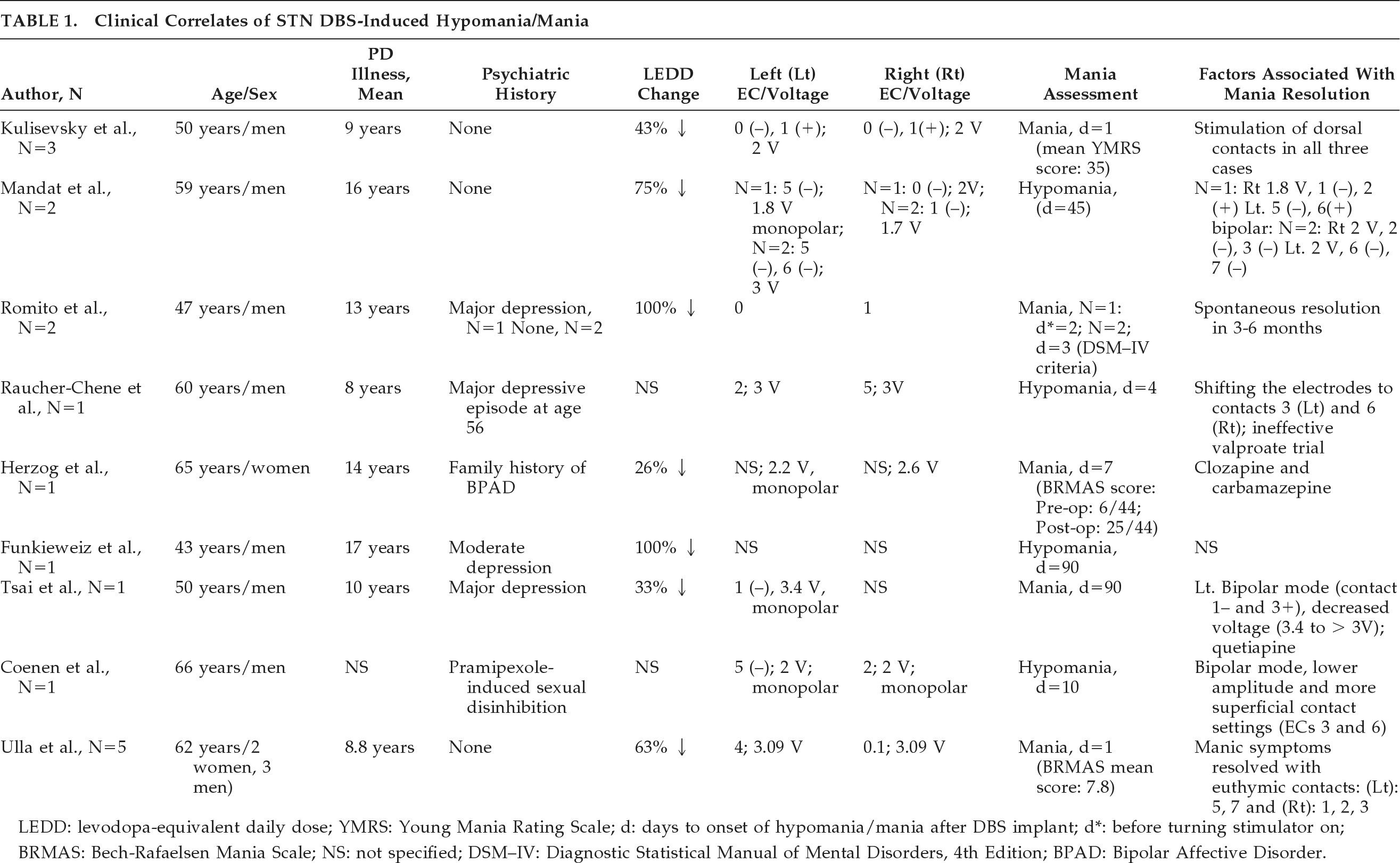
The clinical correlates associated with STN DBS-induced hypomania/mania can be subcategorized into electrode placement, stimulation parameters (unipolar versus bipolar stimulation and voltage), and neurosurgical (i.e., lesioning effect), neurological (age at onset of PD, LEDD change), and demographic factors (Table 1).
TABLE 1. Clinical Correlates of STN DBS-Induced Hypomania/Mania
LEDD: levodopa-equivalent daily dose; YMRS: Young Mania Rating Scale; d: days to onset of hypomania/mania after DBS implant; d*: before turning stimulator on; BRMAS: Bech-Rafaelsen Mania Scale; NS: not specified; DSM–IV: Diagnostic Statistical Manual of Mental Disorders, 4th Edition; BPAD: Bipolar Affective Disorder.
In all, 14 of 17 patients (82%) developed STN DBS-induced hypomania/mania with ventromedial STN electrode placement.1218 This pattern was reproducible (i.e., induction of manic symptoms with ventral stimulation and resolution of manic symptoms with switch to dorsolateral simulation) in five patients.17 Where reported, manic symptoms appeared to resolve in 12 of 14 patients (86%) after switching from ventromedial to dorsolateral STN; this occurred with an electrode configuration switch-alone in 9 of 12 patients (75%) or in combination with changes in stimulation parameters (i.e., voltage and stimulation mode) in 3 of 12 patients (25%).
In two case reports13,18 both voltage >3 V and monopolar stimulation mode were associated with STN DBS-induced mania. Monopolar stimulation produces a current that runs in multiple parallel paths. This kind of stimulation may influence a larger volume of tissue, especially when the current density is relatively high. Bipolar stimulation produces a more concentrated electric field around the electrode pair.20 Both increased voltage and unipolar stimulation can potentially result in enhanced current spread throughout the local tissue (relative to lower voltage or bipolar stimulation) and thus would be expected to influence a greater proportion of STN and nearby cells.21 Lowering voltage, along with switching to bipolar stimulation mode, has been associated with manic symptom resolution.13,18
The clinical correlates associated with STN DBS-induced hypomania/mania in the immediate and early postoperative period may include localized edema, a partial lesioning effect due to the STN DBS implant,11 as well as a possible add-on effect of STN DBS to the L-dopa treatment.5,18,22 The mean duration until onset of hypomania/mania in 17 patients was found to be 17.8 (30.6) days.
Among the demographic and neurological correlates, 14 of 17 patients (82%) with STN DBS-induced hypomania/mania were men (mean age: 55.6 [10.6] years), with relatively early onset of PD (N=13; mean age at PD onset: 43.6 [11.5] years) versus 3 of 17 female patients (17%; N=3; mean age: 58.6 [6.0] years; mean age at PD onset: 48.3 [2.5] years). A history of major depression or dopamine-induced mood disorder was reported in 4 of 17 patients (23%).

DISCUSSION

Despite the increasing use of DBS as a therapeutic tool for PD, the precise mechanism of action of DBS is currently debated. Since the effects of DBS are similar to those of a lesion, DBS has been thought to silence neurons at the site of stimulation. Reviewed extensively elsewhere,23,24 hypotheses concerning the mechanism of action of DBS have focused on a state of depolarization blockade at the stimulation site. This DBS-mediated neural network disruption has been hypothesized to result from preferential activation of GABA-ergic inhibitory neurons or high-frequency stimulation-mediated jamming of normal neuronal communication.2528 However, there is also evidence to suggest that DBS can elicit neurotransmitter-release distal to the site of stimulation2931 and/or effect more enduring plasticity-mediated changes in network function.32

STN DBS Mechanism of Action: Preclinical Models

Bilateral STN DBS typically reduces the need for L-DOPA treatment and recovers striatal dopamine-receptor function, indicating that dopamine levels have increased.33,34 This suggests that clinically effective STN DBS mediates dopamine neurotransmission. In support of this idea, striatal dopamine-release during STN DBS has been demonstrated in both intact and dopamine-depleted preclinical animal models.31,3539 Furthermore, STN DBS-evoked dopamine efflux has recently been reported in a large-animal (pig) model that faithfully replicates the human DBS neurosurgical procedure and stimulation paradigm.39 In this study, DBS-mediated striatal dopamine-release was observed to be voltage-dependent, in that, while maintaining constant frequency (120 Hz), increases in the voltage intensity from 3 V to 7 V resulted in a progressive increase in striatal dopamine-release. Animal studies have also suggested that dopamine synthesis may be upregulated by DBS.37,40
Extrapolation of results from preclinical animal models directly to DBS-mediated effects in PD patients, however, must be done with some caution. Differences in the type of electrodes utilized, as well as the scale and anatomy of the rodent brain, somewhat limit translation of the findings. In particular, specific parameters applied to the DBS electrode and the precise location of active leads within the brain can profoundly influence the biological impact of high-frequency stimulation on neuronal activity and neurotransmitter efflux.41,42
Given the potential for dopamine dysregulation to contribute toward behavioral expression of mania in PD,43,44 observations of up-regulated dopamine receptor function in STN DBS patients and preclinical data demonstrating STN DBS-elicited dopamine-release in motor and limbic basal ganglia circuits,29,31 the potential for regional STN DBS to contribute to the occurrence of manic symptoms needs to be explored more fully. Toward this end, a recent preclinical primate study has demonstrated that reversible, regional dysfunction of the STN selectively induces movement disorders and radical changes in behavior in healthy animals. In particular, the induction of hyperactivity reliably occurred with inhibition of the anteromedial region of the STN.45

STN DBS Mechanism of Action: Clinical Models

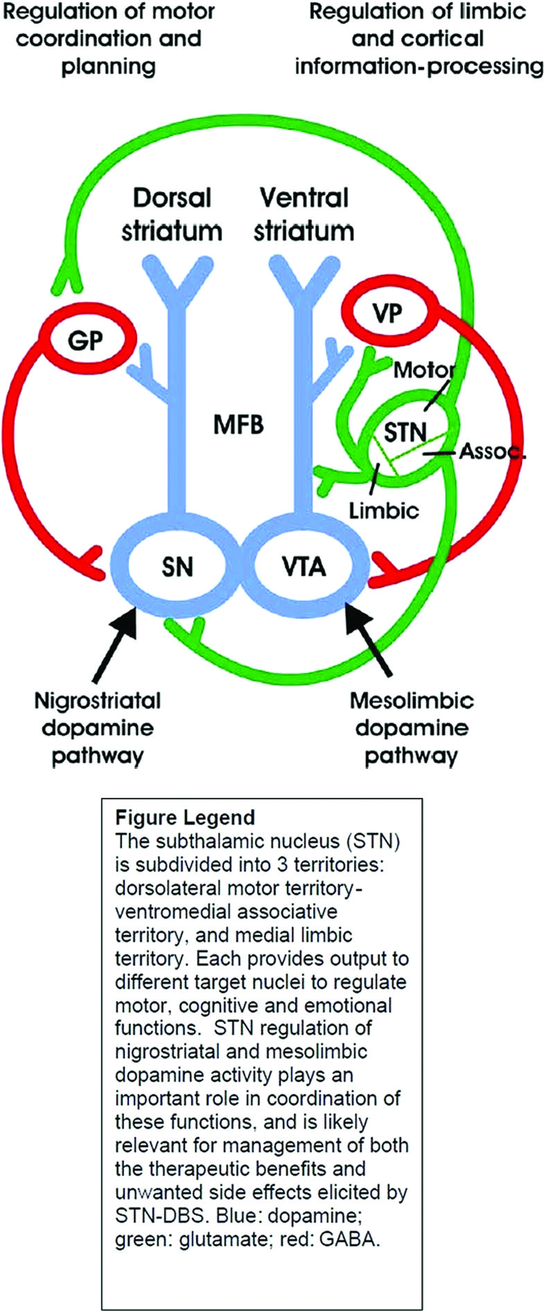
The subthalamic nucleus (STN) is subdivided into three territories: dorsolateral motor territory, ventromedial cognitive territory, and medial limbic territory. Each provides output to different target nuclei to regulate motor, cognitive, and emotional functions. STN regulation of nigrostriatal and mesolimbic dopamine activity helps to coordinate these functions and is likely implicated in both the therapeutic benefits and unwanted PAEs associated with STN DBS. Therapeutic effects of STN DBS in PD are optimized when the electrode is placed in the dorsolateral motor territory. Excitatory glutamatergic efferents project from the dorsolateral motor territory of the STN to the globus pallidus (GP). The GP, in turn, sends an inhibitory GABA-ergic projection to the dopamine cell bodies of the substantia nigra (SN). The SN is also innervated by an excitatory glutamatergic afferent from the ventromedial associative territory of the STN, as depicted in Figure 1.46 Thus, DBS activation of these projections could increase dopamine cell activity in the SN and dopamine release in the dorsal striatum to mediate motor function. Conversely, when the DBS electrode is positioned in the limbic territory, excitatory glutamatergic projections to dopaminergic axons of the median forebrain bundle (MFB) may be activated.15 The axons activated in this instance arise out of dopamine cell bodies in the ventral tegmental area (VTA) and project to the ventral striatum to influence the processing of limbic and cortical information. A glutamatergic projection is also sent from the limbic STN to the ventral pallidum (VP), which in turn sends an inhibitory GABA-ergic projection to the VTA, activation of which would inhibit dopamine cells.
FIGURE 1. Activities of the Subthalamic Nucleus in Various Functions
Given that dysregulation of mesolimbic dopamine is implicated in the expression of manic symptomatology, further investigation of the effects of STN DBS on mesolimbic dopamine is much needed, as is determination of its role in STN DBS-induced hypomania.
The role of the STN in regulation of emotional responses and behavior is becoming clearer, and several neuroimaging studies have reported DBS-induced specific changes at the level of cortical and subcortical associative and limbic regions.4749 Such a potent influence on associative, limbic, and motor functions has not been observed with other targets, like the GPi5053 or the thalamus.54,55 Also, recent controlled data have suggested that PD patient status post-DBS had differential emotional responses based on dorsal or ventral contact STN stimulation.56 Emotional reactivity of 20 PD patients given bilateral STN DBS was quantified by assessing emotional reaction to three different movie clips of different valences (neutral, sad, and amusing); ventral stimulation was associated with significantly increased ratings of positive affect, as compared with dorsal stimulation. Not surprisingly, STN stimulation, particularly when contacts are located ventromedially to the intended motor territory, may result in adverse cognitive and neuropsychiatric outcomes, including transient confusion, apathy, reduced verbal fluency, impaired attention, working memory, response inhibition, depression, anxiety, hypomania, hypersexuality, hallucinations, psychosis, and even suicide.46

STN DBS-Induced Mania: Underlying Neurobiology and Clinical Correlates

Application of DBS to the ventromedial STN, particularly with high voltages and/or unipolar stimulation mode, can potentially stimulate adjacent neural structures, including the substantia nigra, ventral tegmental area, and lateral hypothalamus.14 STN DBS has also been associated with potential stimulation of more formal neural circuits, including orbito-frontal basal ganglia,12 midbrain-orbitofrontal brain regions, anterior cingulate striato-pallido-thalamo-cortical circuits, or fibers from the ventral tegmentum area to the limbic striatum, causing symptoms typical of mania.5759 On the basis of evidence from diffusion tensor imaging (DTI), STN DBS-induced reversible acute hypomania might be elicited by inadvertent and unilateral coactivation of putative limbic STN tributaries to the medial forebrain bundle (MFB), a recently-identified pathway connecting the medial STN to reward circuitry.13
A recent study, focusing on contact-dependent reproducible hypomania in five PD patients, demonstrates the role of subcortical structures, specifically the substantia nigra, in the genesis of STN DBS-induced hypomania. Three-dimensional anatomical reconstruction with multimodal imaging (preoperative stereotactic MRI, postoperative CT scan, oxygen-15 PET scan) of “euthymic” and “manic” contacts revealed that 9 of 10 “manic” contacts were located in ventral substantia nigra, with right anterior cingulate and medial prefrontal cortex activation.17
The observation of bilateral globus pallidus DBS-induced mania in a patient with no previous psychiatric history60 suggests that there may be other stimulation sites outside of STN that have potential to cause this psychiatric adverse event. Because of the much-less-reported burden of PAEs associated with GPi stimulation; it may be an intuitive target choice for DBS stimulation in patients at risk of developing DBS-induced PAEs. However, these data are limited, and a higher rate of suicide in a 10-year meta-analysis has been reported with GPi stimulation.61
The literature reviewed to-date, would suggest that STN DBS-induced hypomania/mania seems to be most consistently associated with ventromedial STN electrode placement. The reproducibility of mania with ventral stimulation, resolution of manic symptoms with dorsal stimulation in 86% of cases (a change that also shifts the stimulated site laterally, given the typical DBS lead trajectory), and persisting manic symptoms despite significant reduction in LEDD further confirm this association. In terms of stimulation parameters, higher stimulation voltage (>3 V) and/or unipolar stimulation mode have been associated with STN DBS-induced mania. The voltage-dependent nature of STN DBS-induced mania10 replicates recently-reported voltage-dependent dopamine efflux in a large-animal model of DBS.39 Men and/or those with early-onset PD may be at significant risk of developing treatment-emergent adverse events to a number of PD treatment interventions. In a multicenter study on suicide outcomes after STN DBS for Parkinson's disease;62 younger age and early-onset PD were significantly associated with STN DBS treatment-emergent suicide attempts. Furthermore, in a cross-sectional evaluation of patients evaluated in a Movement Disorders Clinic, men were more likely to develop dopamine agonist-induced impulse-control disorders.63 These findings may suggest that men and/or early-onset PD may represent the PD population at the most risk of developing dopamine-agonist (whether medication- or DBS-induced) PAEs such as mania and suicidality.
Somewhat surprising, a past psychiatric history of depression or dopamine-induced mood disorder was only present in 4 of 17 reported cases (23%). It may be possible that STN DBS may have unmasked symptoms of preexisting psychiatric illness that were not identified in baseline assessment.64 Patients with preoperative cognitive deficits and affective disorders seem to be at risk of behavioral problems after STN DBS surgery.65,66 A recent small study of 14 PD patients undergoing STN DBS suggested that baseline motor impairment was associated with post-surgical hypomania symptoms,67 as it has already been suggested that sudden improvement in motor function after DBS surgery may have a profound effect on mood.68

Limitations

We acknowledge that psychiatric side effects after STN DBS surgery are published mostly in case-report format; therefore, their frequency might be overestimated.22,69,70 But it might equally well be the case that frequency of such side effects is underestimated, as these side effects are not subjected to systematic assessment. The precise mechanism(s) of action of STN DBS-induced hypomania/mania are currently not well understood. Our findings/proposed mechanisms may still be preliminary and speculative, and this needs further systematic research.

CONCLUSION

STN DBS-induced mania and other adverse events emphasize the need for comprehensive presurgery psychiatric evaluation and close follow-up after implantation. Given the significant psychiatric morbidity and mortality associated with STN DBS surgery in PD patients, there is need for preclinical animal studies and prospective cohort/ longitudinal studies, combined with advanced neuroimaging techniques, to specifically establish psychiatric, neurological, and neurosurgical correlates and elucidate the neurobiological mechanism(s) associated with STN DBS-induced mania. Such working models would serve to further our understanding of the neurobiological underpinnings of mania and contribute valuable new insight toward development of future DBS mood-stabilization therapies.

Acknowledgments

This work was presented in part at the American Neuropsychiatric Association Annual Meeting, Tampa, FL, March 18, 2010.
We thank Ms. Cindy Stoppel for her help in the preparation of this manuscript.
Dr. Tye was supported by a NARSAD Young Investigator Award.
Mark A. Frye, M.D. has grant support from Pfizer, National Alliance for Schizophrenia and Depression (NARSAD), NIMH, National Institute of Alcohol Abuse and Alcoholism (NIAAA), and the Mayo Foundation, and is a consultant to Dainippon Sumittomo Pharma, Merck, and Sepracor. CME-supported activity: Astra-Zeneca, Bristol-Myers Squibb, Eli Lilly and Co., GlaxoSmithKline, Merck, Otsuka Pharmaceuticals, Pfizer, and Sanofi-Aventis.
Dr. Kendall H. Lee has patents pending relating to DBS technology and has industry support from St. Jude Neuromodulation Division. This work was supported by NIH (K08 NS 52232 award) and Mayo Foundation (2008-2010 Research Early-Career Development Award for Clinician-Scientists) to KHL.
Julie A. Fields, Ph.D., is a consultant to Medtronic, Inc.

References

1.
Lauterbach EC: The neuropsychiatry of Parkinson's disease. Minerva Medica 2005; 96:155–173
2.
Parashos SA, Maraganore DM, O'Brien PC, et al.: Medical services utilization and prognosis in Parkinson disease: a population-based study. Mayo Clin Proc 2002; 77:918–925
3.
Weaver FMP, Follett KMDP, Stern MMD, et al.: Bilateral deep brain stimulation vs. best medical therapy for patients with advanced Parkinson disease: a randomized, controlled trial. JAMA 2009; 301:63–73
4.
Witt K, Daniels C, Reiff J, et al.: Neuropsychological and psychiatric changes after deep brain stimulation for Parkinson's disease: a randomised, multicentre study. Lancet Neurol 2008; 7:605–614
5.
Krack P, Batir A, Van Blercom N, et al.: Five-year follow-up of bilateral stimulation of the subthalamic nucleus in advanced Parkinson's disease. New Engl J Med 2003; 349:1925–1934
6.
Follett KA, Weaver FM, Stern M, et al.: Pallidal versus subthalamic deep-brain stimulation for Parkinson's disease. N Engl J Med 2010; 362:2077–2091
7.
Okun MS, Fernandez HH, Wu SS, et al.: Cognition and mood in Parkinson's disease in subthalamic nucleus versus globus pallidus interna deep brain stimulation: The Compare Trial. Ann Neurol 2009; 65:586–595
8.
Temel Y, Kessels A, Tan S, et al.: Behavioural changes after bilateral subthalamic stimulation in advanced Parkinson disease: a systematic review. Parkinson Relat Disord 2006; 12:265–272
9.
Riedel O, Klotsche J, Spottke A, et al. Frequency of dementia, depression, and other neuropsychiatric symptoms in 1,449 outpatients with Parkinson's disease. J Neurol 2010; 257:1073–1082
10.
Chopra A, Tye S, Kendall L, et al.: Voltage-dependent mania status post-subthalamic nucleus deep brain stimulation in Parkinson's disease: a case report. Biol Psychiatry 2011; 70:e5–e7
11.
Romito LM, Raja M, Daniele A, et al.: Transient mania with hypersexuality after surgery for high-frequency stimulation of the subthalamic nucleus in Parkinson's disease. Mov Disord 2002; 17:1371–1374
12.
Herzog J, Reiff J, Krack P, et al.: Manic episode with psychotic symptoms induced by subthalamic nucleus stimulation in a patient with Parkinson's disease. Mov Disord 2003; 18:1382–1384
13.
Coenen VA, Honey CR, Hurwitz T, et al.: Medial forebrain bundle stimulation as a pathophysiological mechanism for hypomania in subthalamic nucleus deep brain stimulation for Parkinson's disease. Neurosurgery 2009; 64:1106–1114; discussion 1114–1105
14.
Mandat TS, Hurwitz T, Honey CR, et al.: Hypomania as an adverse effect of subthalamic nucleus stimulation: report of two cases. Acta Neurochirurg 2006; 148:895–898
15.
Raucher-Chene D, Charrel C-L, de Maindreville AD, et al.: Manic episode with psychotic symptoms in a patient with Parkinson's disease treated by subthalamic nucleus stimulation: improvement on switching the target. J Neurol Sci 2008; 273:116–117
16.
Kulisevsky J, Berthier ML, Gironell A, et al.: Mania following deep brain stimulation for Parkinson's disease. Neurology 2002; 59:1421–1424
17.
Ulla M, Thobois S, Llorca PM, et al.: Contact-dependent reproducible hypomania induced by deep brain stimulation in Parkinson's disease: clinical, anatomical, and functional imaging study. J Neurol Neurosurg Psychiatry 2010; 82:607–614
18.
Tsai S, Lin S, Lin S, et al.: Neuropsychological effects after chronic subthalamic stimulation and topography of the nucleus in Parkinson's disease. Neurosurgery 2007; 61:E1024–E1030
19.
Hobson DE, Lang AE, Martin WR, et al.: Excessive daytime sleepiness and sudden-onset sleep in Parkinson disease: a survey by The Canadian Movement Disorders Group. JAMA 2002; 287:455–463
20.
Temel Y, Visser-Vandewalle V, van der Wolf M, et al.: Monopolar versus bipolar high-frequency stimulation in the rat subthalamic nucleus: differences in histological damage. Neurosci Lett 2004; 367:92–96
21.
McIntyre CC, Mori S, Sherman DL, et al.: Electric field and stimulating influence generated by deep brain stimulation of the subthalamic nucleus. Clin Neurophysiol 2004; 115:589–595
22.
Funkiewiez A, Ardouin C, Krack P, et al.: Acute psychotropic effects of bilateral subthalamic nucleus stimulation and levodopa in Parkinson's disease. Mov Disord 2003; 18:524–530
23.
Kopell BH, Greenberg BD: Anatomy and physiology of the basal ganglia: implications for DBS in psychiatry. Neurosci Biobehav Rev 2008; 32:408–422
24.
Tye SJ, Frye MA, Lee KH: Disrupting disordered neurocircuitry: treating refractory psychiatric illness with neuromodulation. Mayo Clin Proc 2009; 84:522–532
25.
Benazzouz A, Piallat B, Pollak P, et al.: Responses of substantia nigra pars reticulata and globus pallidus complex to high-frequency stimulation of the subthalamic nucleus in rats: electrophysiological data. Neurosci Lett 1995; 189:77–80
26.
Garcia L, D'Alessandro G, Bioulac B, et al.: High-frequency stimulation in Parkinson's disease: more or less? Trends Neurosci 2005; 28:209–216
27.
Lozano AM, Dostrovsky J, Chen R, et al.: Deep brain stimulation for Parkinson's disease: disrupting the disruption. Lancet Neurol 2002; 1:225–231
28.
Magarinos-Ascone C, Pazo JH, Macadar O, et al.: High-frequency stimulation of the subthalamic nucleus silences subthalamic neurons: a possible cellular mechanism in Parkinson's disease. Neuroscience 2002; 115:1109–1117
29.
Lee KH, Blaha CD, Harris BT, et al.: Dopamine efflux in the rat striatum evoked by electrical stimulation of the subthalamic nucleus: potential mechanism of action in Parkinson's disease. Eur J Neurosci 2006; 23:1005–1014
30.
McCracken CB, Grace AA: High-frequency deep brain stimulation of the nucleus accumbens region suppresses neuronal activity and selectively modulates afferent drive in rat orbitofrontal cortex in vivo. J Neurosci 2007; 27:12601–12610
31.
Winter C, Lemke C, Sohr R, et al.: High-frequency stimulation of the subthalamic nucleus modulates neurotransmission in limbic brain regions of the rat. Exp Brain Res 2008; 185:497–507
32.
Xiang-Hong L, Jin-Yan W, Ge G, Jing-Yu C, et al. High-frequency stimulation of the subthalamic nucleus restores neural and behavioral functions during reaction-time task in a rat model of Parkinson's disease. J Neurosci Res 2009;
33.
Blahak C, Bazner H, Capelle H-H, et al.: Rapid response of parkinsonian tremor to STN-DBS changes: direct modulation of oscillatory basal ganglia activity? Mov Disord 2009; 24:1221–1225
34.
Hesse S, Strecker K, Winkler D, et al.: Effects of subthalamic nucleus stimulation on striatal dopaminergic transmission in patients with Parkinson's disease within one-year follow-up. J Neurol 2008; 255:1059–1066
35.
Bruet NM, Windels FM, Bertrand AB, et al.: High-frequency stimulation of the subthalamic nucleus increases the extracellular contents of striatal dopamine in normal and partially dopaminergic-denervated rats. J Neuropathol Exp Neurol 2001; 60:15–24
36.
Meissner W, Harnack D, Paul G, et al.: Deep brain stimulation of subthalamic neurons increases striatal dopamine metabolism and induces contralateral circling in freely moving 6-hydroxydopamine-lesioned rats. Neurosci Lett 2002; 328:105–108
37.
Meissner W, Harnack D, Reese R, et al.: High-frequency stimulation of the subthalamic nucleus enhances striatal dopamine release and metabolism in rats. J Neurochem 2003; 85:601–609
38.
Meissner W, Reum T, Paul G, et al.: Striatal dopaminergic metabolism is increased by deep brain stimulation of the subthalamic nucleus in 6-hydroxydopamine-lesioned rats. Neurosci Lett 2001; 303:165–168
39.
Shon Y-M, Lee KH, Goerss SJ, et al.: High-frequency stimulation of the subthalamic nucleus evokes striatal dopamine release in a large-animal model of human DBS neurosurgery. Neurosci Lett 2010; 475:136–140
40.
Reese R, Winter C, Nadjar A, et al.: Subthalamic stimulation increases striatal tyrosine hydroxylase phosphorylation. Neuroreport 2008; 19:179–182
41.
Maks CB, Butson CR, Walter BL, et al.: Deep brain stimulation activation volumes and their association with neurophysiological mapping and therapeutic outcomes. J Neurol Neurosurg Psychiatry 2009; 80:659–666
42.
Miocinovic S, Lempka SF, Russo GS, et al.: Experimental and theoretical characterization of the voltage distribution generated by deep brain stimulation. Exp Neurol 2009; 216:166–176
43.
Berk M, Dodd S, Kauer-Sant'anna M, et al.: Dopamine dysregulation syndrome: implications for a dopamine hypothesis of bipolar disorder. Acta Psychiatr Scand Supplementum 2007; 41–49
44.
Cousins DA, Butts K, Young AH: The role of dopamine in bipolar disorder. Bipolar Disord 2009; 11:787–806
45.
Karachi C, Grabli D, Baup N, et al.: Dysfunction of the subthalamic nucleus induces behavioral and movement disorders in monkeys. Mov Disord 2009; 24:1183–1192
46.
Benarroch EEMD: Subthalamic nucleus and its connections: anatomic substrate for the network effects of deep brain stimulation. Neurology 2008; 70:1991–1995
47.
Hilker R, Voges J, Weisenbach S, et al.: Subthalamic nucleus stimulation restores glucose metabolism in associative and limbic cortices and in cerebellum: evidence from a FDG-PET study in advanced Parkinson's disease. J Cereb Blood Flow Metab 2004; 24:7–16
48.
Schroeder U, Kuehler A, Haslinger B, et al.: Subthalamic nucleus stimulation affects striato-anterior cingulate cortex circuit in a response-conflict task: a PET study. Brain 2002; 125:1995–2004
49.
Ulrike S, Andreas K, Klaus WL, et al.: Subthalamic nucleus stimulation affects a frontotemporal network: a PET study. Ann Neurol 2003; 54:445–450
50.
Ghika J, Villemure J-G, Fankhauser H, et al.: Efficiency and safety of bilateral contemporaneous pallidal stimulation (deep brain stimulation) in levodopa-responsive patients with Parkinson's disease with severe motor fluctuations: a 2-year follow-up review. J Neurosurg 1998; 89:713–718
51.
Iacono RP, Shima F, Lonser RR, et al.: The results, indications, and physiology of posteroventral pallidotomy for patients with Parkinson's disease. Neurosurgery 1995; 36:1118–1125, discussion 1125–1117
52.
Vingerhoets G, van der Linden C, Lannoo E, et al.: Cognitive outcome after unilateral pallidal stimulation in Parkinson's disease. J Neurol Neurosurg Psychiatry 1999; 66:297–304
53.
Visser-Vandewalle V, Temel Y, van der Linden C, et al.: Deep brain stimulation in movement disorders: the applications reconsidered. Acta Neurol Belgica 2004; 104:33–36
54.
Benabid AL, Benazzouz A, Hoffmann D, et al.: Long-term electrical inhibition of deep brain targets in movement disorders. Mov Disord 1998; 13(Suppl 3):119–125
55.
Pollak P, Fraix V, Krack P, et al. Treatment results: Parkinson's disease. Mov Disord 2002; 17:(Suppl 3):S75–S83
56.
Greenhouse I, Gould M, Houser J, et al.: Stimulation at dorsal and ventral electrode contacts targeted at the subthalamic nucleus has different effects on motor and emotion functions in Parkinson's disease. Neuropsychologia 2011; 49:528–534
57.
Cummings JL, Mendez MF: Secondary mania with focal cerebrovascular lesions. Am J Psychiatry 1984; 141:1084–1087
58.
Kulisevsky J, Avila A, Berthier ML: Bipolar affective disorder and unilateral parkinsonism after a brainstem infarction. Mov Disord 1995; 10:799–802
59.
Kulisevsky J, Berthier ML, Pujol J: Hemiballismus and secondary mania following a right-thalamic infarction. Neurology 1993; 43:1422–1424
60.
Miyawaki E, Perlmutter JS, Troster AI, et al.: The behavioral complications of pallidal stimulation: a case report. Brain Cog 2000; 42:417–434
61.
Appleby B, Duggan P, Regenberg A, et al.: Psychiatric and neuropsychiatric adverse events associated with deep brain stimulation: a meta-analysis of ten years' experience. Mov Disord 2007; 22:1722–1728
62.
Voon V, Krack P, Lang AE, et al.: A multicentre study on suicide outcomes following subthalamic stimulation for Parkinson's disease. Brain 2008; 131(Pt 10):2720–2728
63.
Hassan A, Bower JH, Kumar N, et al.: Dopamine agonist-triggered pathological behaviors: surveillance in the PD clinic reveals high frequencies. Parkinson Relat Disord 2011; 17:260–264
64.
Lilleeng B, Dietrichs E: Unmasking psychiatric symptoms after STN deep brain stimulation in Parkinson's disease. Acta Neurol Scand 2008; 117:(Suppl):41–45
65.
Kumar R, Lozano AM, Kim YJ, et al.: Double-blind evaluation of subthalamic nucleus deep brain stimulation in advanced Parkinson's disease. Neurology 1998; 51:850–855
66.
Rodriguez MC, Guridi OJ, Alvarez L, et al.: The subthalamic nucleus and tremor in Parkinson's disease. Mov Disord 1998; 13:111–118
67.
Schneider F, Reske M, Finkelmeyer A, et al.: Predicting acute affective symptoms after deep brain stimulation surgery in Parkinson's disease. Stereotact Funct Neurosurg 2010; 88:367–373
68.
Berney A, Vingerhoets F, Perrin A, et al.: Effect on mood of subthalamic DBS for Parkinson's disease: a consecutive series of 24 patients. Neurology 2002; 59:1427–1429
69.
Funkiewiez A, Ardouin C, Caputo E, et al.: Long term effects of bilateral subthalamic nucleus stimulation on cognitive function, mood, and behaviour in Parkinson's disease. J Neurol Neurosurgery Psychiatry 2004; 75:834–839
70.
Houeto JL, Mesnage V, Mallet L, et al.: Behavioural disorders, Parkinson's disease, and subthalamic stimulation J Neurol Neurosurg Psychiatry 2002; 72:701–707

Information & Authors

Information

Published In

Go to The Journal of Neuropsychiatry and Clinical Neurosciences
The Journal of Neuropsychiatry and Clinical Neurosciences
Pages: 102 - 110
PubMed: 22450620

History

Received: 15 July 2010
Revision requested: 24 January 2011
Accepted: 31 March 2011
Published online: 1 January 2012
Published in print: Winter 2012

Authors

Affiliations

Amit Chopra, M.B.B.S.
From the Department of Psychiatry and Psychology, Mayo Clinic, Rochester MN, USA (AC, SS, JAF, MAF); the Department of Psychology, Deakin University, Burwood VIC, Australia (SJT); the Departments of Neurosurgery & Physiology and Biomedical Engineering, Mayo Clinic, Rochester MN, USA (KHL); and the Department of Neurology, Mayo Clinic, Rochester MN, USA (JM, AA, BK, MS).
Susannah J. Tye, Ph.D.
From the Department of Psychiatry and Psychology, Mayo Clinic, Rochester MN, USA (AC, SS, JAF, MAF); the Department of Psychology, Deakin University, Burwood VIC, Australia (SJT); the Departments of Neurosurgery & Physiology and Biomedical Engineering, Mayo Clinic, Rochester MN, USA (KHL); and the Department of Neurology, Mayo Clinic, Rochester MN, USA (JM, AA, BK, MS).
Kendall H. Lee, M.D., Ph.D.
From the Department of Psychiatry and Psychology, Mayo Clinic, Rochester MN, USA (AC, SS, JAF, MAF); the Department of Psychology, Deakin University, Burwood VIC, Australia (SJT); the Departments of Neurosurgery & Physiology and Biomedical Engineering, Mayo Clinic, Rochester MN, USA (KHL); and the Department of Neurology, Mayo Clinic, Rochester MN, USA (JM, AA, BK, MS).
Shirlene Sampson, M.D.
From the Department of Psychiatry and Psychology, Mayo Clinic, Rochester MN, USA (AC, SS, JAF, MAF); the Department of Psychology, Deakin University, Burwood VIC, Australia (SJT); the Departments of Neurosurgery & Physiology and Biomedical Engineering, Mayo Clinic, Rochester MN, USA (KHL); and the Department of Neurology, Mayo Clinic, Rochester MN, USA (JM, AA, BK, MS).
Joseph Matsumoto, M.D.
From the Department of Psychiatry and Psychology, Mayo Clinic, Rochester MN, USA (AC, SS, JAF, MAF); the Department of Psychology, Deakin University, Burwood VIC, Australia (SJT); the Departments of Neurosurgery & Physiology and Biomedical Engineering, Mayo Clinic, Rochester MN, USA (KHL); and the Department of Neurology, Mayo Clinic, Rochester MN, USA (JM, AA, BK, MS).
Andrea Adams, M.D.
From the Department of Psychiatry and Psychology, Mayo Clinic, Rochester MN, USA (AC, SS, JAF, MAF); the Department of Psychology, Deakin University, Burwood VIC, Australia (SJT); the Departments of Neurosurgery & Physiology and Biomedical Engineering, Mayo Clinic, Rochester MN, USA (KHL); and the Department of Neurology, Mayo Clinic, Rochester MN, USA (JM, AA, BK, MS).
Bryan Klassen, M.D.
From the Department of Psychiatry and Psychology, Mayo Clinic, Rochester MN, USA (AC, SS, JAF, MAF); the Department of Psychology, Deakin University, Burwood VIC, Australia (SJT); the Departments of Neurosurgery & Physiology and Biomedical Engineering, Mayo Clinic, Rochester MN, USA (KHL); and the Department of Neurology, Mayo Clinic, Rochester MN, USA (JM, AA, BK, MS).
Matt Stead, M.D., Ph.D.
From the Department of Psychiatry and Psychology, Mayo Clinic, Rochester MN, USA (AC, SS, JAF, MAF); the Department of Psychology, Deakin University, Burwood VIC, Australia (SJT); the Departments of Neurosurgery & Physiology and Biomedical Engineering, Mayo Clinic, Rochester MN, USA (KHL); and the Department of Neurology, Mayo Clinic, Rochester MN, USA (JM, AA, BK, MS).
Julie A. Fields, Ph.D.
From the Department of Psychiatry and Psychology, Mayo Clinic, Rochester MN, USA (AC, SS, JAF, MAF); the Department of Psychology, Deakin University, Burwood VIC, Australia (SJT); the Departments of Neurosurgery & Physiology and Biomedical Engineering, Mayo Clinic, Rochester MN, USA (KHL); and the Department of Neurology, Mayo Clinic, Rochester MN, USA (JM, AA, BK, MS).
Mark A. Frye, M.D.
From the Department of Psychiatry and Psychology, Mayo Clinic, Rochester MN, USA (AC, SS, JAF, MAF); the Department of Psychology, Deakin University, Burwood VIC, Australia (SJT); the Departments of Neurosurgery & Physiology and Biomedical Engineering, Mayo Clinic, Rochester MN, USA (KHL); and the Department of Neurology, Mayo Clinic, Rochester MN, USA (JM, AA, BK, MS).

Notes

Correspondence: Mark A. Frye M.D., Mayo Clinic Department of Psychiatry; [email protected] (e-mail).

Metrics & Citations

Metrics

Citations

Export Citations

If you have the appropriate software installed, you can download article citation data to the citation manager of your choice. Simply select your manager software from the list below and click Download.

For more information or tips please see 'Downloading to a citation manager' in the Help menu.

Format
Citation style
Style
Copy to clipboard

There are no citations for this item

View Options

View options

PDF/ePub

View PDF/ePub

Get Access

Login options

Already a subscriber? Access your subscription through your login credentials or your institution for full access to this article.

Personal login Institutional Login Open Athens login
Purchase Options

Purchase this article to access the full text.

PPV Articles - Journal of Neuropsychiatry and Clinical Neurosciences

PPV Articles - Journal of Neuropsychiatry and Clinical Neurosciences

Not a subscriber?

Subscribe Now / Learn More

PsychiatryOnline subscription options offer access to the DSM-5-TR® library, books, journals, CME, and patient resources. This all-in-one virtual library provides psychiatrists and mental health professionals with key resources for diagnosis, treatment, research, and professional development.

Need more help? PsychiatryOnline Customer Service may be reached by emailing [email protected] or by calling 800-368-5777 (in the U.S.) or 703-907-7322 (outside the U.S.).

Media

Figures

Other

Tables

Share

Share

Share article link

Share