Full access
Letters
Published Online: 1 April 2013

Rapidly Progressive Anomic Aphasia: A Rare Presentation of Temporal Lobe Tumor

Publication: The Journal of Neuropsychiatry and Clinical Neurosciences
To the Editor: Brain tumors may demonstrate variable neuropsychiatric symptoms, such as headache, seizure, hemiparesis, and abnormalities of mental status,1 which depend on their neuroanatomic location, and typically have a subacute and progressive course. Herein, we report an elderly patient presenting with rapidly progressive anomic aphasia due to a brain tumor occupying more than three quadrants of the left temporal lobe.

Case Report

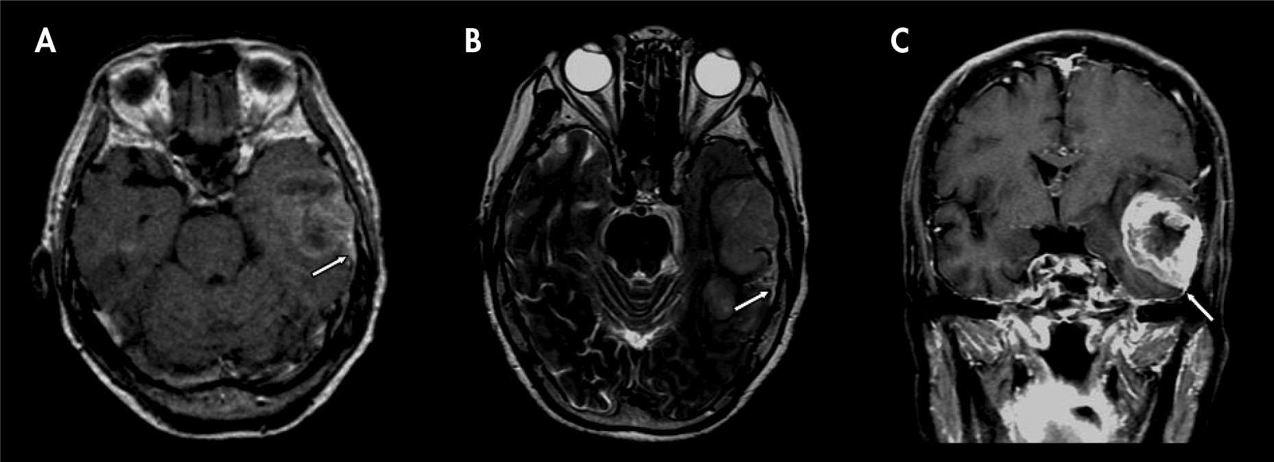
An 86-year-old, right-handed woman was hospitalized because of 1 month of difficulties in naming familiar objects and people. The patient presented circumlocution, mentioning the function and the use of the objects. On neurological assessment, the spontaneous speech was fluent, and auditory comprehension and repetitions were intact. Neither motor symptoms nor sensory deficits were detected. The patient could read and write normally. She had no difficulty with a test that required her to perform a sequential set of hand movements. Also, the patient did not have any discomfort about neurological symptoms, memory loss, bizarre behaviors, or delirium symptoms, except anomic aphasia. A laboratory work-up including complete blood cell counts; electrolyte levels; renal, liver, and thyroid function profiles; vitamin B12 and folic acid; serology for syphilis; and human immunodeficiency virus produced normal results. A bran CT and contrast-enhanced MRI revealed a huge solid tumor over the left temporal lobe, causing apparent perifocal edema, compressing the left lateral ventricle, and pushing the midline to the right (Figure 1). She underwent standard tumor resection and radiotherapy. The pathological result revealed a glioblastoma multiforme. However, the anomic aphasia did not improve postoperatively.
FIGURE 1. Axial CT [A] and Axial T2-Weighed MR [B] Images Show a Huge and Heterogeneous Tumor (4.2×4.3×5.6 cm) Over the Left Temporal Lobe, With a Nonspecific “Dural Tail” Sign (white arrows). Coronal T1-Weighted MR images [C] With Injection of Gadolinium Revealed a Central Necrosis and a Nonspecific “Dural Tail” Sign of the Tumor (white arrows).

Discussion

Anomic aphasia is a special subtype of aphasia characterized by difficulty in naming familiar objects and people. Although aphasia is a common symptom in dementia, anomic aphasia as a core symptom is rarely reported in patients with dementia.2 In the present case, a rapid progression of anomic aphasia without memory impairment excluded the differential diagnosis of Alzheimer's disease. Vascular lesion or other organic problems should be first considered. The CT and MR images revealed that the tumor occupied more than three quadrants of the left temporal lobe (a dominant hemisphere). This result is consistent with studies finding that anomic aphasia almost exclusively occurs with damage to the left hemisphere in right-handed patients.1
Traumatic brain injury, brain surgery, and stroke have been described to be the etiology of secondary anomic aphasia,3,4 which most involved the supramarginal-pulvinar conjunctive area, and was rarely focused in the temporal lobe.4 Furthermore, compared with previous reports of metastatic brain tumor presenting with rapidly progressive dementia,5,6 a solid temporal tumor displaying a rapidly progressive anomic aphasia has never been reported. This report reminds clinicians to be aware that these patients who first display acute anomic aphasia may have more malignant than benign etiology.
Much evidence supports a good outcome of radical resection for either low-grade or high-grade brain tumors.7 However, poor prognostic factors that affecting language deficits, such as old age, female sex, high-grade tumors, and tumors involving two or more quadrants of the temporal lobe, were all present in this patient.7 Consistent with the earlier studies, the anomic aphasia did not improve postoperatively in this patient.

References

1.
DeAngelis LM: Brain tumors. N Engl J Med 2001; 344:114–123
2.
Mesulam M, Rogalski E, Wieneke C, et al.: Neurology of anomia in the semantic variant of primary progressive aphasia. Brain 2009; 132:2553–2565
3.
Miozzo A, Soardi M, Cappa SF: Pure anomia with spared action naming due to a left temporal lesion. Neuropsychologia 1994; 32:1101–1109
4.
Yang ZH, Zhao XQ, Wang CX, et al.: Neuroanatomic correlation of the post-stroke aphasias studied with imaging. Neurol Res 2008; 30:356–360
5.
Weaver JD, Vinters HV, Koretz B, et al.: Lymphomatosis cerebri presenting as rapidly progressive dementia. Neurologist 2007; 13:150–153
6.
Rivas E, Sanchez-Herrero J, Alonso M, et al.: Miliary brain metastases presenting as rapidly progressive dementia. Neuropathology 2005; 25:153–158
7.
Haglund MM, Berger MS, Shamseldin M, et al.: Cortical localization of temporal lobe language sites in patients with gliomas. Neurosurgery 1994; 34:567–576, discussion 576

Information & Authors

Information

Published In

Go to The Journal of Neuropsychiatry and Clinical Neurosciences
The Journal of Neuropsychiatry and Clinical Neurosciences
Pages: E18 - E19
PubMed: 23686047

History

Published online: 1 April 2013
Published in print: Spring 2013

Authors

Affiliations

Hui-Ming Feng, M.D.
Dept. of Otolaryngology–Head and Neck Surgery, Tri-Service General Hospital, National Defense Medical Center, Taipei, Taiwan
Shin-Chang Kuo, M.D.
Dept. of Psychiatry, Tri-Service General Hospital, National Defense Medical Center, Taipei, Taiwan
Chun-Yen Chen, M.D.
Dept. of Psychiatry, Tri-Service General Hospital, National Defense Medical Center, Taipei, Taiwan
Yi-Wei Yeh, M.D.
Dept. of Psychiatry, Tri-Service General Hospital, National Defense Medical Center, Taipei, Taiwan

Notes

Correspondence: Yi-Wei Yeh, M.D., Dept. of Psychiatry, Tri-Service General Hospital, National Defense Medical Center; e-mail: [email protected]

Metrics & Citations

Metrics

Citations

Export Citations

If you have the appropriate software installed, you can download article citation data to the citation manager of your choice. Simply select your manager software from the list below and click Download.

For more information or tips please see 'Downloading to a citation manager' in the Help menu.

Format
Citation style
Style
Copy to clipboard

There are no citations for this item

View Options

View options

PDF/ePub

View PDF/ePub

Get Access

Login options

Already a subscriber? Access your subscription through your login credentials or your institution for full access to this article.

Personal login Institutional Login Open Athens login
Purchase Options

Purchase this article to access the full text.

PPV Articles - Journal of Neuropsychiatry and Clinical Neurosciences

PPV Articles - Journal of Neuropsychiatry and Clinical Neurosciences

Not a subscriber?

Subscribe Now / Learn More

PsychiatryOnline subscription options offer access to the DSM-5-TR® library, books, journals, CME, and patient resources. This all-in-one virtual library provides psychiatrists and mental health professionals with key resources for diagnosis, treatment, research, and professional development.

Need more help? PsychiatryOnline Customer Service may be reached by emailing [email protected] or by calling 800-368-5777 (in the U.S.) or 703-907-7322 (outside the U.S.).

Media

Figures

Other

Tables

Share

Share

Share article link

Share