Positron emission tomographic (PET) imaging of the brain has been used for many years in research and clinical settings. Many factors have contributed to increased demand including improved techniques, wider availability, reimbursement by third-party payers, and clinical application in oncology. PET imaging demonstrated a 57% annual growth rate after 2004.
5 PET neuroimaging has many applications for use in neurodegenerative disorders, movement disorders, and neuropsychiatric conditions. Through examining metabolism, enzyme activity, protein accumulation, and receptor binding, abnormalities on PET imaging may be detected before changes are apparent on structural imaging. As PET brain imaging is applied to clinical practice, it is important for clinicians to understand both the advantages and limitations as a diagnostic tool. Illustrative clinical examples will focus on the most mature application, the evaluation of patients with primary neurodegenerative disorders.
Principles of PET
PET scanners detect and localize radiotracers (ligands labeled with a radioisotope) to produce semiquantitative images of in vivo brain functioning.
6–8 The radioisotopes decay through emitting a positron, which annihilates by colliding with an electron. Two gamma-photons traveling at 180 degrees away from each other result from the annihilation, each with energy of 511 keV. They strike opposite detectors nearly simultaneously resulting in a true coincidence event. The scintillator detector converts the 511 keV photon to light, which is detected by the attached photomultiplier tubes. The path between the two detectors hit in coincidence is termed the line of response (LOR). This is critical for the accuracy of the PET scan because the LOR provides additional information regarding position of the coincidence event compared with single photon imaging. An image reconstruction algorithm is applied to the raw data and the resulting spatial distribution of the radiotracer reflects the functional process. Spatial resolution is typically 5 to 10 mm. Although this is much better than single photon emission tomography (SPECT) imaging, companion structural imaging (magnetic resonance imaging, MRI; computed tomography, CT) is typically obtained for better mapping of function to anatomy.
7There are several methodological issues that can degrade PET images.
9 One of the most important is Compton scatter. Interaction of one or both of the photons with electrons causes varying degrees of change in direction, artifactually shifting the LOR. Since Compton scatter decreases the energy of the photons, some scatter can be removed by setting an energy threshold. However, some scattered photons are able to hit detectors with energy above the threshold. Therefore, mathematical scatter correction is also required. In addition, photons can be lost to photoelectric absorption or severe Compton scatter in the tissue, resulting in attenuation artifact. PET is particularly sensitive to attenuation since two photons must be detected for each coincidence event. Attenuation correction is accomplished by measuring the composition of the body along the LOR, most commonly using CT (MRI attenuation correction is also starting to enter clinical use). Attenuation correction depends on matching two different scans, so it is important to instruct the patient to limit movement during scanning to avoid misregistration.
10 Random coincidence events occur when two photons from different annihilation events strike the detectors within the coincidence timing window. The random rate depends on count rate in the scanner and can be adjusted for mathematically. Previously, detector rings were separated with septa (lead or tungsten annular shields) to reduce scatter coincidence events. However, this also blocks cross plane true coincidence events, decreasing count rate and prolonging scan time. Removing the septa reduced data gaps and permitted the generation cross-plane data, allowing for three dimensions (3D) PET.
9,11Radiotracers are defined as molecules selective for biochemical processes labeled with a radioactive isotope. For PET, the radioactive moiety is commonly fluorine-18 (
18F) or carbon-11 (
11 C).
12–14 These radioisotopes are produced by a cyclotron.
15 The short half-life of these isotopes, 20 min for carbon-11 and 110 min for fluorine-18, limits patients’ exposure to radioactivity, but presents challenges to availability. The very short half-life of
11C limits its use to centers with an on-site cyclotron. The longer half -life of
18F provides sufficient time for regional shipping of radiotracers.
13 There are several requirements for developing a PET radiotracer useful for in vivo imaging.
12,13 The radiotracer should provide sufficient signal at a concentration low enough to prevent pharmacodynamics effects. It is important for the radiotracer to have highly specific binding to the target (high selectivity) and low nonspecific (off target) binding. The radiotracer should localize rapidly to the target. It is preferable that the radiotracer not be metabolized. If it metabolized, metabolites should not bind with the target.
12 For brain imaging, the radiotracer must be able to reach its target by crossing the blood brain barrier. Important factors include molecule size, lipophilicity, protein binding, size, and polarity.
12FDG PET
2-[
18F]-fluoro-2-deoxy-D-glucose (FDG) is the mostly widely used PET ligand. FDG PET provides a measure of cellular glucose metabolism. FDG is a glucose analog that is taken into cells by membrane-bound glucose transporters. FDG undergoes only the first step in glucose metabolism (phosphorylation). It is trapped within the cell until it is dephosphorylated. FDG accumulation in brain is proportional to regional cerebral metabolic rate (rCMR), which is closely related to neuronal and synaptic function.
16,19 FDG PET is FDA approved for the identification of epileptic seizure foci, evaluation of malignancies, and for specific cardiology studies.
20 FDG PET has been in used in studies of many neuropsychiatric conditions including depression, bipolar disorder, schizophrenia, traumatic brain injury, and neurocognitive disorders.
21In 2009, the Society of Nuclear Medicine released guidelines for the use of FDG PET in brain imaging.
17 The guidelines should be reviewed and used consistently to ensure accuracy and reproducibility of imaging. Multiple patient-related factors can alter FDG uptake, including blood glucose level, medications, and emotional state (e.g. angry, agitated, profoundly depressed).
7,18,19 Presence of some psychoactive medications (e.g. barbiturates, benzodiazepines) can alter uptake of FDG. Insulin can alter biodistribution of FDG by promoting uptake into muscle, so it is recommended that patients fast and have intravenous fluid containing dextrose or parenteral feedings withheld for 4–6 hours before administration of FDG. In patients with diabetes, blood glucose levels may become elevated to the point of competing with FDG for uptake into the brain. Blood glucose level should be measured prior to injection of FDG in all patients. If too high, rescheduling of the examination is recommended. Alternatively, short acting insulin can be administered with delay of the FDG injection to allow time for clearance of insulin. Good hydration is important to assure rapid renal clearance, which decreases background activity and patients’ radiation exposure. FDG is taken up gradually, so the patient needs to remain in a stable environment (quiet darkened room, very limited activity) for at least 30 min after administration. Eyes open is preferred, as occipital lobe metabolism will be reduced under eyes closed conditions.
7,18FDG PET can be used clinically to assist in differentiating types of primary neurodegenerative disorders. These conditions affect more than half of individuals greater than 85 years of age and approximately 14% of individuals greater than 65 years.
18 The most prevalent are major neurocognitive disorder due to Alzheimer's disease (AD, 50%−60%), major neurocognitive disorder due to frontotemporal lobar degeneration (FTD, 15%−25%), and major neurocognitive disorder with Lewy bodies (DLB, 15%−25%).
18 FDG PET is currently not FDA approved to assist in the diagnosis of mild neurocognitive disorders or neurodegenerative disorders; thus it is considered an off-label use. However, evidence from clinical trials, national guidelines, and policies from third party payers support its use under defined criteria. In 2001, the Centers for Medicare and Medicaid Services approved coverage for use of FDG PET in patients who meet the minimal requirements of having a decline in cognitive function for at least six months, meeting criteria for both FTD and AD, and cause of clinical symptoms is unclear.
14,22Although presence of characteristic patterns of altered metabolism can improve diagnostic sensitivity, it is important to remember that there is sufficient variability to cause overlap between disease states. The most common pattern in AD is hypometabolism in the posterior cingulate cortex (PCC), precuneus and posterior temporoparietal areas including the angular gyrus (
Figure 1).
16,18,23,24 PCC is often the earliest area affected. In advanced AD, metabolic activity is reduced in the prefrontal cortex with possible involvement of more posterior frontal lobe (sparing the sensorimotor cortex). Metabolic deficits are typically bilateral but may be asymmetric, and unilateral deficits are also seen. Metabolism is usually preserved in anterior cingulate and visual cortices, thalamus, basal ganglia and cerebellum. In contrast, the most common pattern in FTD is hypometabolism in the frontal and anterior temporal cortices. The anterior cingulate cortex is usually affected, whereas the parietal cortex is spared (
Figure 1). The pattern of metabolic impairment in temporoparietal cortices is similar in DLB and AD. PCC is commonly preserved in DLB (although some studies disagree) but impaired in AD. Visual cortex is commonly impaired in DLB but preserved in AD (
Figure 1).
16,18,23,24Multiple studies have evaluated the use of FDG PET in differentiating among dementing disorders. Sensitivity and specificity are generally favorable, but vary across studies and are affected by analytic methodology (e.g. axial sections versus surface projections, visual versus quantitative assessment).
16,18,25,26 The 2011 National Institute on Aging-Alzheimer’s Association guidelines for the diagnosis of dementia due to AD stated that FDG PET and other biomarkers may be used to confirm that the pathophysiological process is consistent with AD in patients who meet the core clinical diagnosis for probable AD, but does not recommend for routine use in diagnosis.
27 FDG PET may be particularly useful when structural imaging is uninformative.
26,28 In some cases frontal and temporoparietal hypometabolism occur equally, complicating the clinical diagnosis. For these patients, β-amyloid PET (
Figure 2) may provide additional guidance (discussed below).
Amyloid and Tau PET
The hallmark pathologies of AD are cerebral β-amyloid plaques (extracellular) and tau neurofibrillary tangles (intracellular).
14,29,30 However, the relationship between these markers, neurodegeneration, and development of dementia remains controversial. Both autopsy and amyloid PET studies have reported that amyloid burden does not correlate with cognitive state, synaptic activity or neurodegeneration.
30,31 Many cognitively healthy elderly individuals have considerable amyloid deposition (
Figure 2).
2–4 Studies have reported that both cognitive state and neurodegeneration do strongly correlate with level of soluble β-amyloid oligomers and the density of tau neurofibrillary tangles.
14,30,31 It has been proposed that presence of β-amyloid facilitates hyperphosphorylation of tau, the first stage in the process that ends with deposition of aggregated tau as neurofibrillary tangles.
30In 2013 The Amyloid Imaging Task Force (a collaboration between the Alzheimer’s Association and the Society for Nuclear Medicine) released appropriate use criteria for amyloid PET imaging: atypical symptom presentation (etiologically mixed or atypical clinical course), atypical age of onset (65 years or less), persistent or progressive unexplained mild cognitive impairment.
32,33 The cost of amyloid PET remains a factor in widespread clinical adoption. In 2013, the Centers for Medicare and Medicaid Services concluded that there was insufficient evidence to approve coverage for use of amyloid PET for the diagnosis of AD. Limited use in research was approved. A single amyloid PET scan is covered provided it is done as part of an approved clinical research study.
34 The most studied amyloid PET radiotracer is Pittsburg Compound B (
11C-PiB).
14,33 However, the short half-life (20 min) prohibited commercial use. There are currently three 18F amyloid PET radiotracers approved by the FDA: florbetaben, florbetapir, and flutemetamol. A recent meta-analysis found that all three tracers had high sensitivity and specificity.
33 There were no differences in diagnostic accuracy. The use of amyloid PET may enhance the development of treatments targeting β-amyloid plaques. Currently florbetapir is being used in a phase 1b clinical trial for adacanumab (a human immunoglobulin γ1 monoclonal antibody selective for aggregated β-amyloid plaques) to identify amyloid positive patients. Of 278 patients evaluated with amyloid PET imaging, 39% were excluded due to a negative scan.
35 Multiple other radiotracers with improved characteristics, such as reduced binding in white matter, are under development.
14Combining information from different ligands may provide a better understanding of some aspects of pathophysiology.
36 A recent longitudinal study obtained both FDG and amyloid (
11C-PiB) PET as well as functional connectivity measures (resting state functional MRI) in patients with probable early stage AD.
37 Hypometabolism in areas of prefrontal cortex free of amyloid was associated with amyloid burden in areas with high functional connectivity to those areas.
37 As noted by the authors of this study, their results are consistent with the hypothesis that functional disconnection is the basis for neuronal dysfunction (as indicated by hypometabolism) in areas with little or no amyloid deposition.
Tauopathies (e.g. AD, progressive supranuclear palsy, corticobasal degeneration, chronic trauma encephalopathy, some FTD variants) are neurodegenerative conditions characterized by the accumulation of misfolded aggregates of the tau phosphoprotein.
29–31 AD is the most common tauopathy. Tau is critical for the assembly and stabilization of the microtubules which support the cytoskeleton and intracellular transport.
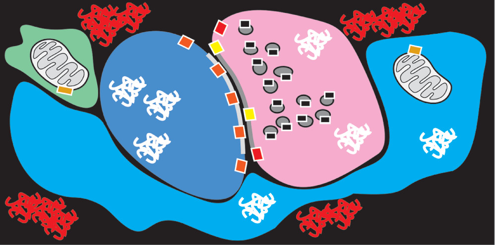
29 As noted above, tau deposition is highly associated with cognitive impairment and neurodegeneration, so early detection is important for developing interventions. However, the characteristics of tau deposition pose many challenges for developing PET tracers in addition to the requirement of crossing the blood brain barrier. Deposition is intracellular, so PET tracers must be able to pass into the cells of the brain (tau aggregates can occur in neurons, astrocytes, and oligodendrocytes;
Figure 3).
38 Further, there are six tau isoforms. Tau aggregates associated with specific tauopathies vary in their ultrastructural conformation and histopathological characteristics.
31 Tau aggregates also have multiple sites available for binding of tracers and conformational differences could result in variable binding affinity.
30,31 Binding to other misfolded proteins in addition to tau is also a concern. The PET tracer 18F-FDDNP, for example, binds to both amyloid plaques and tau neurofibrillary tangles.
30 The development of tau PET tracers is a rapidly growing field, and requires establishment of reference ranges, stability and applicable quantification methods.
30,31 The tau PET tracer [
18F]-T807 (also known as [
18F]-1451) is the most studied.
31 Several candidate ligands have been shown to have unexpectedly high retention in nontarget areas including the striatum, brainstem, fibria and cerebellar white matter.
30 Studies are underway in multiple conditions (e.g. AD, traumatic brain injury, depression).
31Other Targets for PET Radiotracers
In recent decades, a wide array of PET ligands have been developed that provide a basis for assessing many aspects of presynaptic (e.g. neurotransmitter reuptake transporters, vesicular neurotransmitter transporters, neurotransmitter synthesis, modulatory receptors), postsynaptic (e.g. neurotransmitter receptors) and glial (e.g. mitochondrial receptor) functioning (
Figure 3).
14,16,39 Most familiar are the ligands targeting postsynaptic neurotransmitter receptors. Parkinson’s disease (PD) has been established to have decrease in nigrostriatal dopamine functioning, and provides a useful example of the diversity of presynaptic targets for PET ligands.
14,39–41 One is dopamine transporter (DAT) receptors in the striatum. These presynaptic receptors are responsible for dopamine reuptake, and levels decrease in PD due to both loss of terminals and adaptive changes.
39,41 A DAT SPECT tracer (
123I-ioflupane) was approved by the FDA in 2011 as an adjunctive diagnostic tool in the evaluation of adult patients with suspected Parkinsonian syndromes.
40 Several DAT PET tracers are under development including both 11C and 18F forms of PE2I (a cocaine derivative) and
11C methylphenidate.
41,42 Presynaptic dopamine functioning can also be assessed utilizing tracers for vesicular monoamine transporter type 2 (VMAT2) such as
11C or
18F DTBZ.
14,39,41,43 Dihydroxyphenylalanine (DOPA) is a final precursor for monoamine synthesis, so uptake of 18F-DOPA reflects local synthesis of dopamine, norepinephrine and/or serotonin (depending on the region examined).
39,44PET ligands that bind to a similar array of targets are available for many other neurotransmitter systems. For example, PET tracers that bind to serotonin 2A (5-HT
2A) receptors include
18F setoperone,
11C MDL 100907 and
18F altanserin.
14 Decreased 5-HT
2A binding has been reported in mania, aggression, and schizophrenia. Increased binding has been reported in obsessive-compulsive disorder and Tourette syndrome.
14 The endocannabinoid type 1(CB1) receptor is present on all cell types in the brain (presynaptic terminals, astrocytes, microglia, oligodendroglia).
14 Increased binding of 18F-MK-9470 (an inverse agonist) has been reported in PD, anorexia nervosa, migraine, and following stroke.
14,39 Elevated binding of 11C-OMAR (a selective antagonist) has been reported in schizophrenia.
14The translocator protein (TSPO, previously called the peripheral-type benzodiazepine receptor) provides an example of an entirely different type of target. TSPO is an 18 kDa protein located on the outer mitochondrial membrane of microglia and astrocytes.
14,45,46 In healthy brain, expression is quite low. Expression increases with injury-induced microglial and astroglial activation.
45,46 The most widely utilized tracer has been
11C (R)PK11195 (selective antagonist). Multiple new tracers are in development with better characteristics (e.g. higher brain permeability, lower nonspecific binding,
18F for longer half -life).
46 A present limitation is the need to assess a genetic polymorphism that is associated with intrinsic differences in binding status (high, mixed, low).
14,46 Clinical studies suggest potential for evaluating and monitoring therapeutic interventions for brain injuring conditions.
14,45,46 Binding has been reported to be increased in the ischemic penumbra for several days to weeks after stroke onset. Focal increases in both lesions and surrounding white matter have been found in patients with multiple sclerosis. Increases have also been found in several neurodegenerative conditions (e.g. AD, FTD, PD, viral encephalitis). It may also serve as a useful biomarker in other disorders. A recent study reported that patients with schizophrenia had elevated TSPO binding in gray matter compared with healthy controls.
47 In the group with subclinical symptoms (high risk for psychosis), symptom severity was positively correlated with TSPO distribution volume in gray matter, suggesting potential as a biomarker for elevated risk during the prodromal stage. Although TSPO has been considered a marker for neuroinflammation, its presence on multiple cell types including astrocytes suggests a less specific role.


