Full access
Case Reports
Published Online: 3 May 2019

Long-Standing Psychiatric Features as the Only Clinical Presentation of Vanishing White Matter Disease

Publication: The Journal of Neuropsychiatry and Clinical Neurosciences
Vanishing white matter disease (VWMD) is an autosomal recessive leukoencephalopathy caused by mutations in any of the five genes (EIF2B1, EIF2B2, EIF2B3, EIF2B4, and EIF2B5) that encode the subunits of eukaryotic translation initiation factor 2B (eIF2B), which is essential for protein synthesis (1).
VWMD clinically ranges from antenatal or early infantile onset with poor outcome (2) to adult onset with slow progression, where the age at onset is a strong predictor of disease course (3). Affected individuals typically have normal early development, followed by neurological deterioration triggered by stress-provoked episodes of rapid decline. Patients with adolescent or adult onset are more likely to exhibit cognitive problems at the time of or shortly after the disease onset. However, psychiatric disorders have been rarely reported as the only symptoms at the time of diagnosis (37) and usually are followed by neurologic manifestations within a few years (6).
In women, VWMD can present as ovarioleukodystrophy, a condition in which leukodystrophy is associated with primary or secondary ovarian failure (2, 3). While this could help in reaching a diagnosis in females, the diagnosis in adult males may be challenging, given the possible atypical onset and insidious clinical course.
VWMD is a recognizable disorder on brain MRI, usually characterized by diffuse and symmetric white matter T2-hyperintensities and progressive white matter rarefaction. The latter is a hallmark of the disease, both in pediatric- and adult-onset cases (7).
EIF2B5 is by far the most commonly mutated gene among adults with VWMD, with the recurrent c.338G>A(p.Arg113His) mutation accounting for the majority of adult cases (6), while c.260C>T(p.Ala87Val) is a founder EIF2B3 VWMD mutation in the French Canadian population of Quebec (8).
Here, we present the case of an adult male who presented with only psychiatric symptoms for more than 20 years before occurrence of the typical neurological symptoms and deterioration of VWMD.

Case Report

The patient came to our attention at the age of 41 for suspected genetic leukoencephalopathy. He was the second child of French Canadian consanguineous (first cousins) parents, originally from Matapedia, a small region in Quebec. His family history was remarkable for psychiatric illness on the maternal side: two of his mother’s brothers died by suicide in the context of drug addiction, and his grandmother had schizophrenia. There were no neurological disorders segregating in the family.
In terms of development, the patient had normal psychomotor and language acquisitions. During late childhood and adolescence, he started to exhibit severe behavioral problems, which retrospectively fit the criteria for disruptive behavior disorder: he would lose his temper quickly, he was frequently involved in violent fights with his peers, and he was defiant toward authority both at home and in school. In addition, he sexually abused a family member. He experienced his first episode of depression at age 16 when he attempted suicide by driving into the family house, causing considerable property damage. This led to his removal from the family home, and a residential treatment program was started. During this period, he did not receive a clear psychiatric diagnosis. His behavior disorder led to significant impairment in his social interactions and caused him to drop out of high school. He eventually worked as a truck driver until the age of 41 when he was finally diagnosed with VWMD.
Since age 16, the patient presented transient urinary incontinence that continued for many years without any other associated neurological symptoms. He experienced a second episode of depression at age 34 when he started to notice some difficulty concentrating as well as short-term memory impairment. For the depressive symptoms, he was prescribed venlafaxine without significant benefit. Four years later, he was admitted to the hospital for a suicide attempt in the context of a further episode of major depression. At that time, his family noticed, for the first time, mild gait instability, and the patient complained of occasional dizziness and lack of energy.
At age 40, the patient was involved in a truck accident, during which he lost consciousness and required hospital admission. Since the accident, he has experienced cognitive decline, progressive gait deterioration with recurrent falls, increasing fine motor impairment, and daily dizziness.

Neuroradiological Findings

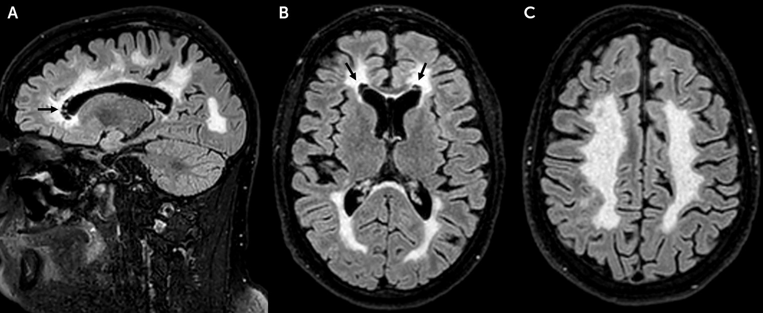
Following the truck accident, a head CT scan revealed the presence of extensive, symmetric white matter disease. Brain MRI (Figure 1) confirmed the presence of bilateral, symmetric, and diffuse white matter abnormalities. Small areas of focal white matter rarefaction were noted bilaterally at the level of the frontal horns of the lateral ventricles. The U fibers, the external rim of the corpus callosum, the internal capsule, and cerebellar white matter were relatively spared.
FIGURE 1. Brain MRI of a patient with vanishing white matter diseasea
a The sagittal (panel A) and axial (panels B and C) fluid-attenuated inversion recovery T2-weighted images are presented, showing the diffuse symmetric white matter abnormalities involving the cerebral hemispheres and sparing the U fibers (panel C) and the outer rim of the corpus callosum and the capsules (panel B). Areas of focal white matter rarefaction are visible around the frontal horns of the lateral ventricles.

Physical Examination

At the time of the patient’s clinical examination, he was still receiving monotherapy with venlafaxine. He was oriented and collaborative. He required help from his parents to recollect his personal and clinical history, he showed detachment from major events of his life (i.e., suicide attempts), and his mood appeared to be depressed. Neurological evaluation revealed mild increased tone, brisk reflexes in the lower limbs, difficulties with rapid alternating movements, and tandem gait. The patient’s gait was mildly ataxic. Given the constellation of the neuroradiological and clinical findings, VWMD was suspected.

Genetic Analysis

A gene panel for leukodystrophies, including the EIF2B1–5 genes, revealed the homozygous mutation c.260C>T(p.Ala87Val) in EIF2B3 (NM_020365.3).

Discussion

The long-standing psychiatric phenotype of our patient, in the absence of any neurological symptoms apart from sphincter dysfunction, represents a unique disease course never before reported, to our knowledge, among EIF2B-related disorders. Psychiatric features and cognitive decline have been described as presenting symptoms in up to 24% of all VWMD patients and 11%−16% of adult-onset VWMD cases, but other neurological symptoms usually occur shortly afterward, within a few years (2, 3, 6, 9). From our patient’s history, his gait and coordination were normal to the extent of working as a truck driver.
Isolated psychiatric symptoms can dominate the presentation of leukodystrophies in patients with onset in adolescence or adulthood (1013). For this reason, we suggest that physicians take into account the possibility of genetic leukoencephalopathies in patients with long-standing psychiatric symptoms, especially if associated with nonspecific neurological symptoms (e.g., sphincter dysfunction). Interestingly, our patient presented with urinary incontinence since age 16, whereas in VWMD, this usually occurs during more advanced disease stages, when other neurological symptoms are already evident (3). The predominant psychiatric presentation of our patient leads us to assume that VWMD may remain undiagnosed during the early stages of the disease, and its frequency may be underestimated among adult patients, especially when neurological symptoms are absent or nonspecific. Stress events, such as our patient’s truck accident, dramatically influence the course of the disease, leading to progressive motor decline.
The identification of a leukoencephalopathy was incidental in this patient, because it was only in the context of the truck accident that he underwent a CT scan, a test that started the neuroradiological assessment that led to the final diagnosis. The usefulness of brain MRI pattern recognition in genetic leukoencephalopathies is well known (14). The neuroradiological phenotype of our patient is consistent with that of adult patients with VWMD in previous studies showing diffuse and symmetric white matter involvement with focal white matter rarefaction (15). As demonstrated in a study of adult genetic leukoencephalopathies, the presence of white matter rarefaction should promptly suggest the diagnosis of VWMD (7). The neuroradiological findings in our patient, in line with previous reports, confirm that anterior periventricular white matter rarefaction is a main distinctive neuroradiological feature in adults with VWMD.
EIF2B regulates protein synthesis rates under basal and cellular stress conditions, facilitating ternary complex formation and translation initiation (1). Failure of the translation process of certain astrocytic mRNAs has been thought to be the underlying mechanism that induces degeneration of white matter through loss of astrocyte function, failure of astrocyte-microglia crosstalk, and secondary effect on both oligodendroglia and axons (16). Several studies have shed light on the pathomechanism of EIF2B, showing deregulation of endoplasmic reticulum function in astrocytes in mutant mice (17) and prolonged state of translational hyperrepression in human VWMD cells that fail to recover from stress (18). Although the pathomechanism underlying VWMD is becoming increasingly recognized, the reason for the broad phenotypic spectrum among EIF2B-related disorders remains largely unknown.
Since the first report of the mutation more than a decade ago, c.260C>T(p.Ala87Val) has been rarely documented (1922). Of note, it has been previously reported in two pediatric cases (8) and in one adult female case (15) in the Quebec population, showing variable disease course according to different age at disease onset. Our report confirms that c.260C>T(p.Ala87Val) in EIF2B3 is a common mutation in the French Canadian population.
In conclusion, we have reported an isolated long-standing psychiatric presentation in an adult carrying an EIF2B3 mutation, broadening the clinical spectrum associated with eIF2B-related disorders. Importantly, we have highlighted the need for clinicians to consider a thorough neurological and neuroradiological assessment in individuals presenting with psychiatric features, especially when accompanied by mild nonspecific neurological symptoms, since the finding of focal white matter rarefaction on brain MRI would lead to a VWMD diagnosis.
Recognizing VWMD at an early stage is crucial in order to avoid stress-provoked episodes of rapid decline and may be critical for future therapeutic strategies.
Acknowledgments

The authors thank the study patient.

Footnote

The authors have confirmed that details of the case have been disguised to protect patient privacy.

References

1.
Pavitt GD: Regulation of translation initiation factor eIF2B at the hub of the integrated stress response. Wiley Interdiscip Rev RNA 2018; 9:e1491
2.
Schiffmann R, Fogli A, van der Knaap MS, et al: Childhood ataxia with central nervous system hypomyelination/vanishing white matter, in GeneReviews, Adam MP, Ardinger HH, Pagon RA, Seattle, University of Washington, 1993–2018
3.
Hamilton EMC, van der Lei HDW, Vermeulen G, et al: VWM Research Group: Natural history of vanishing white matter. Ann Neurol 2018; 84:274–288
4.
Ohtake H, Shimohata T, Terajima K, et al: Adult-onset leukoencephalopathy with vanishing white matter with a missense mutation in EIF2B5. Neurology 2004; 62:1601–1603
5.
van der Knaap MS, Leegwater PA, van Berkel CG, et al: Arg113His mutation in eIF2Bepsilon as cause of leukoencephalopathy in adults. Neurology 2004; 62:1598–1600
6.
Labauge P, Horzinski L, Ayrignac X, et al: Natural history of adult-onset eIF2B-related disorders: a multi-centric survey of 16 cases. Brain 2009; 132:2161–2169
7.
Ayrignac X, Carra-Dalliere C, Menjot de Champfleur N, et al: Adult-onset genetic leukoencephalopathies: a MRI pattern-based approach in a comprehensive study of 154 patients. Brain 2015; 138:284–292
8.
Robinson MÈ, Rossignol E, Brais B, et al: Vanishing white matter disease in French-Canadian patients from Quebec. Pediatr Neurol 2014; 51:225–232
9.
Matsukawa T, Wang X, Liu R, et al: Adult-onset leukoencephalopathies with vanishing white matter with novel missense mutations in EIF2B2, EIF2B3, and EIF2B5. Neurogenetics 2011; 12:259–261
10.
Zheng F, Lin Z, Ye X, et al: Unusual brain images of a boy with adolescent cerebral X-linked adrenoleukodystrophy presenting with exhibitionism: a CARE-compliant case report. Medicine (Baltimore) 2017; 96:e9481
11.
Haag C, Laakmann G, Voderholzer U, et al: Adult form of metachromatic leukodystrophy with predominantly psychotic manifestations. Nervenarzt 1992; 63:444–446
12.
Smith J, Williams MT, Misra VK: Psychiatric disease in an adolescent as a harbinger of cerebral X-linked adrenoleukodystrophy. Pract Neurol 2018; 18:242–245
13.
Rauschka H, Colsch B, Baumann N, et al.: Late-onset metachromatic leukodystrophy: genotype strongly influences phenotype. Neurology 2006; 67:859–863
14.
Schiffmann R, van der Knaap MS: Invited article: an MRI-based approach to the diagnosis of white matter disorders. Neurology 2009; 72:750–759
15.
La Piana R, Vanderver A, van der Knaap M, et al: Adult-onset vanishing white matter disease due to a novel EIF2B3 mutation. Arch Neurol 2012; 69:765–768
16.
Bugiani M, Vuong C, Breur M, et al: Vanishing white matter: a leukodystrophy due to astrocytic dysfunction. Brain Pathol 2018; 28:408–421
17.
Wisse LE, Penning R, Zaal EA, et al: Proteomic and metabolomic analyses of vanishing white matter mouse astrocytes reveal deregulation of ER functions. Front Cell Neurosci 2017; 11:411
18.
Moon SL, Parker R: EIF2B2 mutations in vanishing white matter disease hypersuppress translation and delay recovery during the integrated stress response. RNA 2018; 24:841–852
19.
van der Knaap MS, Leegwater PA, Könst AA, et al: Mutations in each of the five subunits of translation initiation factor eIF2B can cause leukoencephalopathy with vanishing white matter. Ann Neurol 2002; 51:264–270
20.
Carra-Dalliere C, Horzinski L, Ayrignac X, et al: Natural history of adult-onset eIF2B-related disorders: a multicentric survey of 24 cases. Rev Neurol (Paris) 2011; 167:802–811
21.
Herwerth M, Schwaiger BJ, Kreiser K, et al: Adult-onset vanishing white matter disease as differential diagnosis of primary progressive multiple sclerosis: a case report. Mult Scler 2015; 21:666–668
22.
Ghezzi L, Scarpini E, Rango M, et al.: A 66-year-old patient with vanishing white matter disease due to the p.Ala87Val EIF2B3 mutation. Neurology 2012; 79:2077–2078

Information & Authors

Information

Published In

Go to The Journal of Neuropsychiatry and Clinical Neurosciences
The Journal of Neuropsychiatry and Clinical Neurosciences
Pages: 276 - 279
PubMed: 31046592

History

Received: 21 November 2018
Accepted: 12 December 2018
Published online: 3 May 2019
Published in print: Summer 2019

Keywords

  1. Vanishing White Matter disease
  2. Psychiatric Disorders
  3. EIF2B-Related Disorders
  4. EIF2B3
  5. Lukodystrophy

Authors

Details

Andrea Accogli, M.D.
The Departments of Neurology and Neurosurgery and Pediatrics, McGill University, Montreal (Accogli); DINOGMI-Università di Genova, Italy (Accogli); IRCCS Ospedale Policlinico San Martino, Genova, Italy (Accogli); the Laboratory of Neurogenetics of Motion, Montreal Neurological Institute, McGill University, Montreal (Brais, La Piana); the Department of Human Genetics, McGill University, Montreal (Brais); the Department of Diagnostic Radiology, Kingston General Hospital, Queen’s University, Kingston, Ontario, Canada (Tampieri); and the Department of Neuroradiology, Montreal Neurological Hospital and Institute, McGill University, Montreal (La Piana).
Bernard Brais, M.D., Ph.D.
The Departments of Neurology and Neurosurgery and Pediatrics, McGill University, Montreal (Accogli); DINOGMI-Università di Genova, Italy (Accogli); IRCCS Ospedale Policlinico San Martino, Genova, Italy (Accogli); the Laboratory of Neurogenetics of Motion, Montreal Neurological Institute, McGill University, Montreal (Brais, La Piana); the Department of Human Genetics, McGill University, Montreal (Brais); the Department of Diagnostic Radiology, Kingston General Hospital, Queen’s University, Kingston, Ontario, Canada (Tampieri); and the Department of Neuroradiology, Montreal Neurological Hospital and Institute, McGill University, Montreal (La Piana).
Donatella Tampieri, M.D.
The Departments of Neurology and Neurosurgery and Pediatrics, McGill University, Montreal (Accogli); DINOGMI-Università di Genova, Italy (Accogli); IRCCS Ospedale Policlinico San Martino, Genova, Italy (Accogli); the Laboratory of Neurogenetics of Motion, Montreal Neurological Institute, McGill University, Montreal (Brais, La Piana); the Department of Human Genetics, McGill University, Montreal (Brais); the Department of Diagnostic Radiology, Kingston General Hospital, Queen’s University, Kingston, Ontario, Canada (Tampieri); and the Department of Neuroradiology, Montreal Neurological Hospital and Institute, McGill University, Montreal (La Piana).
Roberta La Piana, M.D., Ph.D [email protected]
The Departments of Neurology and Neurosurgery and Pediatrics, McGill University, Montreal (Accogli); DINOGMI-Università di Genova, Italy (Accogli); IRCCS Ospedale Policlinico San Martino, Genova, Italy (Accogli); the Laboratory of Neurogenetics of Motion, Montreal Neurological Institute, McGill University, Montreal (Brais, La Piana); the Department of Human Genetics, McGill University, Montreal (Brais); the Department of Diagnostic Radiology, Kingston General Hospital, Queen’s University, Kingston, Ontario, Canada (Tampieri); and the Department of Neuroradiology, Montreal Neurological Hospital and Institute, McGill University, Montreal (La Piana).

Notes

Send correspondence to Dr. La Piana ([email protected]).

Funding Information

Supported by a doctoral award from the Fonds de recherche du Québec–Santé to Dr. La Piana, and the Canadian Radiological Foundation.Dr. La Piana has received research support from the Fondation Monaco. The other authors report no financial relationships with commercial interests.

Metrics & Citations

Metrics

Citations

Export Citations

If you have the appropriate software installed, you can download article citation data to the citation manager of your choice. Simply select your manager software from the list below and click Download.

For more information or tips please see 'Downloading to a citation manager' in the Help menu.

Format
Citation style
Style
Copy to clipboard

View Options

View options

PDF/EPUB

View PDF/EPUB

Login options

Already a subscriber? Access your subscription through your login credentials or your institution for full access to this article.

Personal login Institutional Login Open Athens login
Purchase Options

Purchase this article to access the full text.

PPV Articles - Journal of Neuropsychiatry and Clinical Neurosciences

PPV Articles - Journal of Neuropsychiatry and Clinical Neurosciences

Not a subscriber?

Subscribe Now / Learn More

PsychiatryOnline subscription options offer access to the DSM-5-TR® library, books, journals, CME, and patient resources. This all-in-one virtual library provides psychiatrists and mental health professionals with key resources for diagnosis, treatment, research, and professional development.

Need more help? PsychiatryOnline Customer Service may be reached by emailing [email protected] or by calling 800-368-5777 (in the U.S.) or 703-907-7322 (outside the U.S.).

Media

Figures

Other

Tables

Share

Share

Share article link

Share