Full access
Articles
Published Online: 19 November 2021

Clinical Features, Immunotherapy, and Outcomes of Anti-Leucine-Rich Glioma-Inactivated-1 Encephalitis

Publication: The Journal of Neuropsychiatry and Clinical Neurosciences

Abstract

Objective:

The investigators aimed to explore the clinical characteristics, immunotherapy, and outcomes of patients with antileucine-rich glioma-inactivated-1 (anti-LGI1) encephalitis.

Methods:

Data on participants’ clinical characteristics, laboratory findings, radiological and electroencephalogram (EEG) features, treatment, and outcomes from January 2012 to December 2016 were collected. Statistical analysis was conducted to assess the factors associated with patient functional outcome. Forty-three patients were enrolled in the study, with a predominance of males (65.1%). The median age at onset was 57 years (interquartile range [IQR]: 44.0–65.0). The median time from onset to diagnosis was 60 days (IQR: 37.0–127.0).

Results:

The main clinical manifestations included epilepsy (100%), faciobrachial dystonic seizures (FBDS; 44.2%), cognitive dysfunction (95.3%), neuropsychiatric disturbances (76.7%), sleep disorders (58.1%), and disturbance of consciousness (48.8%). Twenty-two patients (51.2%) had hyponatremia, 31 (72.1%) had abnormal EEG results, and 30 (69.8%) had abnormal brain MRI scans, mainly involving the hippocampus (76.7%) or temporal lobe (40%). Twenty of 34 patients (58.8%) in a follow-up MRI examination exhibited hippocampal atrophy. Twenty-five patients (58.2%) were administered corticosteroids and intravenous immunoglobulin, whereas 17 patients were treated only with corticosteroids. Forty-one patients (95.3%) had favorable outcomes after a median of 21.5 months (IQR: 7–43) of follow-up. Serum sodium level was a factor associated with a disabled status (odds ratio=0.81, 95% CI=0.66, 0.98, p=0.03). Anti-LGI1 encephalitis patients were characterized by seizures, FBDS, cognitive deficits, neuropsychiatric disturbances, and hyponatremia.

Conclusions:

Most patients with anti-LGI1 encephalitis are nonparaneoplastic, have low recurrence rates, and have favorable prognostic outcomes. Rapid evaluation, prompt immunotherapy, and long-term follow-up are essential in the care of anti-LGI1 encephalitis patients.
Antileucine-rich glioma-inactivated-1 (anti-LGI1) encephalitis is a newly discovered subtype of autoimmune limbic encephalitis. The specific antibody against the LGI1 subunit of the voltage-gated potassium channels (VGKC) complex was identified in 2010 and has been associated with limbic encephalitis (1). LGI1 is a neuronal glycoprotein that is mainly expressed in the hippocampus dentate gyrus, the CA3 region, the lateral temporal cortex, and mossy fibers. As a component of VGKC complexes, LGI1 acts as a transsynaptic link between presynaptic ADAM23 and postsynaptic ADAM22, thereby enabling synaptic transmissions of neuronal signals (2, 3). Anti-LGI1 antibodies can prevent synaptic transmissions involved in the processes of learning, behavior, and memory.
Anti-LGI1 encephalitis has a specific antibody against LGI1 and frequently present medial temporal lobe inflammation (1, 4), resulting in limbic encephalitis, psychiatric symptoms, faciobrachial dystonic seizures (FBDS), and seizures (5, 6). Non-limbic encephalitis patients with anti-LGI1 encephalitis have been shown to present clinical manifestations, such as chorea and ataxia, outside the limbic system (5). Anti-LGI1 encephalitis is a less common autoimmune disease that is rarely associated with tumors. Anti-LGI1 encephalitis patients exhibited good clinical responses to immunotherapy but were more likely to relapse (5, 7, 8). These characteristics may differ from other typical immune-associated limbic encephalitis (5, 9).
The number of reported anti-LGI1 encephalitis patients in various studies is no more than 300, with most of them from Europe and the United States. The clinical course of anti-LGI1 encephalitis is complex, and its diagnosis is challenging. Additionally, therapeutic options have not been standardized, and various outcomes of different treatments have not been fully evaluated. The limited data from a small sample of Chinese patients do not fully elucidate the characteristics and therapeutic outcomes of anti-LGI1 encephalitis (6, 10, 11). This study was aimed to evaluate the clinical characteristics and prognosis of 43 Chinese patients with anti-LGI1 encephalitis.

Methods

Ethical approval for this study was obtained from the Research Ethics Committee of Xuanwu Hospital of Capital Medical University. Patients who agreed to sign an informed consent form were included. Analysis of study data were anonymized to remove patients’ identifying information.

Study Participants

We recruited patients with clinically confirmed LGI1-antibody encephalitis who had been admitted at the Xuanwu Hospital of Capital Medical University. Recruitment was performed from January 1, 2012, to December 31, 2016. Clinical inclusion criteria were as follows: patients who met the anti-LGI1 encephalitis diagnostic criteria (4), which included subacute onset (rapid progression <3 months) of cognitive impairment, seizures, altered mental status, and psychiatric symptoms; reasonable exclusion of alternate disorders; and patients who were positive for LGI1 antibody in serum or in cerebral spinal fluid (CSF).

Data Collection

The following data were recorded: demographic data (gender and age), initial symptoms, clinical symptoms during the disease course, laboratory examination findings, CSF findings, LGI1 antibody levels in serum and CSF, cranial MRI findings, electroencephalogram (EEG) findings, immunotherapeutic methods, and functional status (evaluated by the Modified Rankin Scale [mRS]).

Treatment and Prognostic Evaluation

All patients received symptomatic supportive treatment, immunotherapy, and systemic neoplasm screening. First-line immunotherapy included monotherapy or a combination of intravenous pulse glucocorticoid therapy (methylprednisolone, 1,000 mg, or 500 mg, for 3 or 5 days and tapering oral), intravenous gamma immunoglobulin (IVIg; 0.4 g/kg/day for 5 days), or plasma exchange (3–5 times) (3, 9, 12). Immunosuppressants such as rituximab, cyclophosphamide, azathioprine, or mycophenolate mofetil were used as second-line immunotherapeutic options (7, 10).
Patient follow-up started at 3 months after initial immunotherapy and continued during clinical visits or by telephone. The trained telephone interviewers were blinded to the clinical conditions. Functional status was evaluated using the mRS. On the basis of mRS findings at 6 months, patients were categorized into two groups: functionally independent and disabled. Patients with an mRS score of 0 were considered to have had a complete recovery. Good and poor outcomes were defined as an mRS score of 0–1 and 2–6, respectively, at patients’ last follow-up assessments. Relapse was defined as the appearance of a new symptom or worsening of a preexisting symptom after improvement or stabilization of the disease for at least 1 month. Unfavorable prognostic factors were also evaluated.

Statistical Approach

Categorical variables are expressed as counts (percentages). Nonnormal quantitative variables are expressed using their medians and interquartile ranges (IQRs), while normal quantitative variables are presented as means (and standard deviations). Patients’ baseline variables of demographic and clinical characteristics were analyzed using descriptive statistics and were compared between the functionally independent (mRS score 0–1) and disabled (mRS score 2–6) groups using Student’s t test or Wilcoxon rank sum test (W) for continuous variables and chi-square test for categorical variables. Univariate logistic regression analysis was performed to establish the variables that accounted for disabled outcomes. After adjusting for confounding factors with a p value <0.1 and factors that were clinically considered to be closely related to outcomes, the multivariate logistic regression model was used to assess any independent factors for the disabled outcome. A p value ≤0.05 was considered statistically significant. All statistical analyses were performed using SPSS, version 19.0 (SPSS, Chicago).

Results

Clinical Features

Clinical characteristics of the 43 patients (males, N=28 [65.1%]; females, N=15 [34.9%]) are summarized in Table 1. The median age at onset of anti-LGI1 encephalitis was 57 years (IQR: 44–65 years). The median duration of illness was 60 days (range: 37–127 days).
TABLE 1. Demographic and clinical characteristics of patients with antileucine-rich glioma-inactivated-1 (anti-LGI1) encephalitis (N=43)a
CharacteristicNMedianTotal N%IQR
Age (years) 5743 44–65
Male28 4365.1 
Duration (days) 60 37.0–127.0
Initial reported symptoms    
Seizures18 4341.9 
Memory impairment13 4330.2 
Neuropsychiatric symptoms10 4323.3 
Sleep disturbances1 432.3 
Movement abnormalities1 432.3 
Main clinical features    
 Fever7 4316.3 
 Seizure43 43100.0 
 Status epilepticus10 4323.3 
 Faciobrachial dystonic seizures19 4344.2 
 Cognitive dysfunction41 4395.3 
 Neuropsychiatric symptoms33 4376.7 
 Behavioral dyscontrol18 3354.5 
 Visual and auditory hallucination17 3351.5 
 Nonsensical speech16 3348.5 
 Personality changes8 3324.4 
 Irritability6 3318.2 
 Anxiety5 3315.2 
 Sleep disturbances25 4358.1 
 Disturbance of consciousness21 4348.8 
 Movement abnormalities16 4337.2 
 Autonomic dysfunction14 4332.6 
Serum sodium (mmol/L) 13343 130–138
Hyponatremia (<135 mmol)22 4351.2 
Increased pressure8 4318.6 
CSF pleocytosis (>8 leukocytes/mL)7 4316.3 
Increased CSF protein (>45 mg/dL)9 4320.9 
Intrathecal synthesis of oligoclonal bands8 4318.6 
LGI1 Abs positive in serum and CSF at admission43 43100.0 
LGI1 Abs positive in CSF or serum at follow-up4 2218.2 
LGI1 Abs negative at follow-up18 2281.8 
Abnormal EEG at admission31 4372.1 
 Focal slow activity10 3132.3 
 Diffuse slow activity8 3125.8 
 Epileptic activity13 3141.9 
 Normal12  27.9 
Abnormal MRI at presentation30 4369.8 
 Hippocampal23 3076.7 
 Temporal lobe12 3040.0 
 Basal ganglia6 3020.0 
 Insular cortex3 3010.0 
 Other lesions9 3030.0 
Normal MRI13 4330.2 
MRI at follow-up34 4379.1 
Hippocampal or mesial temporal atrophy20 3458.8 
Hippocampal T2 hyperintensity6 3417.6 
Normal5 3414.7 
Local hypoperfusion on brain SPECT8 100.8 
Time to initiate immunotherapy (days) 64 43.0–127.0
Corticosteroids plus IVIg21 4348.8 
Corticosteroids or IVIg22 4330.2 
Mycophenolate mofetil4 439.3 
mRS score at admission 3 3.3–4.0
mRS score at last follow-up 1 0–1
 ≤241 4395.3 
 3–51 432.3 
 0–115 4334.9 
Deceased at last follow-up1 432.3 
Relapse10 4323.3 
Length of hospitalization (days) 16 13.0–22.0
Period of follow-up (months) 21.5 7.0–43.0
a
CSF=cerebral spinal fluid; EEG=electroencephalogram; FDBS=faciobrachial dystonic seizures; IQR=interquartile range; IVIg=intravenous immunoglobulin; mRS=Modified Rankin Scale; SPECT=single-photon emission computed tomography.
Seven of the 43 patients presented prodromal symptoms, such as fever, headache, dizziness, and stomach discomforts. Initial presentations (Figure 1) were epileptic seizure (N=18, 41.9%), memory impairments (N=13, 30.2%), and neuropsychiatric symptoms (N=10, 23.3%). During the disease course, the most common symptoms among the 43 patients were epileptic seizures, in patients with status epilepticus (N=10, 23.3%), FBDS (N=19, 44.2%), and cognitive dysfunction (N=41, 95.3%), mainly in memory deficits and deterioration of orientation. In addition, 33 patients (76.7%) exhibited neuropsychiatric disturbances, including behavioral dyscontrol (N=18, 54.5%), visual or auditory hallucinations (N=17, 51.5%), nonsensical speech (N=16, 48.5%), personality changes (N=8, 24.4%), anxiety (N=5, 15.2%), and irritability (N=6, 18.2%). Other symptoms in the 43 patients included sleep disturbances (N=25, 58.1%), altered level of consciousness (N=21, 48.8%), movement abnormalities (N=16, 37.2%), and autonomic dysfunction (N=14, 32.6%).
FIGURE 1. Percentage of initial symptoms in 43 patients with antileucine-rich glioma-inactivated-1 encephalitis

Auxiliary Investigations

Laboratory and imaging results are summarized in Table 1. A total of 22 patients (51.2%) had hyponatremia in the acute phase. Increased puncture pressure was observed in eight patients (18.6%). CSF pleocytosis (>8 cells/μL) was reported for seven patients (16.3%), and CSF protein level was elevated in nine (20.9%, mean=36.5 mg/dl [SD=21.3]). Glucose and chloride levels in the CSF were all normal. Other autoantibodies and classical paraneoplastic antibodies in serum and CSF were not detected in any patient. In addition, standard pelvic ultrasound and chest computerized tomography were normal. All patients tested positive for LGI1 antibodies in serum or CSF samples. LGI1 antibodies were retested in 22 patients (51.2%); only four patients (18.2%) presented positive CSF samples during follow-up.
Abnormal EEG results were found in 31 patients (72.1%). The most common EEG abnormalities were focal slow activity (N=10, 32.3%), diffuse slowing (N=8, 25.8%), and epileptiform discharges (N=13, 41.9%).
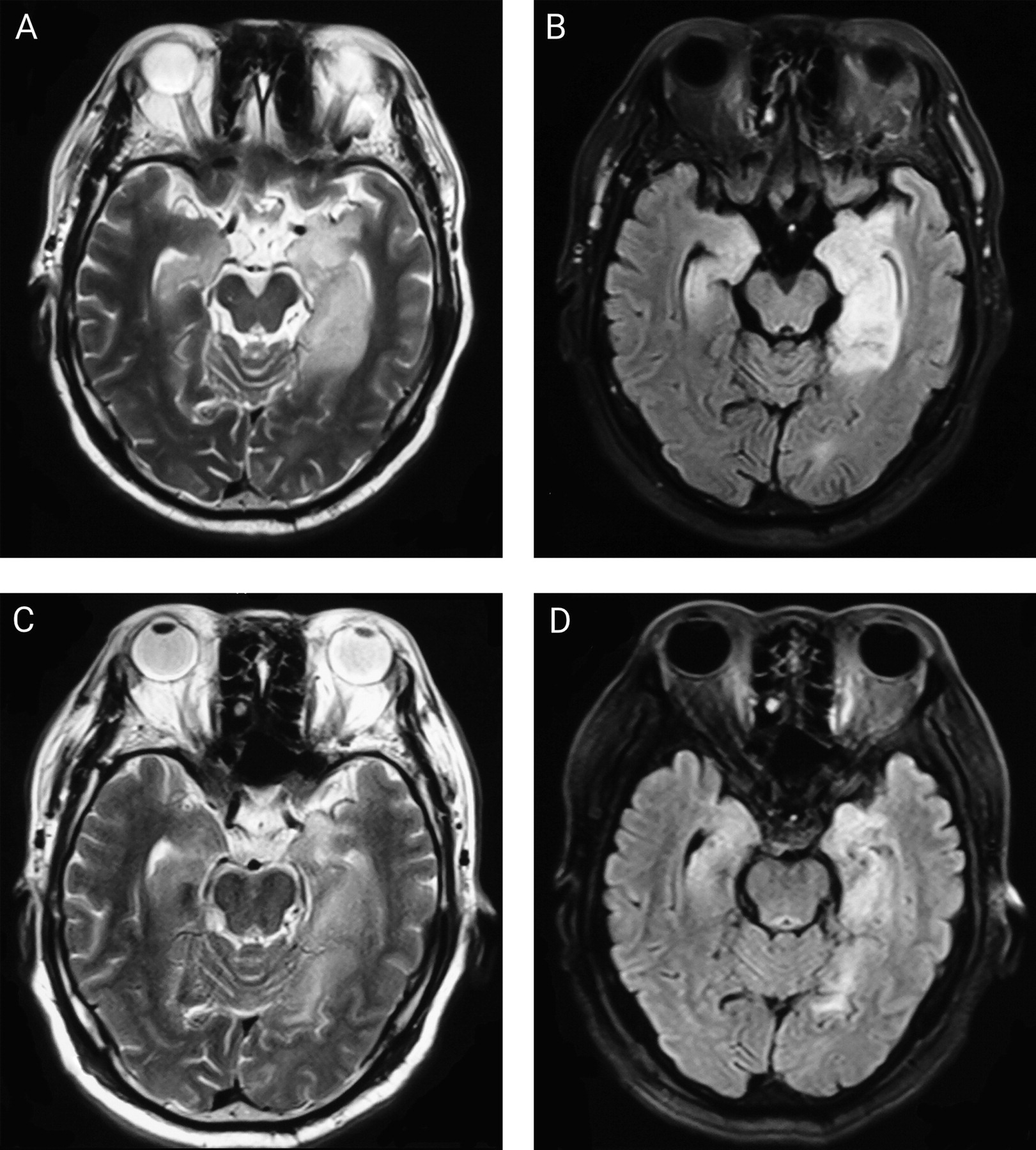
Among the total sample, 30 patients (69.8%) exhibited increased unilateral or bilateral signals on MRI fluid-attenuated inversion recovery or T2 that were thought to be related to encephalitis at admission. MRI detection of one representative patient is presented in Figure 2. Among these 30 patients, involvement of the limbic system, including the hippocampus (N=23, 76.7%), temporal lobe (N=12, 53.5%), and insular cortex (N=3, 10.0%), was observed in 29 patients. Other locations involved white matter (N=8, 26.7%), basal ganglia (N=6, 20.0%), and other lesions (N=9, 30.0%) in the frontal lobe, parietal lobe, and cerebellum. Follow-up MRI scans were performed for 34 patients (79.1%); among these, hippocampal atrophy was shown in 20 (58.8%). Eight of 10 patients showed abnormal local hypoperfusion when compared with the contralateral on single-photon emission tomography (SPECT).
FIGURE 2. Brain imaging of a 65-year-old male patient with antileucine-rich glioma-inactivated-1 encephalitis presenting with paroxysmal unresponsiveness and memory impairment for 25 daysa
aT2-weighted axial (panel A) and fluid-attenuated inversion recovery (FLAIR) axial (panel B) imaging at presentation are shown, demonstrating enlargement and increased signal intensity in the bilateral hippocampi and the bilateral mesial temporal lobe. Follow-up T2-weighted axial (panel C) and FLAIR axial (panel D) imaging after treatment demonstrates smaller lesions compared with previous imaging.

Treatment

Patients with seizures and FBDS were treated with single or combined oral antiepileptic drugs. All patients were treated with immunotherapy consisting of corticosteroids and/or IVIg (Table 1). All patients were initially treated with corticosteroids; 21 (48.8%) also received IVIg (0.4 g/kg daily for 5 days), whereas one patient received only IVIg. Due to relapse, four patients (9.5%) received mycophenolate mofetil. The median time from onset to treatment was 64 days (IQR: 43.0–127.0 days). All patients exhibited good clinical responses to immunotherapy as confirmed by symptomatic improvement (reduced seizures or cognitive improvement). Patients with frequent FBDS and presenting with seizures responded poorly to antiepileptic drugs and improved upon immunotherapy. Ten patients (23.3%) relapsed during corticosteroid reduction. Six of these 10 patients received an initial oral dose of corticosteroids, whereas four patients were treated with mycophenolate mofetil.

Outcomes and Analysis of Factors

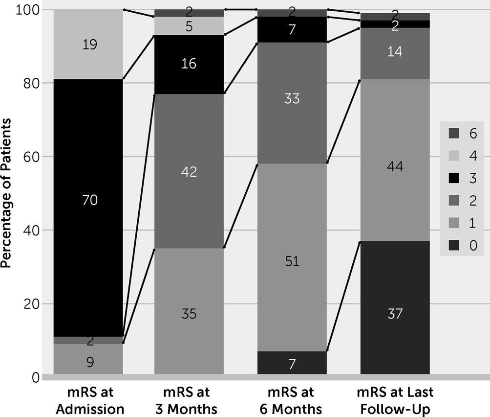
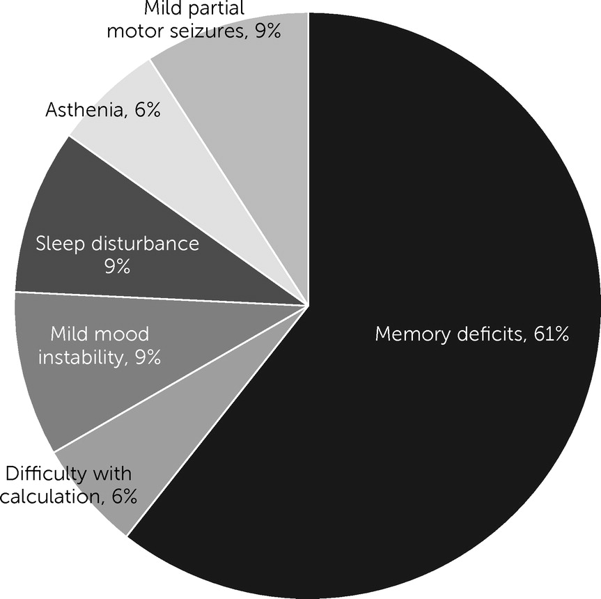
The median length of hospital stay was 16 days (IQR: 13–22). The median follow-up period was 21.5 months (IQR: 7–43). The median mRS score was 3 (IQR: 3.3–4) at admission and 1 (IQR: 0–1) at the last follow-up. Forty-one patients (95.3%) exhibited good clinical outcomes; only two patients (4.7%) exhibited poor clinical outcomes at the last follow-up (Figure 3). Four patients (9.3%) relapsed during follow-up, and one patient died of pulmonary infection during subsequent treatment in the local hospital 1 month after discharge. Fifteen patients were reported to have completely recovered, and 27 exhibited mild residual symptoms. Residual symptoms included memory deficits (N=20), difficulties with calculation (N=2), mild disturbances of emotional regulation (N=3), sleep disturbances (N=3), asthenia (N=2), and mild partial motor occasional seizures (N=3) (Figure 4).
FIGURE 3. Modified Rankin Scale (mRS) scores among patients with antileucine-rich glioma-inactivated-1 encephalitis at hospital admission and the 3-month, 6-month, and last follow-up assessments (N=43)a
a“Good” outcome is defined as an mRS score of 0–2, and a “poor” outcome is defined as an mRS score of 3–6. The numbers in each box denote the percentage of patients.
FIGURE 4. Percentage of residual symptoms in 20 patients with antileucine-rich glioma-inactivated-1 encephalitis
Table 2 shows the association between individual clinical, laboratory, and imaging factors and neurologic function at 6 months. Serum sodium level (p=0.018), abnormal EEG findings (p=0.026), and mRS score on admission (p=0.006) were all associated with poor functional status at 6 months. We performed regression analysis to identify independent predictors of functional status at 6 months. Univariate analysis results are summarized in Table 3. Serum sodium was associated with disabled status (odds ratio=0.81, 95% CI=0.66, 0.98, p<0.05). Abnormal EEG findings (odds ratio=0.11, 95% CI=0.01, 0.10, p<0.1) and mRS score at admission (odds ratio=5.40, 95% CI=1.00, 28.97, p<0.10) were also associated with disabled status. Other factors, such as age, clinical symptoms, time to treatment, CSF findings, MRI, and type of treatment were not significantly associated with prognosis.
TABLE 2. Association of variables with functional status at 6 months of follow-up among patients with antileucine-rich glioma-inactivated-1 encephalitisa
 Functionally independent group (N=25)Disabled group (N=18) 
VariableNMedian%IQRNMedian%IQRp
Age (years) 58.0 44.0–64.5 56.0 45.5–65.50.892
Male15 60.0 9 50.0 0.550
Duration 60.0 38.5–105.0 60.0 36.0–187.50.585
Type of onset (<3 months)16 64.0 11 61.1 1.000
Fever3 12.0 4 22.2 0.427
Seizure21 84.0 17 94.4 0.380
Faciobrachial dystonic seizures13 52.0 6 33.3 0.351
Cognitive dysfunction23 92.0 18 100.0 0.502
Neuropsychiatric symptoms20 80.0 13 72.2 0.717
Disturbances of consciousness11 44.0 10 55.5 0.543
Sleep disturbances14 56.0 11 61.1 0.765
Movement10 40.0 6 33.3 0.755
Autonomic dysfunction8 32.0 7 38.9 0.750
Serum sodium (mmol/L) 137 131.0–139.0 130 126.0–136.50.018b
Hyponatremia (<145 mmol/L)11 44.0 11 61.1 0.358
CSF pleocytosis (>8 cells/dl)7 28.0 5 27.8 1.000
Protein level (mg/dl) 30.0 24.0–41.5 32.5 18.8–48.50.455
Abnormal EEG12 48.0 15 83.3 0.026*
Abnormal MRI15 60.0 13 72.2 0.523
Involvement of limbic system18 72.0 11 61.1 0.335
Hippocampal atrophy14 56.0 9 50.0 0.763
Steroids plus IVIg15 60.0 10 55.6 1.000
Steroids or IVIg10 40.0 8 44.4 1.000
Immunosuppressant2 8.0 2 11.1 1.000
Time to start treat (<1 month)5 20.0 3 16.7 1.000
mRS score on admission 2 1–4 2 0–40.006*
Relapse6 24.0 4 22.2 0.594
a
CSF=cerebral spinal fluid; EEG=electroencephalogram; IQR=interquartile range; IVIg=intravenous immunoglobulins; mRS=Modified Rankin Scale.
b
Serum sodium level, abnormal EEG findings, and mRS on admission were associated with poor functional status at the 6-month follow-up.
*≤0.05.
TABLE 3. Univariate analysis of potential factors for disabled outcomes among patients with antileucine-rich glioma-inactivated-1 encephalitisa
Disability indexOdds ratio95% CIp
Age (years)1.0120.941, 1.0880.749
Sex1.2910.411, 5.4200.543
Type of onset (subacute <3 months)0.1660.009, 3.1700.233
Duration0.1000.996, 1.0120.291
Serum sodium (mmol/L)0.8060.663, 0.9810.031
Protein level of CSF (mg/dl)0.0640.03, 1.6410.097
EEG abnormality0.1090.011, 1.1040.061
MRI abnormality0.3130.046, 2.1430.237
Involvement of limbic system2.3070.47, 11.20.310
Therapy with steroids plus IVIg1.6780.230, 12.2350.610
Time to treatment0.2710.015, 4.8870.376
mRS score on admission5.3981.006, 28.9700.050
a
CSF=cerebral spinal fluid; EEG=electroencephalogram; IVIg=intravenous immunoglobulins; mRS=Modified Rankin Scale.

Discussion

This study evaluated the clinical characteristics and therapeutic outcomes of 43 patients with anti-LGI1 encephalitis. Some of the characteristics of these patients are consistent with those of patients in previously reported studies. For example, seizure and FBDS were the prominent clinical presentation in anti-LGI1 encephalitis patients, followed by cognitive and neuropsychiatric disturbances. This disorder, for the most part, was not a paraneoplastic syndrome. Temporal and/or hippocampal atrophy developed during the later stages of this condition. Seizures responded well to immunotherapy, and cognition gradually improved. However, our findings differ from previous reports in several aspects. First, the median age at onset was 57 years (IQR: 44–65), with a male preponderance (65.1%). This stands in contrast to other autoimmune encephalidities, particularly anti-N-methyl-d-aspartate encephalitis, which has a median age of about 21 years (range of 8 months to 85 years) and a female predominance of 2:1 (4, 13). Second, hyponatremia is a common presentation of anti-LGI1 encephalitis, which is not typical of other autoimmune encephalidities. Third, most patients had more favorable outcomes and lower recurrence and mortality rates than those with anti-LGI1 encephalitis reported elsewhere (5).
The initial and most common presenting symptom in this cohort was frequent multifocal seizures; this symptom was resistant to antiepileptic drugs but responded well to immunotherapy (5, 6, 13). FBDS was common among our patients. Anti-LGI1 encephalitis is associated with FBDS, with incidence of FBDS at approximately 20%−40% (5, 7). FBDS, which is seizure-like episodes, usually presents with brief stereotyped ipsilateral face grimacing and posturing of the upper limb, which may be confounded with myoclonus (14). In 40%−71% of patients, it presents early, before other symptoms (6, 7). Studies have reported on the relationship between anti-LGI1 limbic encephalitis, seizure, and FBDS. However, it has not been established whether FBDS is a seizure or simply an extrapyramidal manifestation (6, 7, 13). Some MRI studies revealed that FBDS duration is associated with reduced pallidum volume (15, 16). Because brain positron emission tomography (17) or SPECT revealed hypermetabolism in the basal ganglia, it is possible that cortical-striatal networks are hyperactive, suggesting a possible subcortical origin for the seizures (6, 13, 17). These observations suggest a role of basal ganglia in FBDS pathophysiology. In addition, magnetoencephalography findings from anti-LGI1 encephalitis patients with FBDS showed that epileptic discharge from the temporal lobe could be transmitted to the insular cortex, thereby causing FBDS (6, 14, 17). Therefore, FBDS might be a unique type of insular seizure between myoclonus and spasm. Further research should aim at elucidating on the mechanisms involved.
There were abnormal EEG findings in 96.8% of the patients in our study. The majority of EEGs showed slow or epileptic activity that is nonspecific in identifying FBDS or establishing disease outcomes. Because of the absence of synchronized EEG abnormalities, it was difficult to recognize these brief seizures. Therefore, there is a need to increase awareness of the multiple semiology of FBDS and follow-up EEG, which may be an early clue for the diagnosis of anti-LGI1 encephalitis.
Most of our patients exhibited rapid progressive memory deficits, which affect recent memory and disorientation. In anti-LGI1 encephalitis associated with cognitive impairment, the hippocampus, temporal lobe, and basal ganglia are the major pathogenetic targets (1619). In this study, unilateral or bilateral hippocampus or basal ganglia hyperintense signals were found in most patients. Moreover, nearly 57% of the patients were found to have developed hippocampal atrophy or sclerosis during follow-up, in tandem with previous studies (15, 16). These findings imply that hippocampal inflammation occurs in the acute phase but is not always detected by routine MRI. Hippocampal atrophy is caused by seizures or inflammation (15, 16, 18). The abnormal local hypoperfusion revealed by SPECT suggests that cortical hypometabolism is associated with synaptic dysfunction as well as hippocampal temporal atrophy and might be an important characteristic of the inflammatory process and temporal damage (1719). There is an association between hippocampal atrophy and cognitive dysfunction in anti-LGI1 encephalitis.
In this study, however, there was no correlation between hippocampal atrophy and clinical outcomes. Instead, the extent of CNS involvement may have influenced cognitive outcomes for patients without temporal hypermetabolism and those with unilateral involvements only (16, 19). On the basis of these preliminary findings, our patients shared similar features. Patients without hypermetabolism were more likely to achieve favorable outcomes than those with mild cognitive sequelae with inflammation and hippocampal damage. Further research is needed to determine the bases for hippocampal atrophy and cognitive impairment among persons with anti-LGI1 encephalitis.
Many (76.5%) of our patients exhibited neuropsychiatric disturbances, a substantial proportion of which resolved with immunotherapy. Neuropsychiatric symptoms that occurred in the early phase led many patients to present for initial evaluation and treatment in psychiatric hospitals, which highlights the importance of early recognition of this disease.
The median time from onset to diagnosis was 2 months, suggesting that early clinical diagnosis remains a challenge in clinical practice. Early diagnosis is often delayed by atypical clinical manifestations, normal early MRI findings, and the lack of evidence of inflammatory changes in the CSF. For early diagnosis and effective immunotherapy, follow-up of suspected encephalitis cases is essential. Abnormalities in temporal lobe MRI scans suggest autoimmune encephalitis, and testing for LGI1 antibodies should be initiated as soon as there is reasonable suspicion of this condition.
Hyponatremia also was a common feature of anti-LGI1 encephalitis, similar to prior reports of this problem among persons with VGKC antibody-associated limbic encephalitis (20). Serum sodium levels were decreased in 51% of patients in the present cohort. Serum sodium levels reflect severity of disease and, accordingly, may be associated with clinical outcomes. After sodium supplementation and immunotherapy, serum sodium levels gradually returned to normal levels. However, hyponatremia is not specific, and its exact mechanisms have not been elucidated. LGI1 is highly expressed in the hypothalamus and specific renal tubules of the kidney. Hyponatremia might result from abnormal secretions of antidiuretic hormones because of hypothalamic-pituitary neuraxis inflammation (16, 21) and the antibody-mediated effect on renal tubules (22).
Although limbic encephalitis may occur as a paraneoplastic complication of small cell lung cancer, thymoma, and testicular cancer (9, 21), no evidence of tumor was found in any patient in the present cohort of patients with anti-LGI1 encephalitis. While anti-LGI1 encephalitis may be associated with tumors (23, 24), the present findings suggest that the strength of this association may be lower than with other forms of limbic encephalitis.
Optimal therapeutic options for anti-LGI1 encephalitis have not been established, and early detection and aggressive immunomodulatory therapy are needed to improve prognosis. Appropriate immunotherapy can reverse the course of this disease; in particular, it may eliminate otherwise refractory seizures (especially for FBDS and those related to electrolyte disturbances), prevent progressive brain atrophy, and mitigate cognitive impairment (24). However, there is at present no widely accepted guideline for the immunotherapy of anti-LGI1 encephalitis. Based on cohort studies and expert opinion, it is suggested that corticosteroids, IVIg, or plasmapheresis be considered as first-line therapeutic options; rituximab and cyclophosphamide be considered as second-line therapeutic options in refractory cases (5, 8).
Most (94%) of the patients in this study responded well to steroids or IVIg treatment. Few developed persistent residual neurological impairments, neuropsychiatric disturbances, or memory impairments, consistent with previous reports (2426). Although a minority (approximately 15%) of patients with anti-LGI1 encephalitis may develop persistent cognitive impairments (26), the persistence and severity of such impairments appears to vary with the timing of immunotherapy (earlier treatment favors better cognitive outcomes). However, the timing of treatment initiation did not predict outcomes in the present cohort, suggesting that this association between treatment timing and clinical outcomes requires further investigation.
Symptomatic relapse was not common in the present cohort. Most of the patients gradually improved when administered the initial dose of oral corticosteroid or mycophenolate mofetil as secondary treatment. However, definitive evidence regarding effective treatment and maintenance of remission has not been reported and require further investigation.
Despite the limitations associated with retrospective and observational studies that are based on medical records in a single center, the characteristics of the present cohort provide useful insights into the clinical features, course, and outcomes of persons with anti-LGI1 encephalitis. However, the relatively small sample size, single center, and short-term follow-up suggest that clinicians should remain circumspect about the relative frequencies of these aspects of anti-LGI1 encephalitis and that further study of this condition is needed to better define its clinical features and natural history.

Conclusions

Anti-LGI1 encephalitis is a subtype of autoimmune limbic encephalitis with common clinical features such as seizures, FBDS, cognitive impairments, neuropsychiatric disturbances, and hyponatremia. Most cases of anti-LGI1 encephalitis are not paraneoplastic, have low recurrence rates, and are associated with relatively favorable long-term outcomes when early, aggressive, and sustained combination immunotherapies are provided. Accordingly, expeditious identification and treatment along with long-term follow-up are essential elements of the care of persons with anti-LGI1 encephalitis.

Footnote

The authors report no financial relationships with commercial interests.

References

1.
Irani SR, Alexander S, Waters P, et al: Antibodies to Kv1 potassium channel-complex proteins leucine-rich, glioma inactivated 1 protein and contactin-associated protein-2 in limbic encephalitis, Morvan’s syndrome and acquired neuromyotonia. Brain 2010; 133:2734–2748
2.
Fukata Y, Lovero KL, Iwanaga T, et al: Disruption of LGI1-linked synaptic complex causes abnormal synaptic transmission and epilepsy. Proc Natl Acad Sci USA 2010; 107:3799–3804
3.
Ohkawa T, Fukata Y, Yamasaki M, et al: Autoantibodies to epilepsy-related LGI1 in limbic encephalitis neutralize LGI1-ADAM22 interaction and reduce synaptic AMPA receptors. J Neurosci 2013; 33:18161–18174
4.
Gastaldi M, Thouin A, Vincent A: Antibody-mediated autoimmune encephalopathies and immunotherapies. Neurotherapeutics 2016; 13:147–162
5.
van Sonderen A, Thijs RD, Coenders EC, et al: Anti-LGI1 encephalitis: clinical syndrome and long-term follow-up. Neurology 2016; 87:1449–1456
6.
Gao L, Liu A, Zhan S, et al: Clinical characterization of autoimmune LGI1 antibody limbic encephalitis. Epilepsy Behav 2016; 56:165–169
7.
Thompson J, Bi M, Murchison AG, et al: The importance of early immunotherapy in patients with faciobrachial dystonic seizures. Brain 2018; 141:348–356
8.
Irani SR, Gelfand JM, Bettcher BM, et al: Effect of rituximab in patients with leucine-rich, glioma-inactivated 1 antibody-associated encephalopathy. JAMA Neurol 2014; 71:896–900
9.
Vincent A, Bien CG, Irani SR, et al: Autoantibodies associated with diseases of the CNS: new developments and future challenges. Lancet Neurol 2011; 10:759–772
10.
Shen CH, Fang GL, Yang F, et al: Seizures and risk of epilepsy in anti-NMDAR, anti-LGI1, and anti-GABABR encephalitis. Ann Clin Transl Neurol 2020; 7:1392–1399
11.
Yao L, Yue W, Xunyi W, et al: Clinical features and long-term outcomes of seizures associated with autoimmune encephalitis: a follow-up study in East China. J Clin Neurosci 2019; 68:73–79
12.
Graus F, Titulaer MJ, Balu R, et al: A clinical approach to diagnosis of autoimmune encephalitis. Lancet Neurol 2016; 15:391–404
13.
Aurangzeb S, Symmonds M, Knight RK, et al: LGI1-antibody encephalitis is characterised by frequent, multifocal clinical and subclinical seizures. Seizure 2017; 50:14–17
14.
Barajas RF, Collins DE, Cha S, et al: Adult-onset drug-refractory seizure disorder associated with anti-voltage-gated potassium-channel antibody. Epilepsia 2010; 51:473–477
15.
Kotsenas AL, Watson RE, Pittock SJ, et al: MRI findings in autoimmune voltage-gated potassium channel complex encephalitis with seizures: one potential etiology for mesial temporal sclerosis. AJNR Am J Neuroradiol 2014; 35:84–89
16.
Miller TD, Chong TT, Aimola Davies AM, et al: Focal CA3 hippocampal subfield atrophy following LGI1 VGKC-complex antibody limbic encephalitis. Brain 2017; 140:1212–1219
17.
Kamaleshwaran KK, Iyer RS, Antony J, et al: 18F-FDG PET/CT findings in voltage-gated potassium channel limbic encephalitis. Clin Nucl Med 2013; 38:392–394
18.
Szots M, Blaabjerg M, Orsi G, et al: Global brain atrophy and metabolic dysfunction in LGI1 encephalitis: A prospective multimodal MRI study. J Neurol Sci 2017; 376:159–165
19.
Finke C, Prüss H, Heine J, et al: Evaluation of cognitive deficits and structural hippocampal damage in encephalitis with leucine-rich, glioma-inactivated 1 antibodies. JAMA Neurol 2017; 74:50–59
20.
Bien CG, Elger CE: Limbic encephalitis: a cause of temporal lobe epilepsy with onset in adult life. Epilepsy Behav 2007; 10:529–538
21.
Lai M, Huijbers MG, Lancaster E, et al: Investigation of LGI1 as the antigen in limbic encephalitis previously attributed to potassium channels: a case series. Lancet Neurol 2010; 9:776–785
22.
Head K, Gong S, Joseph S, et al: Defining the expression pattern of the LGI1 gene in BAC transgenic mice. Mamm Genome 2007; 18:328–337
23.
Szots M, Marton A, Kover F, et al: Natural course of LGI1 encephalitis: 3–5 years of follow-up without immunotherapy. J Neurol Sci 2014; 343:198–202
24.
Qiao S, Wu HK, Liu LL, et al: Clinical features and long-term outcomes of anti-leucine-rich glioma-inactivated 1 encephalitis: a multi-center study. Neuropsychiatr Dis Treat 2021; 17:203–212
25.
Ariño H, Armangué T, Petit-Pedrol M, et al: Anti-LGI1-associated cognitive impairment: presentation and long-term outcome. Neurology 2016; 87:759–765
26.
Hang HL, Zhang JH, Chen DW, et al: Clinical characteristics of cognitive impairment and 1-year outcome in patients with anti-LGI1 antibody encephalitis. Front Neurol 2020; 11:852

Information & Authors

Information

Published In

Go to The Journal of Neuropsychiatry and Clinical Neurosciences
The Journal of Neuropsychiatry and Clinical Neurosciences
Pages: 141 - 148
PubMed: 34794327

History

Received: 14 December 2020
Revision received: 17 April 2021
Accepted: 19 May 2021
Published online: 19 November 2021
Published in print: Spring 2022

Keywords

  1. Autoimmune Encephalitis
  2. Anti-LGI1 Encephalitis
  3. FBDS
  4. Immunotherapy
  5. Prognosis

Authors

Details

Xiaoqin Huang, M.D, Ph.D. [email protected]
Department of Neurology, Xuanwu Hospital, Capital Medical University, Beijing.
Chunqiu Fan, M.D., Ph.D.
Department of Neurology, Xuanwu Hospital, Capital Medical University, Beijing.
Lehong Gao, M.D., Ph.D.
Department of Neurology, Xuanwu Hospital, Capital Medical University, Beijing.
Liping Li, M.D.
Department of Neurology, Xuanwu Hospital, Capital Medical University, Beijing.
Jing Ye, M.D., Ph.D.
Department of Neurology, Xuanwu Hospital, Capital Medical University, Beijing.
Huixin Shen, M.M.
Department of Neurology, Xuanwu Hospital, Capital Medical University, Beijing.

Notes

Send correspondence to Dr. Huang ([email protected]).

Funding Information

Supported by the Capital Medical University in 2017 (grant 17JL-02, 2017-2019) and the National Key Research and Development Program of China (grant 2016YFC1300600, 2017-2020).The authors thank the physicians, patients, and family members who provided clinical information for this study.

Metrics & Citations

Metrics

Citations

Export Citations

If you have the appropriate software installed, you can download article citation data to the citation manager of your choice. Simply select your manager software from the list below and click Download.

For more information or tips please see 'Downloading to a citation manager' in the Help menu.

Format
Citation style
Style
Copy to clipboard

View Options

View options

PDF/EPUB

View PDF/EPUB

Get Access

Login options

Already a subscriber? Access your subscription through your login credentials or your institution for full access to this article.

Personal login Institutional Login Open Athens login
Purchase Options

Purchase this article to access the full text.

PPV Articles - Journal of Neuropsychiatry and Clinical Neurosciences

PPV Articles - Journal of Neuropsychiatry and Clinical Neurosciences

Not a subscriber?

Subscribe Now / Learn More

PsychiatryOnline subscription options offer access to the DSM-5-TR® library, books, journals, CME, and patient resources. This all-in-one virtual library provides psychiatrists and mental health professionals with key resources for diagnosis, treatment, research, and professional development.

Need more help? PsychiatryOnline Customer Service may be reached by emailing [email protected] or by calling 800-368-5777 (in the U.S.) or 703-907-7322 (outside the U.S.).
Media

Figures

Other

Tables

Share

Share

Share article link

Share