Depressive patients display neuropsychological deficits presumably due to a dysfunction of the prefrontal cortex (Wisconsin Card Sorting Test [WCST],
1 Verbal Fluency Test [VFT],
2–4 oculomotoric tasks
5). In accordance with this hypothesis, functional imaging studies have shown a reduction in metabolic rate or local blood flow in the prefrontal cortex of patients suffering from depression.
6–8 These deficits in functional frontal brain activity have been shown to normalize following successful antidepressive treatment
9 and seem to predict the therapeutic outcome.
10–13In recent years a new, optical method of investigating the oxygenation of brain tissue, called near-infrared spectroscopy (NIRS), has been introduced. This method has been successfully employed in the investigation of functional oxygenation changes due to various cognitive tasks.
14–17 As NIRS is very easy to apply, cheap, and without any side effects, it has been used to study changes in functional brain activity in psychiatric patients, e.g., suffering from schizophrenia
18 or Alzheimer’s dementia.
19With regard to the above mentioned frontal deficit in depression there are few published studies using NIRS. One study measured the brain oxygenation by means of NIRS during a mirror-drawing task in depressive patients and found that these subjects exhibited a disturbed activation pattern in the forebrain.
20 Half of the depressed patients showed an activation of the nondominant hemisphere during the task, which was never seen in healthy subjects. Applying the NIRS optodes to the left forehead only—and thus merely measuring left-hemispheric prefrontal activity—a reduced brain activation during a verbal fluency task (VFT) in depressive patients when compared with healthy subjects was reported.
21 It was argued that depressive patients might show this frontal hypoactivity within the left hemisphere due to a non-dominant right hemisphere activation during the VFT.
21 To test this hypothesis, we investigated VFT-induced changes in brain oxygenation in depressive patients by means of two NIRS channels over left and right prefrontal brain areas.
METHOD
Nine patients meeting the ICD-10 criteria for depression (F32.x, F33.x) (four female and five male, age range 18–62 years, mean=37.3, SD=13.8) and nine healthy subjects (four female and five male, age range=27–44 years, mean=35.1, SD=5.5), who were recruited by the hospital staff, took part in the present study.
The patients were all either psychiatric inpatients (n=8) or outpatients (n=1) at the Psychiatric University Hospital in Wuerzburg. Mean illness duration (time since occurrence of the first depressive episode) was 157 months (SD=169, range=1–444), with an average of 3.9 depressive episodes (SD=3.5) in the past and 3.4 inpatient treatments (SD=3.2). Mean duration of the current hospitalization was 20 days (SD=17). Three patients had positive family histories for depressive disorders, and one patient had a history of suicidal actions in the past. In electroencephalographic investigations carried out routinely one of the patients showed a mild slowing of the basic rhythm, whereas routine neuroradiological examination revealed no major structural abnormalities in any of the patients. Patients were all treated with either tricyclic antidepressants (n=8, mean=144 mg/day, SD=106) or lithium (n=1, 800 mg/day hypnorex retard). Patients had been receiving stable antidepressant medication for at least 2 weeks and showed no acute or prior comorbidities with other psychiatric illnesses.
The healthy comparison subjects were without acute or former neurological or psychiatric disorder and were medication-free. All subjects were right-handed.
22 Written informed consent was obtained before the investigation. As expected, the depressive patients (mean=16.56, SD=8.14) reported significantly more depressive symptoms than the healthy subjects (mean=1.56, SD=0.44) (
t=5.46,
P<0.0001) according to the Beck Depression Inventory. Due to the markedly different age ranges in both groups, equality of variances was tested by means of Levene's test, which confirmed unequal variances in both groups of subjects (
F=5.83,
P<0.05). Nevertheless, a two-tailed t test with unequal variances revealed mean ages that were not significantly different between both groups (
t=−0.45, df=10.45,
P=0.664).
Verbal Fluency Task
Following a resting baseline condition during which the subjects were instructed to relax with eyes closed for approximately 60 seconds, the letter version of the VFT was conducted. The verbal fluency task consisted of three different letter tasks that required the participants to pronounce as many nouns as possible beginning with a particular letter (first “A,” then “F” and “S”; no proper nouns, no repetitions). Each of the three task conditions lasted about 60 seconds. The correct verbal responses were recorded and used as a measure of behavioral performance. The duration of the baseline and the different task conditions were marked on the NIRO-300 monitor by the investigator.
NIRS
In contrast to visible light, light from the near-infrared spectrum (700–1000 nanometers wave length) can penetrate the skull and is absorbed mainly by two chromophores (oxygenated hemoglobin [O
2HB] and reduced hemoglobin [HHB]). By using the different absorption spectra of these two chromophores, concentrations of O
2HB and HHB in living brain tissue can be calculated from the amount of absorbed near-infrared light by the use of the Lambert-Beer-Law.
14 The typical activation pattern of a particular brain area was found to consist of a local increase in O
2HB and a corresponding decrease in HHB.
14This characteristic activation pattern in tissue oxygenation has for example been observed during the execution of mathematic tasks in healthy subjects for the prefrontal cortex. In most subjects, a task-induced local increase in O
2HB and total hemoglobin as well as a decrease in HHB were observed.
23–27 During the WCST an increase in O
2HB bilaterally in prefrontal brain areas was reported,
16 without a corresponding significant decrease in HHB. In contrast, the Continuous Performance Test led to a decrease of HHB over the right prefrontal hemisphere, without significant changes in O
2HB.
15In the present study the NIRS measurements were conducted using a NIRO-300 monitor (Hamamatsu) with two channels. Each of these two channels comprised a light emitter and a light detector that were placed on the scalp of the participants using double-faced adhesive tape. The light emitter's exact position—according to the international 10–20 system for EEG electrode placement—was Fp1 and Fp2 respectively (for the left and right forehead). The light detector was placed between F7 and F3 for the left side and between F4 and F8 for the right. The light emitter (8 mm diameter) emitted near infrared light (775 nm, 810 nm, 850 nm and 910 nm wavelength) with an impulse frequency of 2 kHz and an impulse duration of 100 nsec. The light detector had a diameter of 20 mm. The NIRO 300 monitor measured changes of O2HB and HHB, as well as a tissue oxygenation index (TOI) (TOI=O2HB/(O2HB+HHb)*100%) and a tissue hemoglobin index (THI) (THI=O2HB+HHb; arbitrary units). The principles of the NIRO-300 measurements were based on the modified Beer-Lambert-Law and the spatial resolved spectroscopy. With a pathlength of 24 (constant of adult head=5.93, distance between emitter and detector=4 cm), O2HB and HHB were indicated in the unit “μm”=10-6 mol/liter. Data were measured with a sample rate of 2 Hz and online transferred from the NIRO-300 monitor to a PC via the RS232C interface. Finally, data were stored on the PC and further analyzed off-line.
Data Analysis and Statistics
Data were exported into ASCII data format by means of the NIRO-300 PC-program. We normalized the length of each of the four time segments (baseline measurement and three task conditions) according to the respective minimal length for the segment over all subjects. This procedure resulted in normalized duration of 52 seconds for the baseline measurement, 56 seconds for the letter “A” condition, 54 seconds for the letter “F,” and 53 seconds for the letter “S.” After that, the average activation for all four (normalized) segments was calculated for both hemispheres and each subject. For statistical purposes a two-by-four-by-two (two hemispheres, four task conditions, two diagnostic groups) analysis of variance (ANOVA) for repeated measurements was calculated for the variables O2HB and HHB. For post hoc analyses, Student's two-tailed t tests for independent samples was applied.
Because of markedly different age ranges in both groups of subjects and because of the significant influence of the factor age on oxygenation, additional analyses were conducted using the factor age as a covariate.
Power analyses were conducted with a probability level of 5% of error one and two.
RESULTS
VFT Performance During NIRS
The healthy comparison subjects achieved an average of 34.9 correct responses (SD=13.2, range=15–56) for the three letter tasks in the VFT, showing similar performances in each of the three letter conditions (A mean= 11.4, SD=5.1, F mean=11.2, SD=4.3, S mean=12.2, SD=5.6). The depressive patients performed slightly—though not significantly (t=1.69, df=16, n.s.)—worse, with an average of 25.6 correct answers (SD=10.1, range=10–46) in the VFT (A mean=8.1, SD=4.8, F mean=8.2, SD=2.95, S mean=9.2, SD=3.7). A subsequently conducted power analysis revealed a power of 0.34 for this test situation.
NIRS Data
In a two-by-four-by-two (two hemispheres, four task conditions, two diagnostic groups) ANOVA for repeated measurements for the variable “O
2HB concentration” a significant main effect of the factor “condition” was observed (
F=20.15, df=3, 48,
P<0.001), as well as a significant main effect of the factor “diagnosis” (
F=9.33, df=1, 16,
P<0.01) and a significant interaction between these two variables (
F=5.96, df=3, 48,
P<0.01). Subsequently conducted post hoc tests revealed that mean O
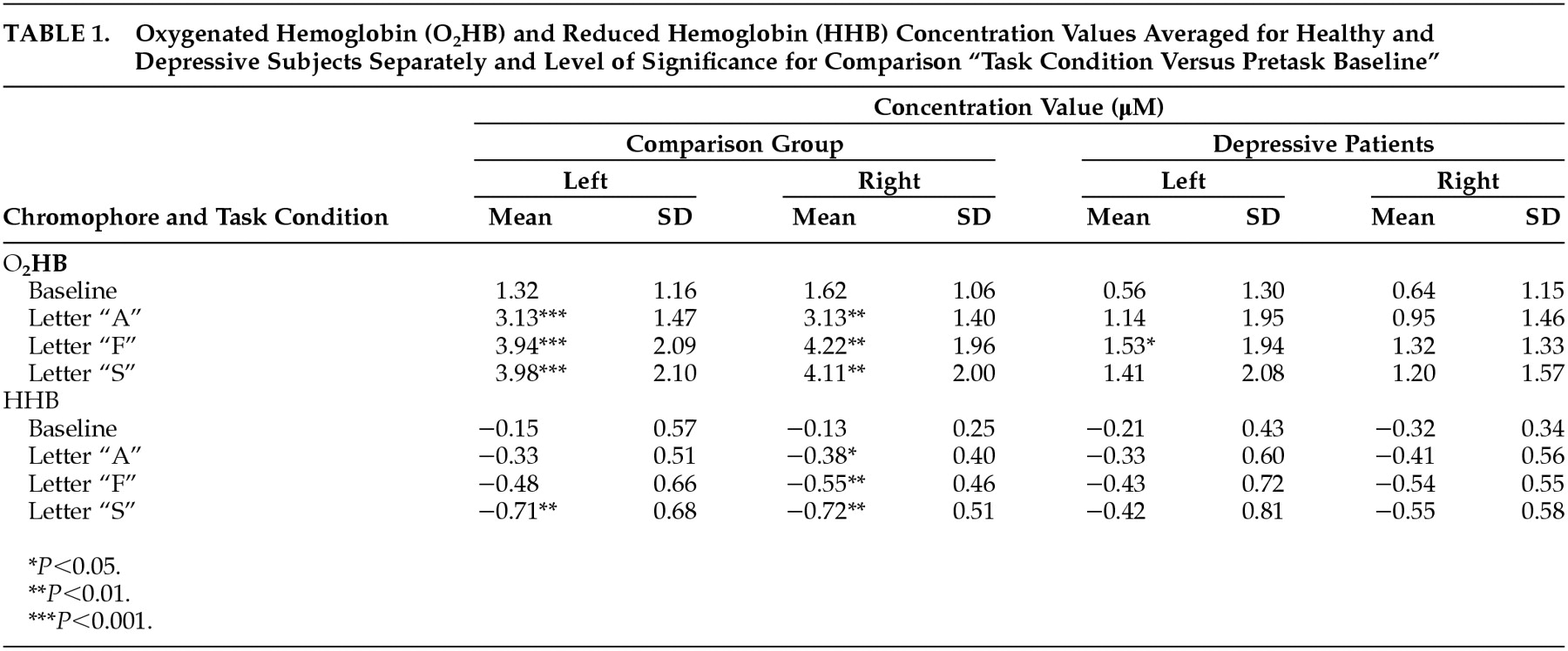
2HB concentration in each of the three letter conditions was significantly increased in the healthy subjects when compared to the depressive subgroup. With regard to the pretask baseline condition the two groups did not differ significantly, although for the right hemisphere the healthy subjects again tended to have increased O
2HB concentrations when compared to the patients (
t=1.88, df=16,
P<0.10). Furthermore, within the healthy subgroup the O
2HB concentrations always increased significantly during each of the three letter tasks when compared to the baseline condition (
Table 1). In the depressive patients this was only the case for left hemispheric values during the letter task “F,” whereas O
2HB concentrations during all the other task conditions did not differ significantly from baseline values within this subgroup. No significant hemispheric differences and no significant hemisphere-by-condition or hemisphere-by-diagnosis interactions were observed.
A corresponding two-by-four-by-two ANOVA for the variable “HHB concentration” similarly revealed a significant main effect of the factor “condition” (
F=10.28, df=3, 48,
P<0.001) with generally lower values in each of the three task conditions than in the pretask baseline measurement (
Table 1). No other significant main effects or interactions were observed for this variable. Although the influence of the factor “diagnosis” did not lead to a significant main effect or interaction in the above mentioned ANOVA, a subsequently conducted post hoc analysis indicated that mean HHB concentration almost consistently decreased significantly during the task conditions but only within the healthy subgroup. In the depressive patients on the other hand, no such task-induced significant decrease of HHB values was observed (
Table 1). However, this effect in the healthy subjects was not found statistically for the left-hemispheric values in all the task conditions (
Table 1). Subsequently conducted power analyses indicated that this might be due to an insufficient power in the letter A and F conditions (power
A=0.101, power
F=0.195).
NIRS Analyses With the Factor Age as Covariate
Using the factor “age” as a covariate in the two-by-four-by-two ANOVA described above, the main effect of the factor “diagnosis” (F=10.27, df=1, 15, P<0.01) as well as the interaction between the variables “diagnosis” and “condition” (F=5.51, df=3, 45, P<0.01) remained significant for the variable “O2HB concentration,” whereas the significant main effect of the factor “condition” was no longer present.
Covarying the factor “age” in the two-by-four-by-two ANOVA for the variable “HHB concentration” eliminated the significant main effect of the factor “condition.”
DISCUSSION
In this study we found a significantly reduced increase of O
2HB in frontal brain areas of depressive patients relative to healthy comparison subjects—as indicated by the above mentioned significant interaction between the factors “diagnosis” and “task condition” for O
2HB—that remained significant after covarying out the factor age. Pointing at a frontal hypoactivity in depressive patients, this finding is in line with several previous studies that have provided evidence for frontal dysfunctions in depression (neuropsychological investigations;
1–4 functional imaging studies
6–8).
Comparing the two groups of participants directly, the healthy subjects exhibited significantly higher O
2HB values than the patients in each of the three task conditions (left and right hemisphere). In contrast, both groups did not differ significantly in the baseline condition. This reduced task-related frontal activation in the depressive patients is in line with previous findings and might be caused by a dysfunction of the prefrontal cortex in depression.
6,7 Furthermore, this dysfunction seems to reflect a
functional deficit rather than a more persistent frontal disorder, as during the resting baseline condition depressive patients did not differ significantly from healthy subjects. Both findings indicate a functional frontal deficit in the depressive patients, which is in line with previous investigations.
Using a one-channel NIRS equipment a significantly decreased left-frontal activation in depressive patients during the VFT has been reported.
21 The authors hypothesized that their finding of a reduced brain activation over the left hemisphere might be caused by a pronounced right hemispherical activation, which would not have been registered due to the unilateral measurement. This hypothesis is not supported by our results, that indicate a reduced activation over
both frontal hemispheres.
On a behavioral level, the group of patients was expected to perform worse than the healthy comparison group, since performance of the VFT is known to involve frontal brain areas and the assumed frontal dysfunction in depressive patients was expected to cause performance deficits in this group. However, the patients of the present study did not differ significantly from the healthy group regarding task performance, although they produced slightly less nouns (mean=25.6, SD=10.1, versus mean=34.9, SD=13.2). This lack of a significant difference may be attributed to an insufficient power (power=0.34) due to the small sample sizes.
To evaluate NIRS methodology we had to compare our results to the results of the two published studies that measured brain activation associated with the VFT by means of NIRS.
19,21 In the first study
19 significantly higher O
2HB values over the left in contrast to the right prefrontal brain were found in healthy subjects, particularly for the letter version of the VFT. As in the statistical analysis only baseline corrected values of O
2HB and HHB were used, the statistical tests did not reveal any information about the significance of the increase in O
2HB and decrease in HHB during the execution of the VFT seen in the figures. In the second study
21 an increase in O
2HB and a decrease in HHB during the VFT compared to baseline were found, but in this study the authors measured with only one sensor over the left hemisphere. In a recently conducted NIRS investigation on healthy subjects not yet published,
28 we found a significant increase in O
2HB and a decrease in HHB during the execution of the VFT in frontal brain regions. For O
2HB no hemispheric differences could be found, whereas the HHB decrease was more pronounced over the right hemisphere. In the present study we again observed a significant increase in O
2HB for both hemispheres in healthy subjects performing the VFT, accompanied by a significant decrease in right-hemispheric HHB, whereas HHB within left prefrontal areas only decreased significantly during the last part of the task.
To sum up, an increase in O
2HB due to activation is a replicated finding with NIRS. In contrast, the effect of a decrease of HHB due to activation seems to be not as stable, probably due to higher inter-individual differences. An important issue is the placement of the NIRS sensors. In three of the four studies cited above different positions were used for the NIRS optodes, which might be a cause for the different hemispherical distribution of the O
2HB increase between studies.
19,28 As in most NIRS studies only a few sensors are used, the exact position of the sensors has to be carefully selected. In contrast to NIRS, other functional imaging procedures have a significantly higher resolution in space, with the possibility to measure the activation of the whole brain. Besides this disadvantage of the poor resolution in space, NIRS is easy to apply, cheap, and non-invasive, and therefore a useful tool for the investigation of psychiatric patients.


