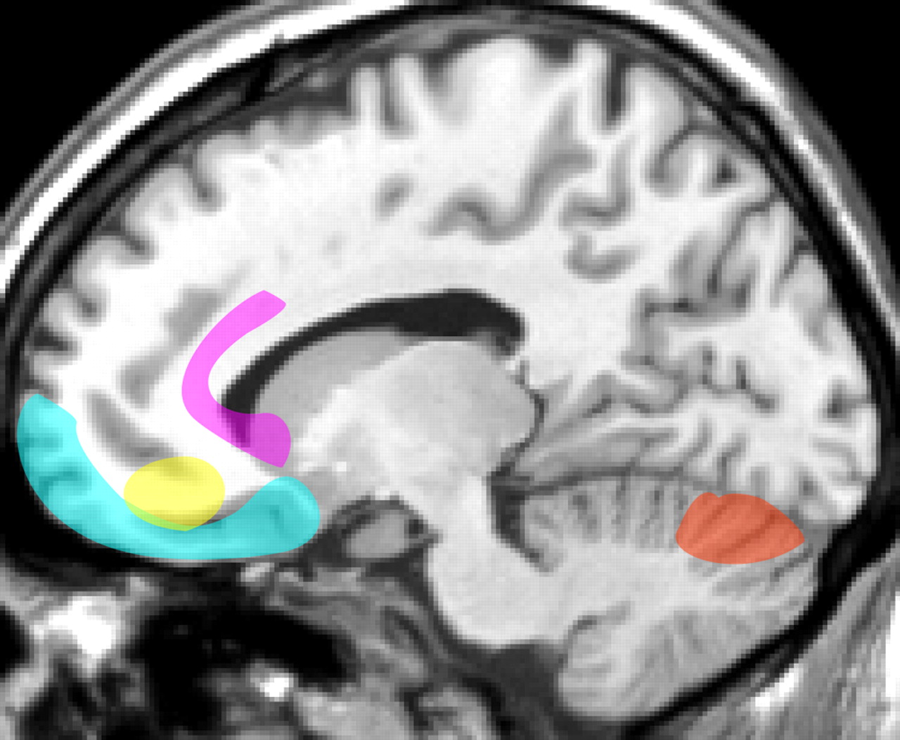
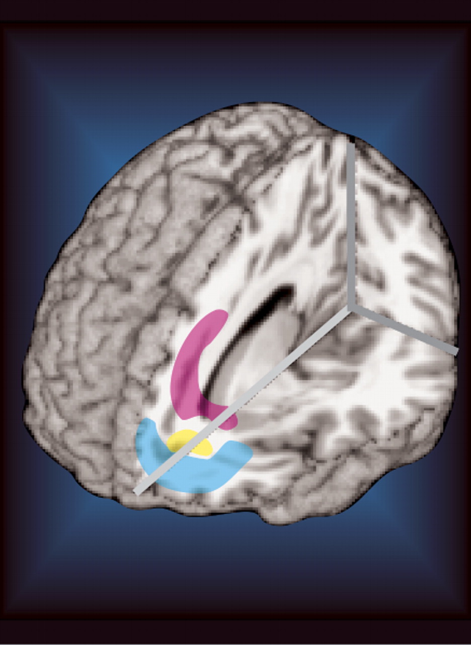
With the above caveats in mind, a few recent positron emission tomography (PET) studies have provided state and trait comparisons in bipolar disorder patients (
Figure 1 ). One study compared regional cerebral blood flow (rCBF) between medicated type I bipolar disorder patients in euthymic and manic states.
1 The only significant differences were higher rCBF on the left in dorsal anterior cingulate cortex and caudate (head) in mania. Two studies (one measuring rCBF, the other measuring regional cerebral metabolic rate, rCMR) have compared bipolar disorder patients in euthymic and depressed states.
4,
5 In both studies, the patient groups were not significantly different. Reduced activity (greater reductions in the depressed state) has been noted in multiple cortical areas (dorsolateral prefrontal, medial prefrontal, orbital prefrontal, parietal) when bipolar disorder patients in all three states were compared to healthy individuals.
2,
4,
5 Studies have reported increased activity in cerebellum in bipolar disorder patients compared to healthy individuals.
4,
5 One study also reported increased subcortical activity (ventral striatum, thalamus, right amygdala) in the depressed state.
5 Subgenual cortex rCBF has also been noted to be decreased in depression (both in bipolar disorder and major depressive disorder) and increased in manic states compared to normal.
3 Euthymic bipolar disorder patients had increased rCBF in dorsal anterior cingulate cortex when compared to healthy individuals in one study.
4 Increased rCMR in the left amygdala has also been reported in bipolar disorder patients in the depressed state, and in the unmedicated euthymic state.
34 Interestingly, this was not the case for euthymic bipolar disorder patients on mood stabilizing medications. From these studies, it appears that the manic state is associated with increased activity in anterior cingulate regions while the depressed state is associated with decreased activity in medial, lateral, and orbital prefrontal regions and increased activity in associated subcortical structures. Studies of the euthymic state suggest a combination of the increased activity in anterior cingulate regions and decreased activity in other prefrontal areas, consistent with the presence of mild symptoms of both mania and depression.
A few studies have compared the depressed and euthymic states in bipolar disorder with major depressive disorder in an effort to detect trait differences. In one study, increased left amygdala metabolism was found in the depressed state for both conditions.
34 Another study used principal component analysis to group symptoms on the Beck Depression Inventory (BDI) into four clusters (negative cognitions, psychomotor-anhedonia, vegetative, somatic).
35 Intercorrelations between these components and correlations between BDI total and component scores with both absolute and normalized rCMR were evaluated for both major depressive disorder and bipolar disorder patients. The components were not intercorrelated for the major depressive disorder group, suggesting that they represent separate aspects of depression. In the bipolar disorder group, the negative cognitions, psychomotor-anhedonia and vegetative components were highly intercorrelated, suggesting a much more unified condition. Consistent with these findings, the brain areas in which both absolute and normalized rCMR correlated with each component were distinctly different in the major depressive disorder group, supporting the involvement of different neuronal networks for each symptom cluster. In contrast, for the bipolar disorder group there were no significant correlations for the negative cognitions and vegetative symptoms components using absolute rCMR. There was considerable overlap in brain areas for all components using normalized rCMR. The psychomotor-anhedonia component score had the strongest correlations with rCMR for the bipolar disorder group (stronger than any other component or the total BDI). The psychomotor-anhedonia component scores correlated with lower absolute metabolism in the insula, anteroventral basal ganglia and temporal and inferior parietal cortices. There was higher normalized metabolism in the anterior cingulate cortex. These results suggest considerably more variability of functioning within the subgroup of individuals diagnosed with major depressive disorder than bipolar disorder, and offer further support that depressive states in bipolar disorder are likely to be neurologically different than depressive states in major depressive disorder though both share clinical similarities.
35


