Full access
Special Article
Published Online: 1 October 1999

Molecular Mechanisms Underlying Mood Stabilization in Manic-Depressive Illness: The Phenotype Challenge

Publication: American Journal of Psychiatry

Abstract

OBJECTIVE: The authors critically examine the evidence supporting the hypothesis that lithium’s therapeutic effects in bipolar affective disorder are mediated by alterations in the expression of specific genes in critical neuronal circuits. METHOD: Using the heuristic “initiation and adaptation paradigm,” the authors appraise the biological effects and underlying molecular and cellular mechanisms of lithium’s action. The evidence is critically reviewed, with special attention to the reductive and integrative approaches necessary for identifying lithium’s clinically relevant cellular and molecular targets. RESULTS: Lithium’s acute effects are mediated through inhibition of specific enzymes involved in two distinct but interacting signaling pathways—the protein kinase C and glycogen synthase kinase 3β signaling ­cascades—that converge at the level of gene transcriptional regulation. The expression of different genes, including transcription factors, is markedly altered by chronic lithium administration. Chronic lithium treatment also robustly increases the expression of the neuroprotective protein Bcl-2, raising the intriguing possibility that some of lithium’s effects are mediated through underappreciated neurotrophic/neuroprotective effects. The importance of lithium’s effect on circadian rhythms and the related methodological problems in validating the role of specific genes in lithium’s therapeutic effects are discussed. CONCLUSIONS: Despite the plethora of lithium effects at the genomic level, direct evidence that the genes identified thus far are responsible for phenotypic changes associated with chronic lithium treatment is still lacking. The combination of sensitive molecular technologies, appropriately designed paradigms, better behavioral analysis, and a chronobiologic approach seems necessary for the future identification of one or more clinically relevant lithium-­target genes.
Bipolar affective disorder (manic-depressive illness) is a common, severe, chronic, and often life-threatening illness (1). Despite the devastating impact of bipolar disorder on the lives of millions, there is still a dearth of knowledge concerning its etiology and pathophysiology. The discovery of lithium’s efficacy as a mood-stabilizing agent revolutionized the treatment of patients with bipolar disorder, and after three decades of use in North America, lithium continues to be the mainstay of treatment for this disorder, both for the acute manic phase and as prophylaxis for recurrent manic and depressive episodes (1, 2). Adequate lithium treatment also reduces the excessive mortality observed in the illness (1, 2). However, the biochemical basis for lithium’s antimanic and mood-stabilizing actions remains to be fully elucidated (36). Understanding the molecular and cellular mechanisms by which lithium stabilizes an underlying dysregulation in critical neuronal circuits not only offers the potential for the development of improved treatments but may also help to delineate the pathophysiology of bipolar disorder (4, 7).
A useful “initiation and adaptation” paradigm for understanding the long-term action of psychotropic drugs has been proposed (8). This paradigm posits that the effect of acute drug administration is mediated through initial direct perturbation of one or more target proteins; repeated administration, by means of the same initial event, over time leads to enduring adaptive changes in critical neuronal networks, thereby resulting in stable long-term effects (8). Based on this model, two experimental approaches to understanding the molecular mechanisms of chronic lithium action can be envisioned. First, the identification of the initial lithium target(s) will facilitate the elucidation of the downstream events mediating its chronic effects. Second, and in a reverse direction, delayed “adaptations” occurring in the context of stable long-term lithium-induced phenotypes should be identified and their relationship with the initial events subsequently established.
Irrespective of the approach, the identification of lithium targets requires an initial reductive step and a subsequent integrative step. The reductive step isolates and characterizes candidate genes and proteins. In the integrative step, the importance of the protein is validated in the original experimental system. For example, if an acute lithium effect is associated with selective enzyme inhibition, the causal relationship between the two events may be established by specific inhibition of the same enzyme with another inhibitor. In the same vein, if a single gene is deemed responsible for some of the long-term lithium-induced changes, a selective manipulation of its protein level should mimic this facet of chronic lithium action. In this context, the search for targets of chronic lithium administration is similar to the task of identifying genes responsible for complex behavioral and physiological traits, such as obesity, hypertension, and circadian rhythms. In all these situations the role of an identified candidate protein is proven by its capacity to correct the abnormal phenotype (for reviews see references 9 and 10). Thus, it is critical to bear in mind that the analysis of the molecular basis of lithium action is meaningful only under conditions of a clear-cut lithium-induced phenotype. This is because the magnitude of the effect, the built-in “redundancy,” and the degree to which the biochemical effect can be compensated for by other pathways all determine whether the molecular or cellular event actually has functional significance. Indeed, any observed biochemical phenomenon may, in fact, even represent a compensatory adaptation to the “true” therapeutic effect. Finally, as lithium targets are inevitably first identified in experimental systems, the identification of genes potentially responsible for lithium’s therapeutic effects involves one additional step—the validation of the role of the gene product in lithium-­induced mood stabilization in patients with bipolar disorder. Thus, although a surfeit of biochemical effects associated with lithium administration have been identified (35), attributing therapeutic relevance to these findings requires extensive validation. In this article we use the initiation-adaptation paradigm to critically assess our current understanding of lithium’s acute and long-term actions.

PHENOTYPIC CHANGES WITH SHORT-TERM AND LONG-TERM LITHIUM APPLICATION

In addition to its beneficial mood-stabilizing effect in bipolar disorder, lithium treatment does produce significant phenotypic changes in a variety of experimental systems. Moreover, in addition to its direct functional effects, lithium treatment markedly modulates the effects of other stimuli. The biological effects of lithium can be divided into short-term effects (manifested soon after lithium’s application and likely mediated through the available cellular machinery) and long-term effects (presumably based on selective changes in gene expression and appearing after a lag period of several days to weeks).
Many of lithium’s short-term effects appear to be cell type or tissue specific. Examples of short-term stimulatory effects include lithium-induced corticotropin secretion by rat anterior pituitary cells (11) and acute glutamate release in lithium-treated brain slices (12). Inhibitory effects of short-term lithium treatment are documented in angiotensin-II-induced aldosterone secretion by adrenal glomerulosa cells (13) and in the relaxation rate following cholinergic induction of smooth muscle contraction (14). A separate group of effects includes the teratogenic effects of short-term lithium exposure in early development. Thus, a brief treatment at a stage of 32–64 cells in a Xenopus embryo leads to an embryo with two dorsal structures, an effect called “dorsalization.” Profound developmental effects of lithium have also been documented in sea urchins, ­zebrafish, and Dictyostelium (15). It should be noted that many of these developmental effects are observed at concentrations that are considerably greater than those attained therapeutically.
Among the long-term phenotypic changes are lithium-induced alterations in circadian rhythmicity (16) and, of course, changes in mood in patients with bi­polar disorder (1), appearing after 2–3 days or weeks, respectively. Chronic lithium treatment in the laboratory rat leads to a persistent performance deficit in some behavioral tests (active avoidance and visually cued maze), whereas the deficit in a spatial memory task is transient. A similar transient deficit is found in the ability of acetylcholine to potentiate N-methyl-d-aspartic acid (NMDA) responses in hippocampal neurons (17). Furthermore, chronic lithium treatment has been reported to inhibit the seizures in a kindling model of epilepsy in the rat (18), although similar treatment potentiates the seizure response to cholinergic agonists (19). Most recently, chronic lithium administration has been shown to exert neuroprotective effects against a variety of insults both in vitro and in vivo (discussed in references 2022), effects that are likely mediated by lithium’s robust increase in the expression of the neuroprotective protein Bcl-2 (discussed later in this article).
Finally, an important (but often overlooked) consideration is that, in addition to the beneficial effects on mood, chronic lithium treatment is associated with many side effects, including polyuria, polydipsia, and clinical or subclinical impairment of thyroid function (1, 3). It is believed that these common side effects are due, at least in part, to lithium-induced inhibition of vaso­pressin- and thyrotropin-sensitive adenylate cyclase in kidney distal tubules and the thyroid gland, respectively (3). In addition, lithium toxicity in yeast is associated with inhibition of RNA-processing enzymes (23). Furthermore, the diabetes-insipidus-like status accompanying chronic administration of dietary lithium to the rat increases the vasopressin mRNA levels in paraventricular and supraoptic hypothalamic nuclei (24). Similarly, thyroid hormones are known to bring about a variety of changes in expression of genes in the central nervous system (CNS). These observations illustrate the multiple causes of differential regulation of gene expression that have to be carefully considered in the search for therapeutically relevant lithium-­target genes.

CURRENT HYPOTHESES ABOUT LITHIUM’S INITIATING EFFECTS

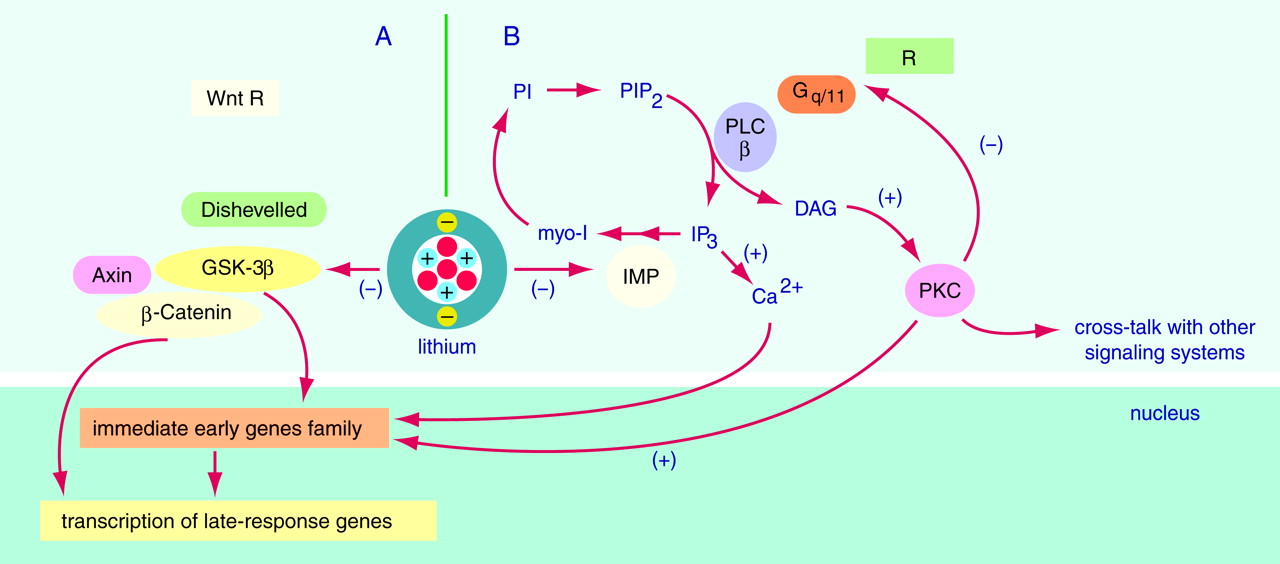
Lithium, whose hydrated ionic radius is similar to that of magnesium, inhibits several enzymes in vitro (15, 25). Most notably, inositol monophosphatases (IMPases) and glycogen synthase kinase 3β (GSK-3β) play important roles in two distinct but interacting signal transduction pathways. The study of these pathways has led to the formulation of two major hypotheses for the cellular basis of lithium’s acute action: the inositol depletion hypothesis (25) and the GSK-3β inhibition hypothesis (15, 26). Here we briefly present these hypotheses, focusing on observations that these acute cellular actions of lithium converge at the level of gene transcription regulation (Figure 1).

LITHIUM AND THE PHOSPHOINOSITIDE CYCLE

Inositol phospholipids play a major role in receptor-mediated signal transduction pathways, and they are involved in diverse responses in the CNS (27). Many cell surface receptors interact with guanine nucleotide binding proteins (G proteins, generally Gq/11), resulting in the generation of two second messengers—inositol-1,4,5-trisphosphate (IP3) and diacylglycerol (DAG) (Figure 1B).
Maintaining sufficient myoinositol supplies is crucial to the resynthesis of phosphoinositides and the efficiency of signaling. Lithium, at therapeutic concentrations, inhibits inositol-1-phosphatase (Ki, 0.8 mM), thereby impairing a major “inositol recycling pathway.” Lithium also inhibits inositol polyphosphate-1-phosphatase, which is involved in recycling inositol polyphosphates to inositol. Since the mode of enzyme inhibition is uncompetitive, lithium’s effects have been postulated to be most pronounced in systems with high rates of phosphoinositide turnover (25, 28). Thus, it was proposed that lithium’s action is mediated through a depletion of free inositol, and its selectivity was attributed to its preferential action on the most active ­receptor-mediated neuronal pathways (25, 29). Since several subtypes of neurotransmitter receptors are coupled to hydrolysis of phosphatidylinositol-4,5-bisphos­phate (PIP2) in the brain, the inositol depletion hypothesis offered a plausible explanation for lithium’s therapeutic efficacy in bipolar disorder and gained wide acceptance.
There is indeed experimental evidence to show that inhibition of IMP does underlie some of lithium’s acute effects. In yeast, for example, acute lithium administration induces a prolongation of the ultradian period (normally around 30 minutes) in a cellular variable called the “septation index.” Overexpression of the human IMP markedly increases the lithium concentration necessary for this effect, suggesting that this lithium effect depends on IMP inhibition (30). However, numerous studies have examined lithium’s effects on CNS-receptor-mediated phosphoinositide responses, and although some have shown a reduction in agonist-stimulated PIP2 hydrolysis in rat brain slices following acute lithium administration, these changes have often been small, inconsistent, and subject to the studies’ numerous methodological differences (see reference 31 for an excellent critique). Most recently, a magnetic resonance spectroscopy study (32) demonstrated that lithium-induced myoinositol reductions are observed in the frontal cortex of patients with bipolar disorder after only 5 days of lithium administration, at a time when the patients’ clinical state is completely unchanged. Consequently, these and other studies suggest that while inhibition of IMP may be an initiating lithium effect, reduction of myoinositol levels itself does not underlie lithium’s therapeutic effects. The hypothesis that these effects initiate a cascade of changes in PKC signaling and gene expression, effects that are ultimately responsible for lithium’s therapeutic effects, is discussed later.

GSK-3b: THERAPEUTICALLY RELEVANT TARGET FOR LITHIUM’S ACTIONS?

In recent years a hitherto unexpected target for lithium’s acute effects has been identified. Klein and Melton (15) were the first to demonstrate that lithium, at therapeutically relevant concentrations, is an inhibitor of GSK-3β in vitro (Figure 1). GSK-3β is an evolutionary highly conserved kinase, originally identified as a regulator of glycogen synthesis. Overexpression of a negative GSK-3β mutant (a biologically inactive version of the enzyme inhibiting the endogenous protein) also induces dorsalization of the Xenopus embryo, indicating the important role of GSK-3β in this teratogenic lithium effect (26). It is now known that GSK-3β plays an important role in the CNS, by regulating various cytoskeletal processes through its effects on Tau and Synapsin I and by inducing long-term nuclear events through phosphorylation of c-Jun and nuclear translocation of β-Catenin (26, 33, 34). As discussed, lithium is known to bring about a variety of biochemical effects, and it is unclear whether inhibition of GSK-3β is a therapeutically relevant effect. We therefore investigated whether the structurally highly dissimilar antimanic (and likely mood-stabilizing) agent valproate also regulates GSK-3β. We found that both lithium and valproate significantly inhibited GSK-3β at therapeutically relevant concentrations (35). Consistent with GSK-3β inhibition, both lithium and valproate produced significant time-dependent increases in cytosolic and nuclear β-Catenin levels in human cells of neuronal origin. These data indicate that GSK-3β is a common initial target for both mood stabilizers and support its implication in lithium’s beneficial long-term action. It is important for the present discussion that GSK-3β is known to phosphorylate the protein c-Jun at three sites adjacent to its DNA binding domain, thereby reducing binding of activator protein 1 (AP-1) (see later discussion) (36). Thus, the acute inhibition of GSK-3β by lithium and valproate has the potential to bring about long-term changes in the CNS through the transcriptional activity of both β-Catenin and AP-1.

LONG-TERM MEDIATORS OF LITHIUM’S THERAPEUTIC EFFECTS: THE ROLE OF THE PKC FAMILY OF ENZYMES

As discussed in the preceding, reduction of myo­inositol levels per se likely does not underlie lithium’s therapeutic effects. However, some of lithium’s initial actions may occur through IMP inhibition, which initiates a cascade of secondary changes in the PKC signaling pathway and gene expression in the CNS, effects that are potentially responsible for lithium’s therapeutic efficacy (reviewed in references 35 and 37). PKC exists as a family of closely related subspecies, has a heterogeneous distribution in the brain, and plays a major role in the regulation of neuronal excitability, neurotransmitter release, and long-term alterations in gene expression and plasticity (3739). Lithium exerts significant effects on PKC in a number of cell systems, including the brain (35). Thus, lithium treatment has complex, biphasic effects on PKC, and chronic lithium administration at therapeutically ­relevant concentrations produces isozyme-specific ­decreases in PKC α and ε in the frontal cortex and ­hippocampus (40, 41). Furthermore, Lenox and associates (42) demonstrated that the levels of a critical PKC substrate, myristoylated alanine-rich C kinase substrate (MARCKS), a key protein implicated in synaptic transmission and neuroplastic events, is also significantly reduced in the hippocampus after chronic lithium exposure. Valproate produces strikingly similar effects on the PKC signaling pathway, including MARCKS (41, 43, 44). These intriguing biochemical observations indicate that the PKC signaling pathway represents a common long-term target for both lithium and valproate, two agents with clinically proven antimanic and likely mood-stabilizing efficacy. It is interesting that preliminary studies suggest that chronic lithium and valproate regimens appear to regulate the PKC signaling pathway by distinct initiating mechanisms (43, 45). This finding is consistent with clinical observations that some patients show preferential response to one or other of the agents and that additive therapeutic effects are sometimes observed in patients.

LITHIUM’S EFFECTS ON IMMEDIATE EARLY GENES

The transcriptional activation of immediate early genes, including the fos and jun families, is a characteristic cellular response to extracellular stimuli such as hormones, growth factors, and neurotransmitters (46). The transcriptional activation is followed by cytoplasmic translation of Fos, Jun, and other proteins, which translocate into the nucleus and form a variety of protein complexes. AP-1 is a collection of homodimeric and heterodimeric complexes composed of products of fos and jun family members. These products bind to a common DNA site—12-O-tetradecanoylphorbol 13-acetate (TPA) response element (TRE)—in the regulatory domain of the gene and activate gene transcription. The genes regulated by AP-1 in the CNS include genes for various neuropeptides, neurotrophins, receptors, transcription factors, enzymes involved in neuro­transmitter synthesis, and proteins that bind to cyto­skeletal elements (46). The final result of this molecular cascade is alteration in the transcription of selected target genes bearing the specific DNA binding site on their regulatory regions.
It is noteworthy that both lithium and valproate, at therapeutically relevant concentrations, produce increases in AP-1 DNA binding activity in rat brain ex vivo and in cultured human neuroblastoma cells in vitro (5, 41, 47, 48). Furthermore, both lithium and valproate increase the expression of a luciferase reporter gene, effects that are markedly attenuated by mutations at the promoter AP-1 sites (41). While the reporter gene effects in vitro suggest that lithium-­induced AP-1 activation does have functional effects, it is clearly necessary to demonstrate that they also occur in critical regions of the CNS in vivo. It is well established that the expression of tyrosine hydroxylase is mediated in large part by the AP-1 family of transcription factors (49). Thus, a study was undertaken to determine whether chronic lithium administration does, in fact, increase the CNS levels of a protein known to be regulated in large part by AP-1 (50). Consistent with AP-1 activation, chronic lithium administration significantly increased the levels of tyrosine hydroxylase in the frontal cortex, hippocampus, and striatum (50), three brain areas implicated in the pathophysiology of bipolar disorder (5153). In view of the key roles of immediate early genes in long-term neuronal plasticity and cellular responsiveness, and the potential to regulate patterns of gene expression in critical neuronal circuits, these effects may play a major role in lithium’s long-term therapeutic effects and are worthy of further study.

IDENTIFICATION OF NOVEL TARGETS

The preceding data suggest that lithium has the potential to regulate CNS gene expression, and indeed many genes have been suggested as targets for the actions of chronic lithium (36). However, the large number of genes expressed in a given cell at any time (10,000–15,000) opens up the opportunity for the identification of novel genes whose expression is affected by chronic drug treatment. In recent years, new methods have evolved to identify the differential expression of multiple genes (e.g., in pathological versus normal tissue or in control versus treated tissue); one such method that is being increasingly used is the reverse transcription polymerase chain reaction mRNA differential display (RT-PCR DD) (54, 55). By using this method, lithium was shown to increase 2′,3′-cyclic nucleotide 3′-phosphodiesterase mRNA levels in C6 glioma cells (56). In order to identify potentially therapeutically relevant changes in gene expression, we have used RT-PCR DD to concurrently investigate the effects of chronic lithium and valproate administration in intact animals (57). One of the transcripts whose levels were markedly increased by both lithium and valproate encodes a transcription factor—the β subunit of the polyomavirus enhancer-binding protein 2 (PEBP2β). In concert, the DNA binding activity of PEBP2αβ also increased in the rat frontal cortex. We therefore next investigated lithium’s effects on the levels of a critical protein known to be regulated by PEBP2β—the major neuroprotective protein Bcl-2. We found that chronic lithium treatment of rats resulted in doubling of Bcl-2 levels in the frontal cortex, an effect accompanied by a marked increase in the number of Bcl-2 immunoreactive cells in frontal cortex layers II and III (22). The robust increase in Bcl-2 levels likely plays a major role in lithium’s neuroprotective effects against a variety of insults both in vitro (including glutamate, calcium, and 1-methyl-4-phenylpiridinium [MPP+]) and ex vivo (including irradiation, kainic acid, and ischemia) (discussed in references 2022).
Are such putative neuroprotective effects relevant for mood disorders? There is now considerable evidence demonstrating significant reductions in regional CNS volume and cell numbers associated with mood disorders from both in vivo brain imaging studies and postmortem studies (5153). It is intriguing that neuronal loss is especially pronounced in layer II of the rostral orbitofrontal region (53), an area with the largest lithium-induced increase in Bcl-2 immunoreactivity. Thus, overall, these data suggest that lithium’s effects on CNS Bcl-2 levels (and accompanying neuro­protective effects) may be of considerable importance in the long-term treatment of mood disorders. It is noteworthy that several antidepressants increase the expression of brain-derived neurotrophic factor, findings that have led to the development of a heuristic molecular and cellular model of depression (58).

LIMITATIONS IN EXPERIMENTAL STUDY OF EFFECTS OF CHRONIC LITHIUM TREATMENT

Although the application of advanced cellular and molecular biologic methods to the study of lithium’s actions has provided intriguing and unexpected new leads, several factors make it difficult to clearly attribute therapeutic relevance to these biochemical findings. To begin with, a suitable experimental model of manic-depression is currently not available. In this context, it is likely that the robust animal models of drug abuse have been instrumental in accelerating the pace of research on the underlying molecular events (8). Another inherent problem in the identification of lithium-target genes is the relative paucity of easily detectable phenotypic changes induced by chronic lithium treatment. This makes the task of ascribing functional significance to the multiple lithium-induced changes at the genomic level quite daunting. Moreover, the genetic basis of mood and affect as quantitative traits is still at its inception (59), and we therefore cannot focus on a group of genes known to be involved in affect regulation. In this regard, the observations that several genes encoding neuromodulatory peptides (prodynorphin, preprotachykinin, neuropeptide Y, and glucocorticoid receptor type II) are markedly affected by lithium treatment in the rat (3, 4) are still awaiting experimental validation of their functional significance in the treatment of mood disorders. In addition, as already discussed, chronic lithium treatment is also associated with side effects that could affect gene expression, including polyuria, polydipsia, and impairment of thyroid function. These observations illustrate the difficulties that the investigator faces in the search for therapeutically relevant lithium-target genes. Thus, overall, a growing body of evidence suggests that chronic lithium action is associated with a number of changes in gene expression. A difficulty arises, however, in testing the hypothesis that the altered expression of any given gene per se is involved in phenotypic changes associated with chronic lithium treatment.
Another factor worthy of consideration is the experimental systems used to study the mechanisms of chronic lithium action—these range from single cells to whole animals. The single cell models are attractive because of easy control over drug concentration and the possibility of intricately addressing cellular and molecular interactions. However, the cells generally do not exhibit readily detectable phenotypic alterations, even at concentrations much above the lithium levels that are toxic for the whole animal. Exceptions to this are the ultradian rhythms in yeast (30) and the protection against NMDA-induced apoptosis in dissociated neurons (20). Although cell culture systems lack normal cell-cell interactions, they offer the potential to identify genes that are direct cellular lithium targets.
Conversely, an example of a protein whose expression is significantly affected by chronic lithium administration in the whole rat but not in a cellular model is Gephyrin. Gephyrin clusters inhibitory postsynaptic membrane receptors (glycine and γ-amino-butyric-acid) in CNS neurons (60). Chronic lithium treatment (5 weeks but not 9 days) significantly increased Gephyrin levels in the frontal cortex but not the hippocampus; moreover, the increase in Gephyrin correlated well with the individual lithium plasma levels (61). By contrast, lithium treatment of human neuroblastoma cells (from 1 to 7 days) did not result in appreciable changes in Gephyrin levels. These data support the view that whole animal models are essential for the detection of certain long-term lithium-target genes whose expression depends on interacting neuronal networks.
However, the use of laboratory rodents also presents us with problems in interpreting the potential changes in gene expression because of chronic lithium-induced alterations in circadian rhythmicity. The latter problem is discussed in the following section.

CHRONIC LITHIUM TREATMENT AND CIRCADIAN RHYTHMS

Rhythmicity is a fundamental characteristic of living matter. When a variable is longitudinally monitored and the data series assessed by inferential statistics, the results are approximated with a sum of several rhythmic components with different periods and amplitudes. The most prominent rhythmic component under normal conditions has a period of 24 hours. Such a rhythm usually reflects a complex interaction between endogenous rhythmic and other (homeostatic, adaptive, pathological, etc.) mechanisms. The 24-hour rhythms with a proven endogenous component are called circadian, a term reflecting the observation that under constant external conditions the rhythms free-run with an endogenous period that is close to but significantly different from 24 hours (62). The circadian period of mammalian locomotor activity is controlled by a circadian clock located in the hypothalamic suprachiasmatic nucleus (63). Under synchronized conditions, all circadian rhythms appear with identical periods but have very different amplitudes and/or peak times (acrophases). Chronic (but not acute) lithium treatment prolongs the free-running period in almost all studied biological systems, including humans (16, 64). Lithium’s effects under synchronized conditions are variable specific, with some variables showing significant acrophase delays and others exhibiting advances (65). The effects also depend on the timing of application, with evening application of lithium hydro­xybutyrate for 10 days destabilizing circadian rhythms in the rat, in contrast to morning administration (66). The mechanism of lithium’s action on the diverse circadian rhythms probably involves combined effects at the level of the circadian clock and at the level of integration of circadian rhythmicity with other regulatory systems. Experiments in a simple circadian model (Neurospora crassa) do not support the potential role of inositol depletion in lithium’s effects on the circadian clock and its light synchronization (67).
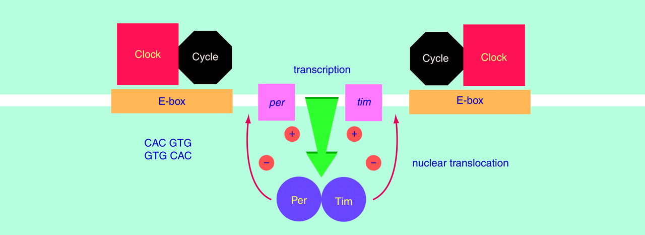
Two recent breakthroughs are relevant to future approaches to the study of molecular and cellular mechanisms of lithium action. First, single dissociated supra­chiasmatic nucleus neurons contain a circadian clock and express circadian rhythm in impulse activity (68). Second, a powerful combination of mutagenesis, isolation of behaviorally arrhythmic mutants, and molecular genetics was used to identify a set of four genes representing the molecular cogwheels of the cellular circadian clock in Drosophila (69) (Figure 2). Two of the genes—period (per) and timeless (tim)—oscillate in a circadian fashion at both the mRNA and protein levels. It is important that the Per and Tim proteins form a cytosolic dimer, which translocates into the cell nucleus and suppresses their own transcription. The other two proteins—Clock and Cycle—also form a dimer in the nucleus. This dimer induces the transcription of per and tim mRNA by binding to E-boxes (5′-CACGTG-3′ sequence) located in the regulatory region of each gene. The activation of per and tim transcription may be suppressed by their own protein dimers, thus creating a molecular circadian clock based on a negative feedback loop (69). It is note­worthy that a mouse arrhythmic mutant has a single base mutation in the mammalian clock gene, which is highly homologous to the clock gene in Drosophila. The same holds true for the hamster BMAL1 gene—a partner of mammalian clock and homologous to the Drosophila cycle gene (69). Finally, mammals have three Drosophila per homologues with conservative E-boxes in their promoters (70). Overall, these observations imply that intracellular circadian clocks are based on evolutionary conserved molecular mechanisms. Together with the evolutionary conserved effects of chronic lithium treatment, this evidence suggests that the genes implicated in circadian clock function may also be targets for chronic lithium action. The potential consequences of chronic lithium treatment for circadian rhythms are illustrated by the following example. If we assume that lithium treatment results solely in a selective 6-hour acrophase delay, then a single-time-point study will be insufficient for properly characterizing the effect and possibly misleading; i.e., is the expression of the gene truly up or down, or simply phase-shifted? This cautionary note suggests that chronobiological studies are needed for many of lithium’s putative molecular targets in order to determine whether the observed effects reflect changes in the spatiotemporal patterns of gene expression in critical neuronal circuits, effects that are likely to underlie the complex behavioral effects of lithium. Indeed, the importance of circadian factors and the sleep-wake cycle in bipolar disorder has been recognized for many years (discussed in ­reference 1).

CONCLUSIONS AND PERSPECTIVES

Lithium’s profound effects on mood stabilization take several weeks to develop, a lag period that is most compatible with changes in the expression of critical neuronal genes. Although the indirect evidence is compelling, we cannot yet fully ascribe the therapeutic changes to alterations in the expression of any sets of genes per se. One of the major limitations is the absence of a good experimental model of bipolar disorder. Thus, the biological effects of lithium have, of necessity, been studied in “normal” experimental systems with the belief that the evolutionary conserved mechanisms of lithium’s action are also relevant in the treatment of bipolar disorder. Indeed, abundant evidence indicates that many of lithium’s effects are mediated through alterations in signal transduction pathways in critical neuronal circuits (35). Many of these signal transduction cascades are known to induce long-term cellular responses by means of regulated gene expression, and indeed, chronic lithium treatment is associated with changes in the expression of multiple genes. However, this reductive analysis is not yet complemented by evidence that these lithium-regulated genes are relevant to a particular lithium-induced phenotype. The few exceptions to this rule were studies that applied acute lithium treatment in early development (26) or in yeast (30). In conclusion, we have recently uncovered exciting new cellular and molecular leads concerning lithium’s long-term therapeutic efficacy, but we await direct experimental evidence that a lithium-induced phenotypic change is based on selective alterations in gene expression. The additional use of systems with marked chronic lithium-induced phenotypic changes will greatly enhance the identification of experimentally tractable candidate lithium-target genes. The future approaches should also involve the evolutionary conserved effect of lithium on circadian rhythms. It is our firm belief that the use of a marked phenotype and chronobiologic approach, combined with the recent technological advances in the systems for identifying discordantly regulated genes and manipulating gene expression (including microarrays of DNA and oligo­nucleotides), holds much promise in the validation of chronic lithium-target genes in clinically relevant experimental systems and, ultimately, for the development of improved therapeutics.
FIGURE 1. Cellular and Molecular Mechanisms of Action of Lithiuma
aAt therapeutic concentrations lithium directly inhibits (arrows) two enzymes—glycogen synthase kinase 3β (GSK-3β) and inositol monophosphate phosphatase (IMP). In certain systems (see part A), GSK-3β is in a cytosolic complex with other proteins—Axin and β-Catenin. β-Catenin is a transcription factor and the effector of the Wnt signaling pathway, which also involves extracellular ligands (encoded by the Wnt gene family, not shown), Wnt membrane receptors (encoded by the Frizzled gene family), and Dishevelled proteins. The Wnt signaling activation leads to a nuclear translocation of β-Catenin and activation of c-Jun. Short lithium exposure blocks the tonic inhibitory action of GSK-3β and activates the Wnt signaling pathway, an effect that can be imitated by a dominant negative GSK-3β mutant. In the brain (see part B), lithium inhibits IMP, resulting in a lowering of myoinositol (myo-I). This acute effect has the potential to induce a cascade of effects in the DAG/PKC limb of the signal transduction pathway (DAG, diacylglycerol; PKC, protein kinase C). The effect of lithium is complex and involves biphasic changes—an initial activation of PKC followed by a decrease in PKC α and ε. Lithium, through its effects on PKC, also regulates the expression of the important neuronal phosphoprotein MARCKS (myristoylated alanine-rich C kinase substrate). Increased intracellular Ca2+ and PKC activate the transcription of immediate early genes, including the fos and jun families. Similar to the effects observed with PKC, lithium induces complex, biphasic effects on c-fos expression. Multiple late-response genes have been shown to be significantly affected by lithium treatment. Following are definitions of other terms appearing in the figure: PLC, phospholipase C; PIP2, phosphatidylinositol-4,5-bisphosphate; IP3, inositol-1,4,5-trisphosphate; PI, phosphoinositide; R, membrane receptor; Gq/11, G protein subtype.
FIGURE 2. Molecular Model of Negative Feedback in the Circadian Clock of a Single Drosophila Cella
aTwo proteins—Per and Tim—accumulate rhythmically (owing to rhythmicity at the level of their transcription), form heterodimers, and repress their own transcription when they reach a threshold level in the cell nucleus. This suppression is mediated by yet unknown interactions with the heterodimer of two other proteins—Clock and Cycle—shown to induce the transcription of the per (period) and tim (timeless) genes through interactions with E-boxes in their promoters. The model thus provides an explanation of the self-sustained circadian oscillations of Per and Tim, which are currently considered as a molecular basis of the evolutionary conserved cellular circadian clock.

Footnote

Received Aug. 12, 1998; revision received March 29, 1999; accepted May 11, 1999. From the Laboratory of Molecular Pathophysiology, Department of Psychiatry and Behavioral Neurosciences, the Department of Pharmacology, and the Cellular and Clinical Neurobiology Program, Wayne State University School of Medicine. Address reprint requests to Dr. Ikonomov, Laboratory of Molecular Pathophysiology, Department of Psychiatry and Behavioral Neurosciences, Wayne State University School of Medicine, 2309 Scott Hall, 540 East Canfield Ave., Detroit, MI 48201; [email protected] (e-mail). Supported by NIMH grant MH-57743, the Theodore and Vada Stanley Foundation, the National Alliance for Research on Schizophrenia and Depression, and the Joseph Young Senior Foundation. The authors thank Ms. Celia Knobelsdorf for editorial assistance. A complete list of the studies on which this article is based can be obtained on request.

References

1.
Goodwin FK, Jamison KR: Manic-Depressive Illness. New York, Oxford University Press, 1990
2.
Baldessarini RJ, Tondo L, Hennen J: Effects of lithium treatment and its discontinuation in bipolar manic-depressive disorders. J Clin Psychiatry 1999; 60(suppl 2):77–84
3.
Lenox RH, Manji HK: Lithium, in American Psychiatric Press Textbook of Psychopharmacology, 2nd ed. Edited by Schatzberg AF, Nemeroff CB. Washington, DC, American Psychiatric Press, 1998, pp 379–429
4.
Manji HK, Potter WZ, Lenox RH: Signal transduction pathways: molecular targets for lithium’s actions. Arch Gen Psychiatry 1995; 52:531–543
5.
Jope RS: Anti-bipolar therapy: mechanism of action of lithium. Mol Psychiatry 1999; 4:117–128
6.
Wang JF, Young LT, Li PP, Warsh JJ: Signal transduction abnormalities in bipolar disorder, in Bipolar Disorder: Biological Models and Their Clinical Applications. Edited by Young LT, Joffe RT. New York, Marcel Dekker, 1997, pp 41–80
7.
Lenox RH: Role of receptor coupling to phosphoinositide metabolism in the therapeutic action of lithium. Adv Exp Biol Med 1987; 221:515–530
8.
Hyman SE, Nestler EJ: Initiation and adaptation: a paradigm for understanding psychotropic drug action. Am J Psychiatry 1996; 153:151–162
9.
Lifton RP: Genetic determinants of human hypertension. Proc Natl Acad Sci USA 1995; 92:8545–8551
10.
Takahashi JS, Kornhauser JM, Koumenis C, Eskin A: Molecular approaches to understanding circadian oscillators. Annu Rev Physiol 1993; 55:729–754
11.
Reisine T, Zatz M: Interactions among lithium, calcium, diacylglycerides, and phorbol esters in the regulation of adrenocorticotropin hormone release from AtT-20 cells. J Neurochem 1987; 49:884–889
12.
Hokin LE, Dixon JF, Los GV: A novel action of lithium: stimulation of glutamate release and inositol 1,4,5 trisphosphate accumulation via activation of the N-methyl d-aspartate receptor in monkey and mouse cerebral cortex slices. Adv Enzyme Regul 1996; 36:229–244
13.
Balla T, Enyedi P, Hunyady L, Spat A: Effects of lithium and angiotensin-stimulated phosphatidylinositol turnover and aldosterone production in adrenal glomerulosa cells: a possible causal relationship. FEBS Lett 1984; 171:179–182
14.
Menkes HA, Baraban JM, Freed AN, Snyder SH: Lithium dampens neurotransmitter response in smooth muscle: relevance to action in affective illness. Proc Natl Acad Sci USA 1986; 83:5727–5730
15.
Klein PS, Melton DA: A molecular mechanism for the effect of lithium on development. Proc Natl Acad Sci USA 1996; 93:8455–8459
16.
Klemfuss H: Rhythms and the pharmacology of lithium. Pharmacol Ther 1992; 56:53–78
17.
Richter-Levin G, Markram H, Segal M: Spontaneous recovery of deficits in spatial memory and cholinergic potentiation of NMDA in CA1 neurons during chronic lithium treatment. Hippocampus 1992; 2:279–286
18.
Schmidt J: Lithium suppresses seizure susceptibility in pentylenetetrazol-kindled rats. Biomed Biochim Acta 1986; 45:1167–1172
19.
Honchar MP, Olney JW, Sherman WR: Systemic cholinergic agents induce seizures and brain damage in lithium-treated rats. Science 1983; 220:323–325
20.
Nonaka S, Hough CJ, Chuang DM: Chronic lithium treatment robustly protects neurons in the central nervous system against excitotoxicity by inhibiting N-methyl-d-aspartate receptor-mediated calcium influx. Proc Natl Acad Sci USA 1998; 95:2642–2647
21.
Chen RW, Chuang DM: Long term lithium treatment suppresses p53 and Bax expression but increases Bcl-2 expression: a prominent role in neuroprotection against excito­toxicity. J Biol Chem 1999; 274:6039–6042
22.
Manji HK, Moore GJ, Chen G: Lithium at 50: have the neuroprotective effects of this unique cation been overlooked? Biol Psychiatry (in press)
23.
Dichtl B, Stevens A, Tollerey D: Lithium toxicity in yeast is due to the inhibition of RNA processing enzymes. EMBO J 1997; 16:7184–7195
24.
Anai H, Ueta Y, Serino R, Bomura M, Kabashima N, Shibuya I, Takasugi M, Nakashima Y, Yamashita H: Upregulation of the expression of vasopressin gene in the paraventricular and supraoptic nuclei of the lithium-induced diabetes insipidus rat. Brain Res 1997; 772:161–166
25.
Berridge MJ, Downes CP, Hanley MR: Neuronal and developmental actions of lithium: a unifying hypothesis. Cell 1989; 59:411–419
26.
Hedgepeth CM, Conrad LJ, Zhang J, Huang HC, Lee VM, Klein PS: Activation of the Wnt signaling pathway: a molecular mechanism for lithium action. Dev Biol 1997; 185:82–91
27.
Fisher SK, Heacock AM, Agranoff BW: Inositol lipids and signal transduction in the nervous system: an update. J Neurochem 1992; 58:18–38
28.
Nahorski SR, Ragan CI, Challiss RAJ: Lithium and the phosphoinositide cycle: an example of uncompetitive inhibition and its pharmacological consequences. Trends Pharmacol Sci 1991; 12:297–303
29.
Kofman O, Belmaker RH: Biochemical, behavioral and clinical studies of the role of inositol in lithium treatment and depression. Biol Psychiatry 1993; 34:839–852
30.
Kippert F: Lithium affects the ultradian clock of Schizosaccaromyces pombe by inhibition of inositol monophosphatase. Biochem Soc Trans 1997; 25:S602
31.
Jope RS, Williams MB: Lithium and brain signal transduction systems. Biochem Pharmacol 1994; 47:429–441
32.
Moore GJ, Bebchuk JM, Parrish JK, Faulk MW, Arfken CL, Strahl-Bevacqua J, Manji HK: Temporal dissociation between lithium-induced frontal myo-inositol changes and clinical response in manic-depressive illness. Am J Psychiatry (in press)
33.
Stambolic V, Ruel LR, Woodgett JR: Lithium inhibits glycogen synthase kinase-3 activity and mimics wingless signaling in intact cells. Curr Biol 1996; 6:1664–1668; correction, 1997; 7:196
34.
Lucas FR, Goold RG, Gordon-Weeks PR, Salinas PC: Inhibition of GSK-3beta leading to the loss of phosphorylated MAP-1B is an early event in axonal remodelling induced by WNT-7a or lithium. J Cell Sci 1998; 111:1351–1361
35.
Chen G, Huang LD, Jiang YM, Manji HK: The mood stabilizing agent valproate inhibits the activity of glycogen synthase ­kinase-3. J Neurochem 1999; 72:1327–1330
36.
Lin A, Smeal T, Binetruy B, Deng T, Chambard JC, Karin M: Control of AP-1 activity by signal transduction cascades. Adv Second Messenger Phosphoprotein Res 1993; 28:255–260
37.
Manji HK, Lenox RH: Long-term action of lithium: a role for transcriptional and posttranscriptional factors regulated by protein kinase C. Synapse 1994; 16:11–28
38.
Nishizuka Y: Intracellular signaling by hydrolysis of phospholipids and activation of protein kinase C. Science 1992; 258:607–613
39.
Stabel S, Parker PJ: Protein kinase C. Pharmacol Ther 1991; 51:71–95
40.
Manji HK, Etcheberrigaray R, Chen G, Olds JL: Lithium decreases membrane-associated protein kinase C in hippocampus: selectivity for the alpha isozyme. J Neurochem 1993; 61:2303–2310
41.
Manji HK, Bebchuk JM, Moore GJ, Glitz D, Hasanat K, Chen G: Modulation of CNS signal transduction pathways and gene expression by mood stabilizing agents: therapeutic implications. J Clin Psychiatry 1999; 60(suppl 2):27–39
42.
Lenox RH, Watson DG, Patel J, Ellis J: Chronic lithium administration alters a prominent PKC substrate in rat hippocampus. Brain Res 1992; 570:333–340
43.
Lenox RH, McNamara RK, Watterson JM, Watson DG: Myristoylated alanine-rich C kinase substrate (MARCKS): a molecular target for the therapeutic action of mood stabilizers in the brain? J Clin Psychiatry 1996; 57(suppl 13):23–31
44.
Chen G, Manji HK, Hawver DB, Wright CB, Potter WZ: Chronic sodium valproate selectively decreases protein kinase C α and ε in vitro. J Neurochem 1994; 63:2361–2364
45.
Manji HK, Bersudsky Y, Chen G, Belmaker RH, Potter WZ: Modulation of PKC isozymes and substrates by lithium: the role of myo-inositol. Neuropsychopharmacology 1996; 15:370–382
46.
Morgan JI, Curran TE: Proto-oncogenes: beyond second messengers, in Psychopharmacology: The Fourth Generation of Progress. Edited by Bloom FE, Kupfer DJ. New York, Raven Press, 1995, pp 631–642
47.
Asghari V, Wang JF, Reiach JS, Young LT: Differential effects of mood stabilizers on Fos/Jun proteins and AP-1 DNA binding activity in human neuroblastoma SH-SY5Y cells. Brain Res Mol Brain Res 1998; 58:95–102
48.
Ozaki N, Chuang DM: Lithium increases transcription factor binding to AP-1 and cyclic AMP-responsive element in cultured neurons and rat brain. J Neurochem 1997; 69:2336–2344
49.
Kumer SC, Vrana KE: Intricate regulation of tyrosine hydroxylase activity and gene expression. J Neurochem 1996; 67:443–461
50.
Chen G, Yuan PX, Jiang Y, Huang LD, Manji HK: Lithium increases tyrosine hydroxylase levels both in vivo and in vitro. J Neurochem 1998; 70:1768–1771
51.
Drevets WC, Price JL, Simpson JR Jr, Todd RD, Reich T, Vannier M, Raichle ME: Subgenual prefrontal cortex abnormalities in mood disorders. Nature 1997; 38:824–827
52.
Ketter TA, George MS, Kimbrell TA, Willis MW, Benson BE, Post RM: Neuroanatomical models and brain-imaging studies, in Bipolar Disorder: Biological Models and Their Clinical Application. Edited by Young LT, Joffe RT. New York, Marcel Dekker, 1997, pp 179–217
53.
Rajkowska G, Miguel-Hidalgo JJ, Wei J, Dilley G, Pittman SD, Meltzer HY, Overholser JC, Roth BL, Stockmeier CA: Morphometric evidence for neuronal and glial prefrontal cell pathology in major depression. Biol Psychiatry 1999; 45:1085–1098
54.
Liang P, Pardee AB: Differential display of eukaryotic messenger RNA by means of the polymerase chain reaction. Science 1992; 257:967–971
55.
Ikonomov OC, Jacob MH: Preferential identification of differentially expressed mRNAs of moderate to low abundance in a microscopic system using selected primers, in Gene Cloning and Analysis by RT-PCR. Edited by Siebert P, Larrick JW. Natick, Mass, BioTechniques Books (Eaton Publishing), 1998, pp 171–181
56.
Wang JF, Young LT: Differential display PCR reveals increased expression of 2′,3′-cyclic nucleotide 3′-phosphodiesterase by lithium. FEBS Lett 1996; 386:225–229
57.
Manji HK, Zeng WZ, Huang LD, Ikonomov OC, Chen G: A new common transcriptional target for the mood-stabilizing agents lithium and valproic acid. Abstracts of the Society for Neuroscience 1997; 23:796.12
58.
Duman RS, Heninger GR, Nestler EJ: A molecular and cellular theory of depression. Arch Gen Psychiatry 1997; 54:597–606
59.
Flint J, Corley R: Do animal models have a place in the genetic analysis of quantitative human behavioral traits? J Mol Med 1996; 74:515–521
60.
Kirsch J, Meyer G, Betz H: Synaptic targeting of ionotropic neurotransmitter receptors. Mol Cell Neurosci 1996; 8:93–98
61.
Ikonomov OC, Zeng WZ, Jiang Y, Chen G, Manji HK: Gephyrin and ETO mRNA levels are differentially affected by chronic lithium treatment. Abstracts of the Society for Neuroscience 1997; 23:796.12
62.
Ikonomov OC, Stoynev AG, Shisheva AC: Integrative coordination of circadian mammalian diversity: neuronal networks and peripheral clocks. Prog Neurobiol 1998; 54:87–97
63.
Ralph MR, Foster RG, Davis FC, Menaker M: Transplanted suprachiasmatic nucleus determines circadian period. Science 1990; 247:975–978
64.
Healy D, Waterhouse JM: The circadian system and the therapeutics of the affective disorders. Pharmacol Ther 1995; 65:241–263
65.
Subramanian P, Menon VP, Arokiam FV, Rajakrishnan V, Balamurugan E: Lithium modulates biochemical circadian rhythms in Wistar rats. Chronobiol Int 1998; 15:29–38
66.
Zamoshchina TA, Matveenko AV, Saratikov AS: Effects of lithium hydroxybutyrate on circadian rhythms of rest-activity and sleep structure in experimental animals. Biull Eksp Biol Med 1992; 113:152–154
67.
Lakin-Thomas PL: Evidence against a direct role for inositol phosphate metabolism in the circadian oscillator and the blue-light signal transduction pathway in Neurospora crassa. Biochem J 1993; 292:813–818
68.
Welsh DK, Logothetis DE, Meister M, Reppert SM: Individual neurons dissociated from rat suprachiasmatic nucleus express independently phased circadian firing rhythms. Neuron 1995; 14:697–706
69.
Schibler U: Circadian rhythms: new cogwheels in the clockworks. Nature 1998; 393:620–621
70.
Zylka MJ, Shearman LP, Weaver DR, Reppert SM: Three period homologs in mammals: differential light responses in the suprachiasmatic circadian clock and oscillating transcripts outside of brain. Neuron 1998; 20:1103–1110

Information & Authors

Information

Published In

Go to American Journal of Psychiatry
American Journal of Psychiatry
Pages: 1506 - 1514
PubMed: 10518159

History

Published online: 1 October 1999
Published in print: October 1999

Authors

Details

Ognian C. Ikonomov, M.D., Ph.D.
Husseini K. Manji, M.D.

Metrics & Citations

Metrics

Citations

Export Citations

If you have the appropriate software installed, you can download article citation data to the citation manager of your choice. Simply select your manager software from the list below and click Download.

For more information or tips please see 'Downloading to a citation manager' in the Help menu.

Format
Citation style
Style
Copy to clipboard

View Options

View options

PDF/EPUB

View PDF/EPUB

Get Access

Login options

Already a subscriber? Access your subscription through your login credentials or your institution for full access to this article.

Personal login Institutional Login Open Athens login
Purchase Options

Purchase this article to access the full text.

PPV Articles - American Journal of Psychiatry

PPV Articles - American Journal of Psychiatry

Not a subscriber?

Subscribe Now / Learn More

PsychiatryOnline subscription options offer access to the DSM-5-TR® library, books, journals, CME, and patient resources. This all-in-one virtual library provides psychiatrists and mental health professionals with key resources for diagnosis, treatment, research, and professional development.

Need more help? PsychiatryOnline Customer Service may be reached by emailing [email protected] or by calling 800-368-5777 (in the U.S.) or 703-907-7322 (outside the U.S.).

Media

Figures

Other

Tables

Share

Share

Share article link

Share