Full access
Images in Neuroscience
Published Online: 1 April 1999

Brain Development, XII: Maturation in Brain Activation
Publication: American Journal of Psychiatry
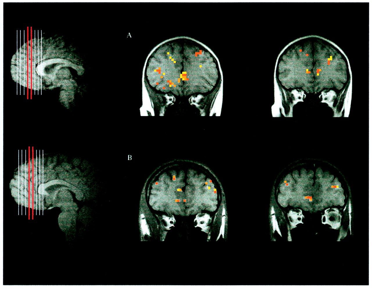
Advances in neuroimaging technology now make it possible to examine the developing human brain in vivo. Functional magnetic resonance imaging can be used as a research tool in children to study patterns of brain activity because the technique delivers no ionizing radiation while providing unique brain performance data. Maturation of the prefrontal cortex, including its dorsolateral and orbital frontal regions, is assumed to correspond to the development of higher-level cognitive processes, like working memory and response inhibition, throughout childhood and adolescence. The study associated with the above figure probed the development of different patterns of activation in the prefrontal cortex during response inhibition by using a modified version of the “Go–No-Go” task. The goal was to examine whether brain circuitry underlying inhibitory mental processes is the same in children (ages 7–12) and adults (ages 21–24) during the performance of a task requiring behavioral inhibition. This study found that the location of the activation in the prefrontal cortex is not different between children and adults (figure). However, the study also found that the volume of the activation was greater for children than for adults, especially in the dorsolateral prefrontal cortex, when performing the inhibitory part of the task (figure). These data suggest that children may have to activate (i.e., utilize) more of the dorsolateral prefrontal area to maintain a representation of (i.e., remember) the task-relevant information. In adults, greater efficiency (i.e., less activation volume) at representing task-relevant information may correspond to the greater neural selectivity characteristic of the mature brain. Maturation, as demonstrated here by task-associated differences in brain activation, may correspond to the molecular and cellular changes (including neuronal and synaptic pruning) that are going on in the neocortex during these years of development.
The midsagittal images to the left of panels A and B depict the prescribed slice locations. Overlays of activation on two T1-weighted coronal images located approximately 40 and 45 mm anterior to the anterior commissure for a right-handed 9-year-old male (panel A) and a right-handed 24-year-old male (panel B).

Footnote

Figure compliments of Dr. Casey.

Information & Authors

Information

Published In

Go to American Journal of Psychiatry
American Journal of Psychiatry
Pages: 504
PubMed: 10200727

History

Published online: 1 April 1999
Published in print: April 1999

Authors

Metrics & Citations

Metrics

Citations

Export Citations

If you have the appropriate software installed, you can download article citation data to the citation manager of your choice. Simply select your manager software from the list below and click Download.

For more information or tips please see 'Downloading to a citation manager' in the Help menu.

Format
Citation style
Style
Copy to clipboard

There are no citations for this item

View Options

View options

PDF/ePub

View PDF/ePub

Get Access

Login options

Already a subscriber? Access your subscription through your login credentials or your institution for full access to this article.

Personal login Institutional Login Open Athens login
Purchase Options

Purchase this article to access the full text.

PPV Articles - American Journal of Psychiatry

PPV Articles - American Journal of Psychiatry

Not a subscriber?

Subscribe Now / Learn More

PsychiatryOnline subscription options offer access to the DSM-5-TR® library, books, journals, CME, and patient resources. This all-in-one virtual library provides psychiatrists and mental health professionals with key resources for diagnosis, treatment, research, and professional development.

Need more help? PsychiatryOnline Customer Service may be reached by emailing [email protected] or by calling 800-368-5777 (in the U.S.) or 703-907-7322 (outside the U.S.).

Media

Figures

Other

Tables

Share

Share

Share article link

Share