Full access
Clinical Case Conference
Published Online: 1 June 2007

Treatment of Psychiatric Symptoms Associated With a Frontal Lobe Tumor Through Surgical Resection

Publication: American Journal of Psychiatry
The frontal lobes of the brain underlie judgment, foresight, motivation, and personality and allow us to behave as socially appropriate human beings (1, 2) . Our understanding of frontal lobe function comes from animal experiments (e.g., the chimpanzee experiments of Jacobsen and Fulton that were the impetus for Moniz’s human lobotomies) and human neuropsychological studies, as well as case reports involving traumatic brain injuries. These case reports have shown that frontal lobe damage often results in antisocial behavior, apathy, disinhibition, and emotional lability. Discrete prefrontal cortex lesions are sometimes associated with unique behavioral profiles. Although numerous exceptions exist, frontal lobe dysfunction exhibits laterality: left hemisphere lesions are more typically associated with depression, whereas right hemisphere lesions are associated with impulsivity and manic-like symptoms (3, 4) .
Brain tumors are a well-known cause of frontal lobe dysfunction. One type is the rare dysembryoplastic neuroepithelial tumor, a benign supratentorial neoplasm seen primarily in children and young adults (5, 6) . Approximately one-third of dysembryoplastic neuroepithelial tumors occur in the frontal lobes, and some become large enough to compress regions of the cortex and white matter to the point of disrupting behavior (79) . In addition to mass effects, dysembryoplastic neuroepithelial tumors are a source of epileptogenic activity, which may lead to neuropsychiatric sequelae when there is frontal lobe involvement (10, 11) . Surgical resection, which characteristically yields an excellent prognosis, is the recommended treatment for these tumors (12, 13) . Even with partial tumor removal, there is little evidence of recurrence, and most patients make a complete but slow neuropsychiatric recovery. This case study is one of a few to report the virtually instantaneous neurosurgical cure of a psychiatric illness in a patient with a frontal lobe dysembryoplastic neuroepithelial tumor. We also provide a summary and review of the literature on psychiatric disturbances associated with various frontal lobe lesions.

Case Presentation

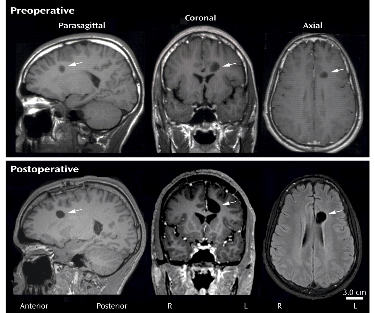
“Jimmy,” a 16-year-old right-handed African American boy with a 9-year history of progressively worsening conduct was referred to our inpatient medical psychiatric unit after the surgical removal of a brain tumor. At age 7, the patient had begun exhibiting mood swings, behavioral problems, and decreased academic performance. According to his mother, his initial symptoms consisted of totally unpredictable and inconsolable “crying jags” that lasted more than an hour. Over the next 5 years, his behavior had deteriorated further, and he became violent with suicidal ideation. During this time, he was given multiple diagnoses: attention deficit hyperactivity disorder, bipolar disorder, and conduct disorder. Eventually his mother placed him in a residential treatment facility because she could no longer manage him at home. At age 12, a magnetic resonance imaging (MRI) study revealed a nearly spherical 0.8-cm-in-diameter mass located medially within the white matter of the left frontal lobe superior to the anterior horn of the lateral ventricle ( Figure 1, top). Based on the indistinct radiographic signature of the mass, a clinical decision was made to treat the patient with mood stabilizers and antipsychotic drugs.
Figure 1. Preoperative Magnetic Resonance Imaging (MRI) (top) (1.5 T magnet, T 1 weighted) Showing a Dysembryoplastic Neuroepithelial Tumor (hypoattenuated mass, arrows) in the Paraventricular White Matter in Three Planes of Section; Postoperative MRI (bottom) (3.0 T, T 1 weighted) Showing Residual Cavity (arrows) in the White Matter 5 Months After Tumor Removal in Approximately the Same Planes of Section
His mother reported that Jimmy’s teenage years were characterized by extreme irritability and that “the sound of my voice seemed to anger him.” Over the next 4 years, the patient was poorly controlled with varying combinations and doses of lithium, methylphenidate, quetiapine, and risperidone. Higher dosing and polypharmacy were at times associated with increasingly violent behavior. EEG studies recorded symmetric 9–10-Hz alpha rhythms anteriorly with faster overlying frequencies but failed to reveal frank seizure activity. At age 16, a second MRI showed that the mass had grown to 1.3×1.4×1.0 cm and was now within 0.1 cm of the adjacent anterior cingulate cortex, but there was no neurological evidence of a mass effect. Around this time, the patient refused to take lithium and risperidone; despite the discontinuation of pharmacological treatment, his behavior did not worsen in the 3 weeks before surgery. Postoperative imaging studies ( Figure 1, bottom) revealed transection of the left cingulum and total removal of the mass, which was histologically identified as a dysembryoplastic neuroepithelial tumor.
Postoperatively, the patient’s behavioral recovery, as described by his mother, was “nearly instantaneous,” although he did have mild weakness and paresthesias of the right lower leg that lasted several weeks. During a 4-week postoperative observation period, he exhibited none of his earlier aggressive behaviors or violent outbursts and was easily managed on the inpatient medical psychiatric unit. Upon discharge, he returned to foster care for an additional 6 months before being released to his mother. At home, his mother reported that her son’s behavior was dramatically improved, and a written evaluation by a school psychologist indicated that our patient had no trouble concentrating and exhibited no noteworthy behavioral problems in the classroom. Psychological testing (see Tables 1 and 2 ) failed to reveal any permanent cognitive sequelae of the neurosurgical procedure. Outside the classroom, the patient had a renewed interest in sports and was forming adequate peer relationships. At a 1-year follow-up visit, the patient and his mother reported that he continued to do well socially, and although he decided to leave school for personal reasons, he had completed his General Equivalency Diploma (GED) and was planning a career in the military.

Discussion

Many studies of humans and nonhuman primates have established that lesioning the frontal lobes can result in abnormal behaviors (14, 15) . Depending on the affected hemisphere and the location within the hemisphere, human frontal lobe lesions can result in alterations in attention, insight, mood, planning, and interpersonal communication—changes that are often permanent (15) .
In each hemisphere, the bulk of the nonmotor frontal lobe is subdivided into the prefrontal anterior cingulate and the dorsolateral, orbitofrontal, and ventromedial cortices. The dorsolateral cortex receives the majority of its distant afferent inputs by means of the superior longitudinal and uncinate fasciculi, and short-range association fibers (U-fibers) mediate local prefrontal connections. Medially, the orbitofrontal cortex connects to limbic structures by means of the uncinate fasciculus and to the ventromedial cortex through U-fibers. The orbitofrontal and ventromedial cortices are reciprocally interconnected, and it is likely that both are connected with the anterior cingulate by means of fibers of the rostral cingulum. Communication between the prefrontal cortices of the cerebral hemispheres is mediated by reciprocal callosal projections, but these are generally much sparser than ipsilateral corticocortical connections and are usually limited to homotopic areas (1618) . In addition to corticocortical connections, all prefrontal areas receive robust input from the limbic channel of the basal ganglia.
The dorsolateral prefrontal cortex plays an essential role in weighting and integrating impulses from various sensory and limbic channels for the purpose of generating goal-directed behaviors (19) . This brain region is also active in working memory tasks (2022) . Thus, major features of dorsolateral lesions regularly include labile affect, depression, and decreased executive function (19, 2325) . Functional imaging studies have linked the negative symptoms of schizophrenic patients to perturbations of dorsolateral cortical metabolism (26, 27) .
Although our patient’s decreased academic performance might suggest a lesion of the dorsolateral prefrontal cortex and/or the underlying white matter, other functions subserved by the dorsolateral prefrontal cortex, such as decision making, working memory, and capacity to plan (1, 2, 2022 ), were largely spared. Thus, we surmise that our patient’s dorsolateral cortex remained intact, a conclusion most strongly supported by serial imaging studies that showed that the dysembryoplastic neuroepithelial tumor was many centimeters away from the dorsolateral prefrontal cortex. We believe our patient’s decreased school performance was more directly related to the tumor’s effects on other prefrontal areas and to the side effects of the polypharmacy intended to treat his psychiatric symptoms.
Intact orbitofrontal cortices are required for normal judgment and socialization. Patients with orbitofrontal lesions tend to be disinhibited (24, 28) . Unlike dorsolateral lesions, orbitofrontal lesions generally spare cognitive abilities and volition, but they can significantly contribute to the genesis of antisocial behaviors, prompting some authors to rename the orbitofrontal syndrome acquired “pseudopsychopathy” (28, 29) . Indeed, the outbursts, mood fluctuations, self-mutilation, and splitting behaviors seen in borderline personalities may be related to a hypofunctioning orbitofrontal cortex (30, 31) . Neuroanatomically, the explosive antisocial traits associated with orbitofrontal lesions have been postulated to result from a loss of inhibitory control over the amygdala (28, 32, 33) .
Although our patient exhibited many antisocial behaviors strongly associated with an orbitofrontal lesion, his tumor was not located within the orbitofrontal cortex. This incongruency might be explained by the fact that his tumor was strategically situated in the white matter just superior to the anterior horn of the left lateral ventricle. Tumors in this location can compress corticofugal orbitofrontal axons as they join the uncinate fasciculus. Thus, in our patient, corticofugal orbitofrontal fibers may have been compromised enough to partially denervate the amygdala and uncover behaviors that are normally suppressed.
The ventromedial prefrontal cortex is believed to regulate empathy, foresight, and reversal learning (14, 34) . In reversal learning tasks mediated by the ventromedial cortex, subjects are trained to respond differentially to two stimuli under reward and punishment conditions, and later they are trained to reverse the reward values (35) . Reversal learning deficits cause patients to persist in simple tasks or deleterious high-risk behaviors that in the past paid dividends, as, for example, in pathological gambling, where high-risk behaviors persist despite reduced payoffs (36, 37) .
Our patient exhibited significant signs of reversal learning impairment as evidenced by his repeatedly engaging in high-stakes behaviors that had been rewarded on the streets but resulted in harsh punishment in the residential treatment facility. These negative behaviors are consistent with dysfunction of the ventromedial cortex, an interpretation further supported by MRI studies showing that the dysembryoplastic neuroepithelial tumor encroached on the caudal boundary of the ventromedial cortex. Based on this proximity, it is very likely that the dysembryoplastic neuroepithelial tumor, by either compression or ephaptic activation of corticofugal axons, perturbed ventromedial efferent impulses and mimicked features of a ventromedial cortical lesion.
The anterior cingulate cortex borders the genu of the corpus callosum and merges into the posterior cingulate. Functionally, the anterior cingulate can be divided into two regions, a rostral affective division and a caudal cognitive division, although recent functional MRI studies suggest even greater functional subdivisions probably exist (38, 39) . The rostral anterior cingulate receives robust input from the amygdala, whereas the caudal anterior cingulate receives projections from the entorhinal cortex and parahippocampal/hippocampal regions (40) . Connecting these areas is the cingulum, a fiber tract embedded within the anterior cingulate gyrus and terminating in more rostral prefrontal cortices. Functionally, the anterior cingulate may underlie drive (motivated attention) and concentration (attention allocation), and it may recognize affect-mood conflicts and other processing errors requiring heightened awareness (cognitive and limbic error detection) (38, 39, 41, 42) . Anterior cingulate lesions interfere with these functions, whereas anterior cingulate hyperactivity has been described in most anxiety disorders, including obsessive-compulsive disorder (OCD) as well as schizophrenia and chronic pain syndromes (42) .
Our patient’s clinical and MRI findings strongly suggest the tumor had the greatest effect on the anterior cingulate. Clinically, Jimmy was apathetic and had difficulty sustaining attention, and he often would respond inappropriately to another person’s affect; both of these behavioral deficits are associated with anterior cingulate dysfunction. Multiple MRIs revealed that of all the prefrontal regions, the dysembryoplastic neuroepithelial tumor was closest to the anterior cingulate (approximately 1 mm of separation), necessitating a mid-rostral cingulotomy for surgical access.
Frontal lobe lesions exhibit lateralization with respect to psychiatric or behavioral disturbances. Left hemisphere lesions are more likely to be associated with depression, particularly if the lesion involves the dorsolateral portion of prefrontal cortex. By contrast, right hemisphere lesions are associated with impulsivity and manic behaviors (3, 4) . Our patient’s dysembryoplastic neuroepithelial tumor was located in the left frontal lobe, but his behaviors were more consistent with right frontal lobe dysfunction, e.g., aggressiveness and disinhibition. This discrepancy could be explained by several neuropsychiatric observations. In children and young adolescents, symptoms of depression can manifest as irritability and psychomotor agitation (43, 44) . Our patient periodically exhibited these traits, but they were consistently overshadowed by his more extreme behaviors. Another explanation is centered on the observation that dysembryoplastic neuroepithelial tumors can be epileptogenic foci (10, 11, 13) . Although this patient’s EEG did not show frank epileptic activity, abnormal electrical activity associated with seizures was detected on at least one occasion, and at some point the tumor may have affected the activity of neurons in the left frontal cortex and contributed to his abnormal behaviors.
This case supports other studies demonstrating that neurosurgical cure of a psychiatric disorder is feasible (45) . Probably the most remarkable aspect of this case is the fact that the worst of our patient’s long-standing conduct disorder traits resolved within hours of neurosurgery. Our patient had a remarkable recovery, but several key issues need to be addressed. The impetus for neurosurgery in our patient was tumor growth, not cure of his behavioral problems. Forecasting neuropsychiatric cure after frontal lobe surgery for tumor removal is difficult at best as differences in tumor size, subtleties in surgical technique, and susceptibilities of different brain regions to operative trauma have left patients like ours with a worsening clinical course, such as postoperative psychosis or epilepsy (46, 47) . The prognosis is worse when bulky tumors destroy a larger volume of the brain parenchyma, fundamentally altering the neuroanatomical substrates of behavior. Fortunately, our patient’s tumor was relatively small, well circumscribed, and surgically accessible, requiring only partial removal of the overlying anterior cingulate gyrus and cingulum. Partial cingulotomy may have even contributed to our patient’s psychiatric improvement, as several reports have indicated that cingulotomy is an effective treatment for chronic pain, intractable depression, and medication-resistant OCD (48, 49) .
Pharmacological management of psychiatric illnesses is the cornerstone of modern psychiatry, but this case shows that medication failures may occur in the setting of an underlying brain tumor. Our patient was treated with a number of different medication combinations over the course of 9 years. Both by the patient’s report and according to his mother and professional staff at the residential treatment facility, the side effects of polypharmacy made him very uncomfortable and probably did little to control his extreme behavior. In fact, 3 weeks before surgery, our patient elected to discontinue all psychiatric medications in favor of reduced side effects, yet his behavior did not worsen during that time. These clinical observations suggest that the deleterious effects of the dysembryoplastic neuroepithelial tumor were far beyond the reach of the then-current neuropsychiatric pharmacopeia.
Conclusion

This case study is one of a handful of reports of near-instantaneous cure of a psychiatric disorder following the removal of a frontal lobe tumor. It illustrates how a more thorough understanding of regional frontal lobe function as it pertains to behavior, cognition, and mood may be helpful when we attempt to use clinical observations to deduce the location of a putative frontal lobe mass. Although exceptions exist, dorsolateral frontal lobe lesions shape mood, volition, and executive function; orbitofrontal lesions affect socialization; ventromedial lesions have an effect on aspects of foresight and reversal learning; and anterior cingulate lesions influence awareness, concentration, error detection, and affect-mood congruence. Small lesions in the frontal white matter, which can affect efferent impulses originating from widespread prefrontal areas, may produce a wider array of aberrant behaviors than similarly sized lesions confined to a single area of the overlying cortex. Furthermore, this case emphasizes that frontal lobe masses causing severe psychiatric disturbances may benefit from evaluation for neurosurgery. Such an evaluation may not be prioritized when there is no evidence of malignancy or significant mass effect, but our case suggests that surgery may potentially lead to significant clinical improvement even in the context of longstanding psychiatric symptoms. In cases in which neurosurgery might damage critical anatomy, drug therapy is usually warranted, but we should expect more frequent medication treatment failures in this patient population.

Footnotes

Received Sept. 19, 2006; revision received Jan. 30, 2007; accepted Feb. 9, 2007. From Drexel University College of Medicine, Philadelphia; and the Department of Psychiatry, Hahnemann University Hospital. Address correspondence and reprint requests to Dr. Mathews, Department of Psychiatry, Hahnemann University Hospital, Broad and Vine St., Philadelphia, PA 19102; [email protected] (e-mail).The authors thank Drs. Richard Roth, Myrna Miller, and Sandra Koffler for criticisms of earlier versions of the article.
CME Disclosure: The authors report no competing interests. APA policy requires disclosure by CME authors of unapproved or investigational use of products discussed in CME programs. Off-label use of medications by individual physicians is permitted and common. Decisions about off-label use can be guided by scientific literature and clinical experience.

References

1.
Chayer C, Freedman M: Frontal lobe functions. Curr Neurol Neurosci Rep 2001; 1:547–552
2.
Koechlin E, Ody C, Kouneiher F: The architecture of cognitive control in the human prefrontal cortex. Science 2003; 302:1181–1185
3.
Clark L, Manes F, Antoun N, Sahakian BJ, Robbins TW: The contributions of lesion laterality and lesion volume to decision-making impairment following frontal lobe damage. Neuropsychologia 2003; 41:1474–1483
4.
Aron AR, Robbins TW, Poldrack RA: Inhibition and the right inferior frontal cortex. Trends Cogn Sci 2004; 8:170–177
5.
Daumas-Duport C, Scheithauer BW, Chodkiewicz JP, Laws ER Jr, Vedrenne C: Dysembryoplastic neuroepithelial tumor: a surgically curable tumor of young patients with intractable partial seizures. report of thirty-nine cases. Neurosurgery 1988; 23:545–556
6.
Cabiol J, Acebes JJ, Isamat F: Dysembryoplastic neuroepithelial tumors. Crit Rev Neurosurg 1999; 9:116–125
7.
Sato T, Takeichi M, Abe M, Tabuchi K, Hara T: Frontal lobe tumor associated with late-onset seizure and psychosis: a case report. Jpn J Psychiatry Neurol 1993; 47:541–544
8.
Taylor DC, Neville BG, Cross JH: Spectrum disorders in childhood epilepsy surgery candidates. Eur Child Adolesc Psychiatry 1999; 8:189–192
9.
Escosa Bage M, Villarejo Ortega FJ, Perez Jimenez MA, Gonzalez Mediero I: Psychosis in a case of temporal lobe epilepsy associated with a dysembryoplastic neuroepithelial tumour. Rev Neurol 2004; 38:643–646
10.
Weissman Z, Michowitz S, Shuper A, Kornreich L, Amir J: Dysembryoplastic neuroepithelial tumor: a curable cause of seizures. Pediatr Hematol Oncol 1996; 13:463–468
11.
Sisodiya SM: Malformations of cortical development: burdens and insights from important causes of human epilepsy. Lancet Neurol 2004; 3:29–38
12.
Nolan MA, Sakuta R, Chuang N, Otsubo H, Rutka JT, Snead OC III, Hawkins CE, Weiss SK: Dysembryoplastic neuroepithelial tumors in childhood: long-term outcome and prognostic features. Neurology 2004; 62:2270–2276
13.
Sandberg DI, Ragheb J, Dunoyer C, Bhatia S, Olavarria G, Morrison G: Surgical outcomes and seizure control rates after resection of dysembryoplastic neuroepithelial tumors. Neurosurg Focus 2005; 18:E5
14.
Shamay-Tsoory SG, Tomer R, Berger BD, Aharon-Peretz J: Characterization of empathy deficits following prefrontal brain damage: the role of the right ventromedial prefrontal cortex. J Cogn Neurosci 2003; 15:324–337
15.
Mah L, Arnold MC, Grafman J: Impairment of social perception associated with lesions of the prefrontal cortex. Am J Psychiatry 2004; 161:1247–1255
16.
Cavada C, Reinoso-Suarez F: Topographical organization of the cortical afferent connections of the prefrontal cortex in the cat. J Comp Neurol 1985; 242:293–324
17.
Marconi B, Genovesio A, Giannetti S, Molinari M, Caminiti R: Callosal connections of dorso-lateral premotor cortex. Eur J Neurosci 2003; 18:775–788
18.
Barbas H, Hilgetag CC, Saha S, Dermon CR, Suski JL: Parallel organization of contralateral and ipsilateral prefrontal cortical projections in the rhesus monkey. BMC Neurosci 2005; 6:32
19.
Colvin MK, Dunbar K, Grafman J: The effects of frontal lobe lesions on goal achievement in the water jug task. J Cogn Neurosci 2001; 13:1129–1147
20.
Levy R, Goldman-Rakic PS: Segregation of working memory functions within the dorsolateral prefrontal cortex. Exp Brain Res 2000; 133:23–32
21.
Petrides M: The role of the mid-dorsolateral prefrontal cortex in working memory. Exp Brain Res 2000; 133:44–54
22.
van’t Wout M, Kahn RS, Sanfey AG, Aleman A: Repetitive transcranial magnetic stimulation over the right dorsolateral prefrontal cortex affects strategic decision-making. Neuroreport 2005; 16:1849–1852
23.
Cummings JL: Frontal-subcortical circuits and human behavior. Arch Neurol 1993; 50:873–880
24.
Duffy JD, Campbell JJ: The regional prefrontal syndromes: a theoretical and clinical overview. J Neuropsychiatry Clin Neurosci 1994; 6:379–387
25.
Schutter DJ, van Honk J: Increased positive emotional memory after repetitive transcranial magnetic stimulation over the orbitofrontal cortex. J Psychiatry Neurosci 2006; 31:101–104
26.
Friston KJ: The dorsolateral prefrontal cortex, schizophrenia and PET. J Neural Transm Suppl 1992; 37:79–93
27.
Bertolino A, Esposito G, Callicott JH, Mattay VS, Van Horn JD, Frank JA, Berman KF, Weinberger DR: Specific relationship between prefrontal neuronal N-acetylaspartate and activation of the working memory cortical network in schizophrenia. Am J Psychiatry 2000; 157:1–2
28.
Brower MC, Price BH: Neuropsychiatry of frontal lobe dysfunction in violent and criminal behaviour: a critical review. J Neurol Neurosurg Psychiatry 2001; 71:720–726
29.
Mitchell DG, Avny SB, Blair RJ: Divergent patterns of aggressive and neurocognitive characteristics in acquired versus developmental psychopathy. Neurocase 2006; 12:164–178
30.
Berlin HA, Rolls ET, Kischka U: Impulsivity, time perception, emotion and reinforcement sensitivity in patients with orbitofrontal cortex lesions. Brain 2004; 127:1108–1126
31.
Berlin HA, Rolls ET, Iversen SD: Borderline personality disorder, impulsivity, and the orbitofrontal cortex. Am J Psychiatry 2005; 162:2360–2373
32.
Davidson RJ, Putnam KM, Larson CL: Dysfunction in the neural circuitry of emotion regulation—a possible prelude to violence. Science 2000; 289:591–594
33.
Best M, Williams JM, Coccaro EF: Evidence for a dysfunctional prefrontal circuit in patients with an impulsive aggressive disorder. Proc Natl Acad Sci U S A 2002; 99:8448–8453
34.
Fellows LK, Farah MJ: Dissociable elements of human foresight: a role for the ventromedial frontal lobes in framing the future, but not in discounting future rewards. Neuropsychologia 2005; 43:1214–1221
35.
Fellows LK, Farah MJ: Ventromedial frontal cortex mediates affective shifting in humans: evidence from a reversal learning paradigm. Brain 2003; 126:1830–1837
36.
Cavedini P, Riboldi G, Keller R, D’Annucci A, Bellodi L: Frontal lobe dysfunction in pathological gambling patients. Biol Psychiatry 2002; 51:334–341
37.
Fellows LK, Farah MJ: Different underlying impairments in decision-making following ventromedial and dorsolateral frontal lobe damage in humans. Cereb Cortex 2005; 15:58–63
38.
Bush G, Luu P, Posner MI: Cognitive and emotional influences in anterior cingulate cortex. Trends Cogn Sci 2000; 4:215–222
39.
Davis KD, Taylor KS, Hutchison WD, Dostrovsky JO, McAndrews MP, Richter EO, Lozano AM: Human anterior cingulate cortex neurons encode cognitive and emotional demands. J Neurosci 2005; 25:8402–8406
40.
Mesulam MM: Behavioral neuroanatomy: large-scale networks, association cortex, frontal syndromes, the limbic system, and hemispheric specialization, in Principles of Behavioral and Cognitive Neurology. Edited by Mesulam MM. New York, Oxford University Press, 2000, pp 41–52
41.
Carter CS, Botvinick MM, Cohen JD: The contribution of the anterior cingulate cortex to executive processes in cognition. Rev Neurosci 1999; 10:49–57
42.
van Veen V, Carter CS: The anterior cingulate as a conflict monitor: fMRI and ERP studies. Physiol Behav 2002; 77:477–482
43.
Kanner AM, Dunn DW: Diagnosis and management of depression and psychosis in children and adolescents with epilepsy. J Child Neurol 2004; 19:S65–S72
44.
Sheikh RM, Weller EB, Weller RA: Prepubertal depression: diagnostic and therapeutic dilemmas. Curr Psychiatry Rep 2006; 8:121–126
45.
Anderson CA, Arciniegas DB: Neurosurgical interventions for neuropsychiatric syndromes. Curr Psychiatry Rep 2004; 6:355–363
46.
Andermann LF, Savard G, Meencke HJ, McLachlan R, Moshe S, Andermann F: Psychosis after resection of ganglioglioma or DNET: evidence for an association. Epilepsia 1999; 40:83–87
47.
Hennessy MJ, Elwes RD, Binnie CD, Polkey CE: Failed surgery for epilepsy: a study of persistence and recurrence of seizures following temporal resection. Brain 2000; 123:2445–2466
48.
Ballantine HT Jr, Bouckoms AJ, Thomas EK, Giriunas IE: Treatment of psychiatric illness by stereotactic cingulotomy. Biol Psychiatry 1987; 22:807–819
49.
Cosgrove GR, Rauch SL: Stereotactic cingulotomy. Neurosurg Clin N Am 2003; 14:225–235

Information & Authors

Information

Published In

Go to American Journal of Psychiatry
American Journal of Psychiatry
Pages: 877 - 882
PubMed: 17541045

History

Published online: 1 June 2007
Published in print: June, 2007

Authors

Details

Zachary S. Hoffer, Ph.D.
Shannon L. Allen, B.S.
Maju Mathews, M.D., M.R.C.Psych.

Metrics & Citations

Metrics

Citations

Export Citations

If you have the appropriate software installed, you can download article citation data to the citation manager of your choice. Simply select your manager software from the list below and click Download.

For more information or tips please see 'Downloading to a citation manager' in the Help menu.

Format
Citation style
Style
Copy to clipboard

View Options

View options

PDF/EPUB

View PDF/EPUB

Get Access

Login options

Already a subscriber? Access your subscription through your login credentials or your institution for full access to this article.

Personal login Institutional Login Open Athens login
Purchase Options

Purchase this article to access the full text.

PPV Articles - American Journal of Psychiatry

PPV Articles - American Journal of Psychiatry

Not a subscriber?

Subscribe Now / Learn More

PsychiatryOnline subscription options offer access to the DSM-5-TR® library, books, journals, CME, and patient resources. This all-in-one virtual library provides psychiatrists and mental health professionals with key resources for diagnosis, treatment, research, and professional development.

Need more help? PsychiatryOnline Customer Service may be reached by emailing [email protected] or by calling 800-368-5777 (in the U.S.) or 703-907-7322 (outside the U.S.).

Media

Figures

Other

Tables

Share

Share

Share article link

Share