Abstract

OBJECTIVE: Disturbed neuronal interactions may be involved in schizophrenia because it is without clear regional pathology. Aberrant connectivity is further suggested by theoretical formulations and neurochemical and neuroanatomical data. The authors applied to schizophrenia a recently available functional neuroimaging analytic method that permits characterization of cooperative action on the systems level. METHOD: Thirteen medication-free patients and 13 matched healthy comparison subjects performed a working memory (n-back) task and sensorimotor baseline task during positron emission tomography. “Functional connectivity” patterns, reflecting distributed correlated activity that differed most between groups, were extracted by a canonical variates analysis. RESULTS: More than half the variance was explained by a single pattern showing inferotemporal, (para-)hippocampal, and cerebellar loadings for patients versus dorsolateral prefrontal and anterior cingulate activity for comparison subjects. Expression of this pattern perfectly separated all patient scans from comparison scans, thus showing promise as a trait marker. This result was validated prospectively by successfully classifying unrelated scans from the same patients and data from a new cohort. An additional 19% of variance corresponded to the pattern activated by the working memory task. Expression of this pattern was more variable in patients during working memory but not the control condition, suggesting inability to sustain a task-adequate neural network, consistent with the disconnection hypothesis. CONCLUSIONS: Pronounced disruptions of distributed cooperative activity in schizophrenia were found. A pattern showing disturbed frontotemporal interactions showed promise as a trait marker and may be useful for future investigations.
Brain function is characterized by both regional differentiation of function and the necessity to coactivate functionally appropriate cooperative networks for all but the most trivial of tasks (1). Mapping regional deficits with neuroimaging has been successfully used to study circumscribed lesions, such as stroke. However, neuropsychiatric disorders such as schizophrenia exhibit a devastating syndrome despite only subtle localizable functional aberrations. In this disorder, investigation of abnormal functional connectivity (i.e., the cooperative action of neural systems) appears promising (2) since many studies show low neuropil levels, abnormalities in synaptic, dendritic, axonal, and white matter tract organization, and abnormalities of glutamatergic neurotransmission, which are consistent with disturbed intracortical connectivity (36).
Despite this interest in connectivity, most neuroimaging studies of schizophrenia have continued to examine circumscribed abnormalities. In the present study, we directly tested for the presence and potential relevance of disturbed connectivity in schizophrenia by using blood flow data obtained during a test of working memory, the capacity to keep information “on-line” as necessary for an ongoing task (7). We chose working memory because it is disturbed in schizophrenia and has been linked to abnormal blood flow (8).
As an operational definition, we regarded brain regions as functionally connected if their activities were correlated (1). We characterized patterns of correlated activity that were most different between patient and comparison groups by using canonical variates analysis (9). These patterns were prospectively validated in two separate data sets, one from the same subjects and one from a different subject group.

Method

Subjects and Task

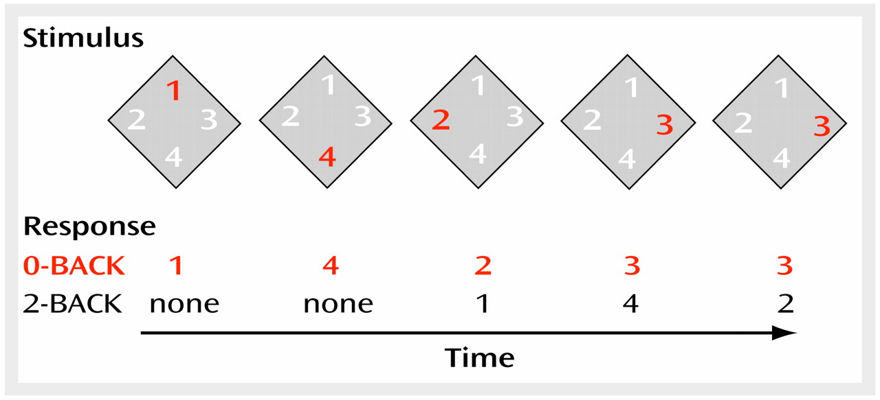
We studied 13 medication-free patients with DSM-IV schizophrenia (paranoid subtype, N=6; undifferentiated subtype, N=7) and 13 age- and sex-matched healthy comparison subjects. The patients’ mean age was 32.5 years (SD=8.2), their mean education level was 13 years (SD=2.9), and the group contained three women and one left-handed subject. The mean age of the comparison subjects was 30.4 years (SD=7.9), their mean education level was 17 years (SD=3.1), and the group contained four women and one left-handed subject. The patients were withdrawn from medication at least 2 weeks before the study. Subjects were excluded if they had a history of medical illness or treatment relevant to regional cerebral blood flow (rCBF). Comparison subjects were excluded if they had a first-degree relative with schizophrenia. After complete description of the study, the subjects provided written informed consent according to National Institutes of Health guidelines. During scanning, the subjects performed the n-back task (Figure 1). The numerals 1, 2, 3, and 4 were presented at the rate of one every 1.8 seconds in a diamond-shaped array by means of a computer. The subjects held a button-box with four buttons arranged similarly. In the 0-back (control) condition, the button corresponding to the current number was to be pressed. In the 2-back (working memory) condition, the button to press corresponded to the number seen two presentations before.

Data Acquisition and Analysis

Positron emission tomography (PET) scans were obtained over 1 minute on a GE Advance (Milwaukee) three-dimensional scanner after injection of 10 mCi [15O]H2O. For each subject, seven images each during the 0-back and 2-back conditions and two to four scans during rest were acquired, corrected for attenuation, and reconstructed (resolution: 6.5 mm full width at half maximum). After subtraction of background activity and registration (10), the images were template-normalized and smoothed with an isotropic 10-mm3 full width at half maximum Gaussian kernel by using SPM 97 (Wellcome Department of Cognitive Neurology). Scan-to-scan variation in global counts was removed by using proportional scaling. Main effects (condition, group) and interactions (condition by group) were assessed with contrasts of the adjusted means by using t statistics transformed into the z statistic. Values significant at p<0.001 were corrected for multiple comparisons on the cluster level (11). Locations of maxima are reported as millimeters relative to the anterior commissure, which were determined after registration to the SPM 97 population-derived template, which is larger than the commonly used single brain depicted by the Talairach-Tournoux atlas (12). The indicated Brodmann’s areas are approximate and were found by affine transformation of the SPM 97 coordinates to the Talairach-Tournoux atlas.

Analysis of Functional Connectivity Patterns

Functional connectivity patterns differing most between groups were extracted by using a canonical variates analysis (9). By this method, similar to partial least squares (13), one first computes a normalized correlation between the data and a set of regressors (contained in the design matrix). This correlation matrix is then decomposed in a series of “eigenimages” and “scan loadings” that best represent the variance (or information) in this correlation. As such, these eigenimages reflect the functional connectivity of brain regions relative to the experiment. Detailed instructions for applying this method to neuroimaging data and the statistical testing (Anderson statistic, including levels of freedom) are given by Worsley et al. (9); the software is available from the second author. Since this method operates on voxel-by-voxel correlation matrices, the extracted eigenimages reflect patterns of correlated activity and, by our operational definition, functional connectivity. Since in our case the design matrix contains no regressors other than those needed to model the group-by-time interactions, there are no specific information and no prior assumptions about the task design, and the method can thus be seen as data driven. It yields an assessment of the variance explained by a given pattern, as well as a test of significance based on a multivariate linear model. For each voxel, the resultant patterns contain a positive or negative value depending on how activity at this voxel contributes to the given pattern (similar to a loading in a factor analysis). The expression of the pattern is calculated for every given scan and is expressed as a positive or negative coefficient. Inferences about the functional relevance of the connectivity patterns can then be made, provided that this profile of pattern expression can be linked to specific disease states or task conditions. Also, an analysis of scan-to-scan variability not previously feasible in PET can be performed. However, inferences are valid only for the entire pattern and not for individual regions. It is possible that a given brain region contributes to several eigenimages (e.g., dorsolateral prefrontal cortex activity in our data).

Results

Behavior

As expected, the patients’ performance during the working memory task was significantly worse than that of the comparison subjects: 53% correct (range=20%–90%) versus 77% correct (range=59%–99%) (t=3.41, df=24, p<0.05). No performance differences were apparent during the control task.

Activation Analysis

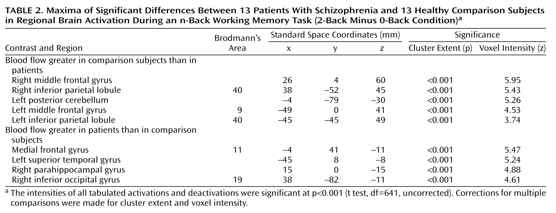
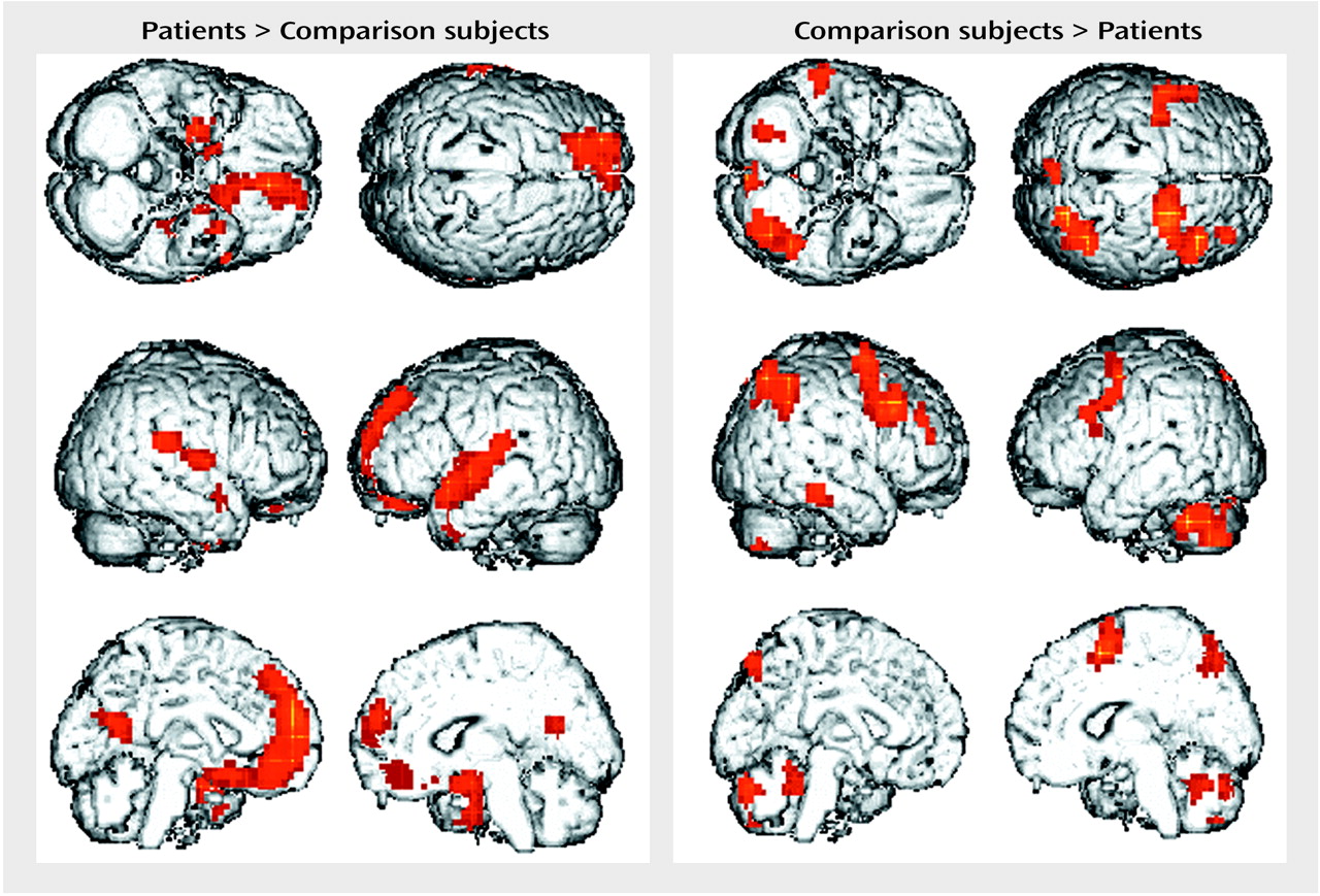
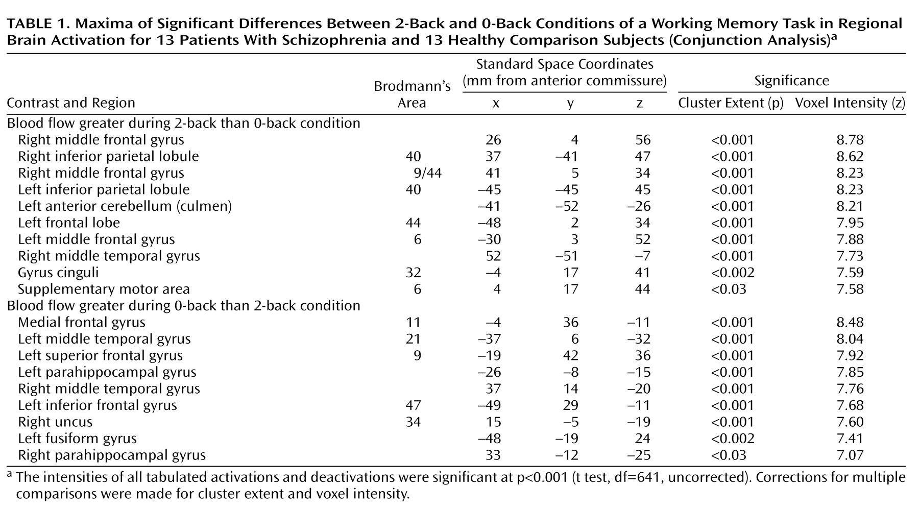
During the working memory task, as compared to the control task, both patients and comparison subjects activated similar networks encompassing the bilateral dorsolateral prefrontal cortex, the inferior parietal lobule, and the cerebellar hemispheres, while deactivating medial frontal, middle temporal, and parahippocampal areas (Table 1). The between-group comparison (condition-by-group interaction) showed less activation in the dorsolateral prefrontal cortex and inferior parietal lobule in the patients than in the comparison subjects (Table 2 and Figure 2, right), as well as less deactivation in the medial frontal gyrus, left superior temporal gyrus, right parahippocampal gyrus, and right inferior occipital gyrus (Table 2 and Figure 2, right). The main findings remained valid when performance differences were taken into account as a confounding covariate (data not shown).

Functional Connectivity

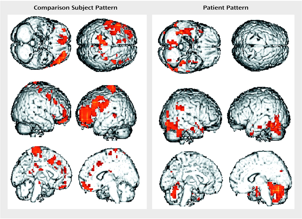
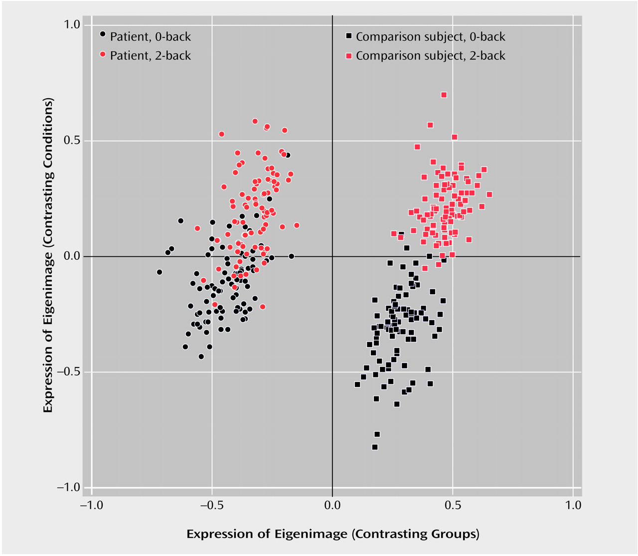
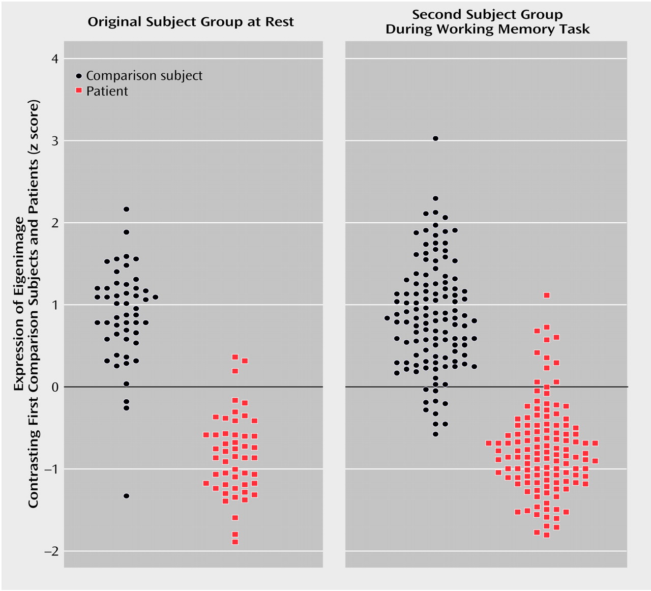
More than 69% of the overall variance could be attributed to two patterns significant at the p<0.0001 level (Anderson statistic [9]). The first pattern (Figure 3) explained more than 50% of the total variance. This pattern perfectly separated all patient scans from those of the comparison subjects: pattern expression was always negative for the patients and positive for the comparison subjects (Figure 4, x axis). The patients’ pattern was characterized by loadings in the temporal lobe, especially the inferior temporal lobe and hippocampus, and the cerebellum, whereas the pattern expressed by the comparison subjects loaded on the dorsolateral prefrontal cortex and cingulate gyrus bilaterally. Since this was true regardless of experimental condition (0-back or 2-back), we tested whether this pattern could be used prospectively to indicate the presence or absence of the disease. First, the expression of the pattern was used to classify 100 scans of the subjects at rest, acquired from the same subjects but not entered into the original analysis used to derive the pattern. Ninety-four percent of these scans (47 of 50 patient scans and 47 of 50 comparison subject scans) were correctly classified (Figure 5, left). As further validation, we used this same pattern derived from the original cohort to classify 252 scans from 13 new comparison subjects (mean age=30.4 years [SD=7.2], four women, two left-handers, mean education level=17 years [SD=3]) and nine new patients (mean age=35.6 years [SD=8.3], two women, three left-handers, mean education level=14 years [SD=2]) acquired during the 2-back and 0-back conditions. Again, 94% of these scans were correctly classified (Figure 5, right).
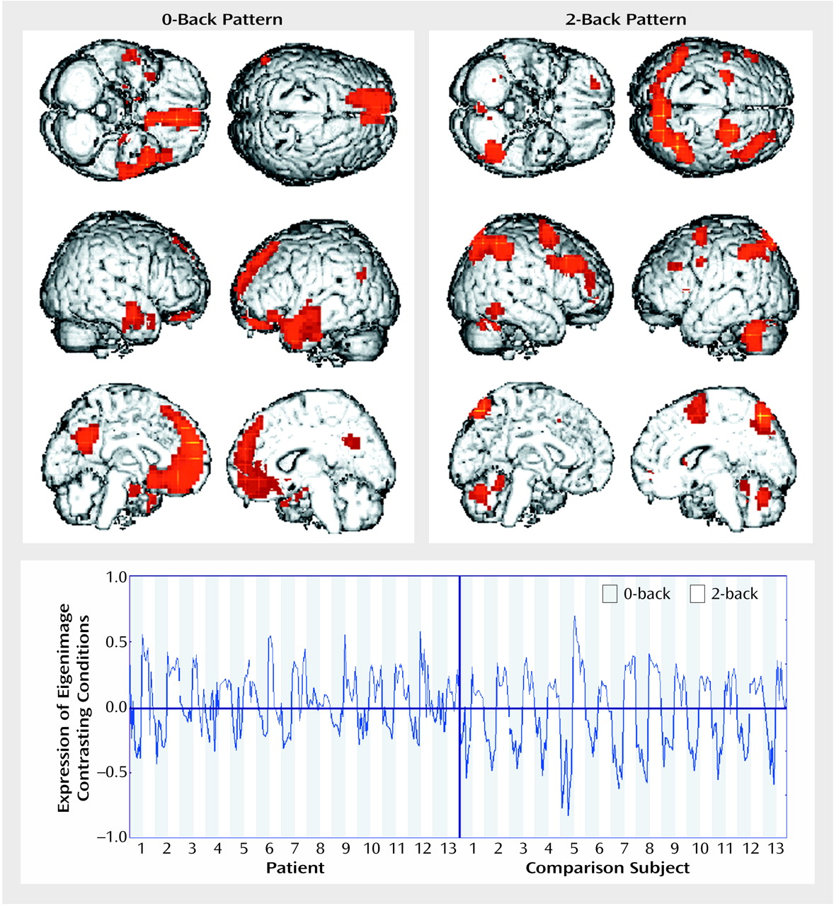
The second pattern (explaining 19% of total variance) closely resembled the activation-deactivation seen during the working memory task relative to the control task (Figure 6, top; compare with Figure 2). Whereas the pattern almost perfectly distinguished between task conditions in the comparison subjects (2.7% misclassified scans), the difference between the 2-back and 0-back tasks was much less clear-cut in the patients, 18.8% of whose scans were misclassified (Figure 4, y axis). Our method also enabled us to compare the variability of the expression of this pattern across scans and groups. The expression of the second pattern was more variable in the patients (Figure 6, bottom). Variability was significantly increased between groups (F=12.5, df=1, 180, p<0.001, Levene test for homogeneity of variance) only during the working memory task (2-back condition), however; no difference (F=0.1, df=1, 180, p>0.75) between groups existed during the control condition.

Discussion

The present analysis used a canonical variates method that uncovered highly significant differences in putative functional connectivity patterns between patients and comparison subjects. Several methods for data-driven extraction of connectivity patterns are available (13, 14). Our method addresses only linear dependencies of the data, whereas others also ensure independence of higher statistical moments (15). However, this is unlikely to be a problem since the distribution of the present data is already close to Gaussian owing to the acquisition mode and preprocessing (smoothing); the higher moments can thus be derived by the first two.

First (Group-Separating) Eigenimage

Both task-dependent and task-independent patterns were found. The main contribution to the group difference was (relatively) independent of task, with predominant weightings in distributed networks that differentiated comparison and patient subjects. The opposite poles of this pattern are the bilateral frontal, especially dorsolateral prefrontal, frontal cortex and parietal areas (at the “comparison subject” endpoint) and temporal, especially inferotemporal and hippocampal, as well as cerebellar areas (at the “patient” pole).
Both structural (16) and functional (8) abnormalities of the dorsolateral prefrontal cortex in schizophrenia have been repeatedly described. The fact that the “comparison subject pole” of this eigenimage is characterized by relatively more frontal activity is thus consistent with previous findings and extends them toward the involvement of the dorsolateral prefrontal cortex in a neural network relevant to the disease. The prominent contribution of the anterior cingulate gyrus to the comparison subject pattern is of interest in view of recent theories about error monitoring or regulatory functionality in this area that may be deficient in schizophrenia (17, 18).
Regions prominently associated with the “patient pole” of the pattern included inferotemporal areas, especially parahippocampal areas and the hippocampus, and the cerebellum. Major roles for hippocampal-limbic areas in the disease have long been proposed (19). It is interesting that most of the available functional neuroimaging data show inappropriate hyperperfusion of hippocampal areas in schizophrenia (20, 21), consistent with the positive association of this area with the pattern’s patient pole. Involvement of the cerebellum, as in our data, has been hypothesized to lead to “cognitive dysmetria” in schizophrenia (22). The face validity of our findings is substantiated by the fact that its prominent contributors are the major regions posited to play a role in schizophrenia, whereas primary sensory and motor regions do not appear to contribute to our group difference, a finding also in agreement with the literature.
Our findings regarding disturbed connectivity proper show that complete group separation between schizophrenic patients and comparison subjects can be achieved, and prospectively validated, by examining differences in functional connectivity, thus capturing an important neurobiological aspect of the disorder on the systems level. Several recent theories about disturbed connectivity underlying schizophrenia implicate specific patterns and can be compared to our findings. Friston and Frith (23), using a different analytical approach (generalized eigenvalue solution), found a connectivity pattern in comparison subjects that was underexpressed in patients, reflecting disturbed frontotemporal connectivity in patients. A separate study confirmed disruption of frontotemporal interactions in schizophrenia (18). Consistent with this was our finding that frontal and temporal structures are prominently involved at opposing ends of the group separation pattern but differ in the preferential inferotemporal/hippocampal involvement within the temporal lobe. Therefore, our data are in good agreement with the proposal that disturbed hippocampal-dorsolateral-prefrontal interactions may underlie schizophrenia (24).

Prospective Validation

To our knowledge, this is the first instance in which the utility of a specific neuroimaging finding to distinguish between diagnostic groups was prospectively examined in an independent group of data. Our ability to use the first eigenimage to classify unrelated scans from the same study group as well as data from a new group of subjects strengthens the assumption that this pattern may reflect a trait marker. The fact that scans obtained at rest could be successfully classified shows that the classification is not likely to be based on a differential response to specific aspects of the task-control conditions (including performance-related effects); rather, it reflects disease-related impairment of cortical connectivity. However, other explanations remain possible, such as nonspecific effects of the scanning environment that affect patients and comparison subjects differently. Also, although our patients were medication free during the experiment, they had all received psychoactive drugs previously, and long-term effects of such treatment on functional connectivity cannot be ruled out.
The success in prospective validation should also encourage use of this approach to investigate other diagnostically related groups, such as patients with schizotypal personality disorder or schizoaffective psychoses, to more clearly delineate the specificity of disturbed connectivity in schizophrenia. Another promising application would involve relatives of schizophrenic patients in an attempt to characterize an intermediate phenotype and facilitate genetic linkage analyses (25).

Second (Task-Related) Eigenimage

The second extracted eigenimage can be confidently linked to the activation-deactivation network recruited by the working memory task both by its scan-by-scan pattern expression and the virtually identical regional distribution. The fact that this pattern was retrieved in the present analysis demonstrates that patients and comparison subjects differ significantly in the expression of their task-associated connectivity pattern. This finding is consistent with work demonstrating abnormal activation during working memory in schizophrenia (8), particularly in the dorsolateral prefrontal cortex (26). Our data extend this work in two ways. First, by assessing scan-by-scan expression, we ascertained that, relative to comparison subjects, patients showed greater variability in recruiting the identified neural system during working memory, but not during the 0-back condition. The fact that this group difference appeared only when the system was stressed may reflect an inability to sustain a task-adequate working memory network. Presumably related to this disease-associated disturbance is the finding that the distinction between the task and control conditions, almost perfect in the comparison subjects, was less clear in the patients. It is possible that this greater variability in state-dependent activation was due to the trait-related functional abnormalities, especially in the dorsolateral prefrontal cortex, reflected in the first pattern.

Assessment of Variance Explained

The second way this work extends previous findings is that we could directly compare the amounts of variance explained by the first and second eigenimages. While differences in neural response to working memory explained 19% of the variance, capturing an important aspect of disordered function in schizophrenia, the main group difference was captured by the first, relatively task-independent pattern. Thus, while cognitive subtraction paradigms (comparing a task to a matched control condition) provide better experimental control than studies at rest and confer some ability to isolate cognitive subcomponents, these approaches may miss important aspects of the pathology if a brain structure is affected but not differentially involved in the compared conditions. A data-driven method, such as the present one, that does not have this limitation can identify relevant neuroimaging patterns that do not fit the partitioning of the data into presupposed experimental conditions.
The significant performance difference between our studied groups may be viewed as a consequence of the neurophysiological alterations demonstrated, or as a confound. This complex issue has been reviewed elsewhere (27). Here we found no evidence for an effect of performance on the neuroimaging variance, since the first eigenimage was not only relatively task independent but also classified resting subjects, and the second eigenimage’s expression was not related to performance.

Limitations of Functional Connectivity Approaches
As stated, functional connectivity is an operational definition capturing only one—albeit important—aspect of disturbed neuronal interactions. The relationship of this measure to neuronal firing, a phenomenon on an entirely different time scale, is likely complex (28). Regional interactions are not necessarily linear (29). Furthermore, linear correlation does not imply causality. For example, a visuomotor task requiring synchronous activation of visual and motor cortices does not argue for anatomical or causal connections of these areas. Thus, the fact that the variances of subpopulations of the data are best described by different eigenvectors does not immediately mean that these regions are “disconnected” if that implies causal or anatomical connections. The present findings should therefore be extended by methods that examine factors underlying the observed relationships between brain areas, such as structural equation modeling (30) or Volterra-kernel series (31). The results of the one such report of which we are aware (32) are in good agreement with the present findings in identifying disturbances in frontotemporal and intrafrontal interactions in a PET study of schizophrenic patients during semantic processing.
TABLE 1
TABLE 2
Figure 1. The n-Back Working Memory Taska
aOne diamond-shaped stimulus was presented every 1.8 seconds. In the 0-back condition, a button corresponding to the number currently displayed was to be pressed; in the 2-back condition, the button corresponding to the number presented two trials before was to be pressed. The 0-back and 2-back conditions were repeated seven times (nine stimuli each).
Figure 2. Brain Regions Showing Significant Differences Between 13 Patients With Schizophrenia and 13 Healthy Comparison Subjects in Regional Brain Activation During an n-Back Working Memory Task (2-Back Minus 0-Back Condition)a
aCorresponding values are presented in Table 2. Images were thresholded at p<0.001 (uncorrected).
Figure 3. Eigenimage Rendering on a Representative MRI Image the 30% Largest Loadings for Brain Activity During an n-Back Working Memory Task in 13 Healthy Comparison Subjects (Positive) and 13 Patients With Schizophrenia (Negative)
Figure 4. Expression Values (Unitless) of Eigenimages for Contrasts Between 13 Healthy Comparison Subjects and 13 Patients With Schizophrenia and Between 2-Back and 0-Back Conditions of a Working Memory Taska
aNote the perfect separation of the patients and comparison subjects and the distinction between the 2-back and 0-back conditions (almost perfect in the comparison subjects, 20% misclassification in the patients).
Figure 5. Post Hoc Classification of PET Scans of the First Group of Healthy Comparison Subjects and Patients With Schizophrenia at Rest (left) and Scans During a Working Memory Task of a Second, Unrelated Group (right), Based on the Eigenimage for the Contrast Between the First Comparison Subjects and Patients
Figure 6. Eigenimage Rendering on a Representative MRI Image the 30% Largest Loadings for Brain Activity During the 2-Back Condition (Positive) and 0-Back Condition (Negative) of a Working Memory Task in 13 Healthy Comparison Subjects and 13 Patients With Schizophreniaa
aBottom panel: expression values (unitless) of this eigenimage for all studied scans of the subjects in the original group. For each subject, there were seven scans during the 0-back condition (gray) and seven scans during the 2-back condition (white).

Footnote

Received July 20, 2000; revisions received March 13 and May 30, 2001; accepted June 6, 2001. From the Unit on Integrative Neuroimaging, Intramural Research Program, NIH; and the Commisériat Energie Atomique, Département de Recherche Médicale, Service Hospitalier Fréderic Joliot, Orsay, France. Address reprint requests to Dr. Meyer-Lindenberg, Unit on Integrative Neuroimaging, Intramural Research Program, NIH, Rm. 4C101, Bldg. 10, 9000 Rockville Pike, Bethesda, MD 20892-1365, [email protected] (e-mail). The authors thank John D. van Horn for reading the manuscript and Timothy Brown, Neha Dixit, and Brooke Gerton for technical assistance.

References

1.
Friston KJ, Frith CD, Liddle PF, Frackowiak RS: Functional connectivity: the principal-component analysis of large (PET) data sets. J Cereb Blood Flow Metab 1993; 13:5-14
2.
Weinberger DR, Berman KF, Suddath R, Torrey EF: Evidence of dysfunction of a prefrontal-limbic network in schizophrenia: a magnetic resonance imaging and regional cerebral blood flow study of discordant monozygotic twins. Am J Psychiatry 1992; 149:890-897
3.
Arnold SE: Neurodevelopmental abnormalities in schizophrenia: insights from neuropathology. Dev Psychopathol 1999; 11:439-456
4.
Selemon LD, Goldman-Rakic PS: The reduced neuropil hypothesis: a circuit based model of schizophrenia. Biol Psychiatry 1999; 45:17-25
5.
Lim KO, Hedehus M, Moseley M, de Crespigny A, Sullivan EV, Pfefferbaum A: Compromised white matter tract integrity in schizophrenia inferred from diffusion tensor imaging. Arch Gen Psychiatry 1999; 56:367-374
6.
Goff DC, Wine L: Glutamate in schizophrenia: clinical and research implications. Schizophr Res 1997; 27:157-168
7.
Baddeley A: Recent developments in working memory. Curr Opin Neurobiol 1998; 8:234-238
8.
Berman KF, Zec RF, Weinberger DR: Physiologic dysfunction of dorsolateral prefrontal cortex in schizophrenia, II: role of neuroleptic treatment, attention, and mental effort. Arch Gen Psychiatry 1986; 43:126-135
9.
Worsley KJ, Poline JB, Friston KJ, Evans AC: Characterizing the response of PET and fMRI data using multivariate linear models. Neuroimage 1997; 6:305-319
10.
Woods RP, Grafton ST, Holmes CJ, Cherry SR, Mazziotta JC: Automated image registration, I: general methods and intrasubject, intramodality validation. J Comput Assist Tomogr 1998; 22:139-152
11.
Friston KJ, Holmes A, Poline JB, Price CJ, Frith CD: Detecting activations in PET and fMRI: levels of inference and power. Neuroimage 1996; 4(3, part 1):223-235
12.
Talairach J, Tournoux P: Co-Planar Stereotaxic Atlas of the Human Brain: Three-Dimensional Proportional System. New York, Thieme Medical, 1988
13.
McIntosh AR, Bookstein FL, Haxby JV, Grady CL: Spatial pattern analysis of functional brain images using partial least squares. Neuroimage 1996; 3(3, part 1):143-157
14.
Strother SC, Kanno I, Rottenberg DA: Commentary and opinion, I: principal component analysis, variance partitioning, and “functional connectivity.” J Cereb Blood Flow Metab 1995; 15:353-360
15.
McKeown MJ, Sejnowski TJ: Independent component analysis of fMRI data: examining the assumptions. Hum Brain Mapp 1998; 6:368-372
16.
Harrison PJ: The neuropathology of schizophrenia: a critical review of the data and their interpretation. Brain 1999; 122:593-624
17.
Carter CS, Braver TS, Barch DM, Botvinick MM, Noll D, Cohen JD: Anterior cingulate cortex, error detection, and the online monitoring of performance. Science 1998; 280:747-749
18.
Fletcher P, McKenna PJ, Friston KJ, Frith CD, Dolan RJ: Abnormal cingulate modulation of fronto-temporal connectivity in schizophrenia. Neuroimage 1999; 9:337-342
19.
Weinberger DR: Cell biology of the hippocampal formation in schizophrenia. Biol Psychiatry 1999; 45:395-402
20.
Silbersweig DA, Stern E, Frith C, Cahill C, Holmes A, Grootoonk S, Seaward J, McKenna P, Chua SE, Schnorr L, Jones T, Frackowiak RSJ: A functional neuroanatomy of hallucinations in schizophrenia. Nature 1995; 378:176-179
21.
Heckers S, Rauch SL, Goff D, Savage CR, Schacter DL, Fischman AJ, Alpert NM: Impaired recruitment of the hippocampus during conscious recollection in schizophrenia. Nat Neurosci 1998; 1:318-323
22.
Andreasen NC, Nopoulos P, O’Leary DS, Miller DD, Wassink T, Flaum M: Defining the phenotype of schizophrenia: cognitive dysmetria and its neural mechanisms. Biol Psychiatry 1999; 46:908-920
23.
Friston KJ, Frith CD: Schizophrenia: a disconnection syndrome? Clin Neurosci 1995; 3:89-97
24.
Egan MF, Weinberger DR: Neurobiology of schizophrenia. Curr Opin Neurobiol 1997; 7:701-707
25.
Weinberger DR: The biological basis of schizophrenia: new directions. J Clin Psychiatry 1997; 58(suppl 10):22-27
26.
Carter CS, Perlstein W, Ganguli R, Brar J, Mintun M, Cohen JD: Functional hypofrontality and working memory dysfunction in schizophrenia. Am J Psychiatry 1998; 155:1285-1287
27.
Weinberger DR, Berman KF: Prefrontal function in schizophrenia: confounds and controversies. Philos Trans R Soc Lond B Biol Sci 1996; 351:1495-1503
28.
Gerstein GL, Bedenbaugh P, Aertsen MH: Neuronal assemblies. IEEE Trans Biomed Eng 1989; 36:4-14
29.
Friston KJ: The labile brain, I: neuronal transients and nonlinear coupling. Philos Trans R Soc Lond B Biol Sci 2000; 355:215-236
30.
McIntosh AR: Understanding neural interactions in learning and memory using functional neuroimaging. Ann NY Acad Sci 1998; 855:556-571
31.
Friston KJ, Josephs O, Rees G, Turner R: Nonlinear event-related responses in fMRI. Magn Reson Med 1998; 39:41-52
32.
Jennings JM, McIntosh AR, Kapur S, Zipursky RB, Houle S: Functional network differences in schizophrenia: a rCBF study of semantic processing. Neuroreport 1998; 9:1697-1700

Information & Authors

Information

Published In

Go to American Journal of Psychiatry
American Journal of Psychiatry
Pages: 1809 - 1817
PubMed: 11691686

History

Published online: 1 November 2001
Published in print: November 2001

Authors

Details

Andreas Meyer-Lindenberg, M.D., Ph.D.
Jean-Baptiste Poline, Ph.D.
Daniel R. Weinberger, M.D.
Karen Faith Berman, M.D.

Metrics & Citations

Metrics

Citations

Export Citations

If you have the appropriate software installed, you can download article citation data to the citation manager of your choice. Simply select your manager software from the list below and click Download.

For more information or tips please see 'Downloading to a citation manager' in the Help menu.

Format
Citation style
Style
Copy to clipboard

View Options

View options

PDF/EPUB

View PDF/EPUB

Login options

Already a subscriber? Access your subscription through your login credentials or your institution for full access to this article.

Personal login Institutional Login Open Athens login
Purchase Options

Purchase this article to access the full text.

PPV Articles - American Journal of Psychiatry

PPV Articles - American Journal of Psychiatry

Not a subscriber?

Subscribe Now / Learn More

PsychiatryOnline subscription options offer access to the DSM-5-TR® library, books, journals, CME, and patient resources. This all-in-one virtual library provides psychiatrists and mental health professionals with key resources for diagnosis, treatment, research, and professional development.

Need more help? PsychiatryOnline Customer Service may be reached by emailing [email protected] or by calling 800-368-5777 (in the U.S.) or 703-907-7322 (outside the U.S.).

Media

Figures

Other

Tables

Share

Share

Share article link

Share