Site maintenance Wednesday, November 13th, 2024. Please note that access to some content and account information will be unavailable on this date.
Skip to main content
Full access
Article
Published Online: 1 August 2001

Superior Temporal Gyrus Abnormalities in Early-Onset Schizophrenia: Similarities and Differences With Adult-Onset Schizophrenia

Publication: American Journal of Psychiatry

Abstract

OBJECTIVE: The superior temporal gyrus is associated with developmental mechanisms of brain lateralization and the pathogenesis of language-related schizophrenic symptoms. It therefore lends itself to investigation of developmental deviance in the early onset of schizophrenia. METHOD: Using stereological methods, the authors obtained bilateral measurements of the superior temporal gyrus (total, gray matter, and white matter volumes) from 40 adolescents with recent-onset schizophrenia and an equal number of matched healthy volunteers. Symptoms were rated by using the Positive and Negative Syndrome Scale. RESULTS: The total and gray matter volume of the right superior temporal gyrus was significantly lower in patients with early-onset schizophrenia than in the healthy volunteers, even after differences in whole brain volume were controlled. Bilateral superior temporal gyrus volumes were positively correlated with the age at onset of psychosis, while severity of thought disorder and hallucinations were inversely related to right superior temporal gyrus volume. CONCLUSIONS: In patients with early-onset schizophrenia, the predominantly right-sided volumetric abnormalities found in the superior temporal gyrus may reflect a particularly early neurodevelopmental disruption. The relationship between language-related symptoms and superior temporal gyrus volume is similar to that seen in adult-onset cases but not as lateralized.
The superior temporal gyrus has been of particular interest in schizophrenia research since Barta et al. (1) and Shenton et al. (2) observed that it is associated with hallucinations and thought disorder. The involvement of the superior temporal gyrus in language-related schizophrenic symptoms has been confirmed by subsequent structural and functional imaging studies (37). Furthermore, the presence of premorbid speech abnormalities suggests the possibility of superior temporal gyrus dysfunction predating the onset of schizophrenia. Premorbid language problems in schizophrenic subjects are three times more likely than in healthy subjects (8), and their prevalence may be higher in patients with early-onset (during childhood and adolescence) schizophrenia (9).
It is not clear whether the degree or nature of superior temporal gyrus abnormalities is modified by the age at onset of the disorder. Morphometric studies in patients with adult-onset schizophrenia have reported mainly left-sided volume deficits compared to healthy subjects (1, 2, 4, 5, 1012). In contrast, relative to healthy comparison subjects, Marsh et al. (11) reported lower bilateral gray matter volume of the superior temporal gyrus in 56 male patients with chronic schizophrenia with early onset, and Jacobsen et al. (13) found superior temporal gyrus enlargement in 21 adolescents with childhood-onset schizophrenia. Jacobsen et al. later reported a bilateral reduction in superior temporal gyrus volume in a group of 10 patients from their initial cohort rescanned after an average interval of 2 years (14).
In the present study, we compared superior temporal gyrus volumes in adolescents with recently diagnosed schizophrenia to those of matched healthy volunteers. Our main aims were to examine 1) whether the earlier age at illness onset was associated with greater deviance of superior temporal gyrus volume and 2) whether the role of the superior temporal gyrus in the pathogenesis of language-related positive symptoms was similar to that seen in patients with adult-onset schizophrenia.

Method

Subjects

Forty adolescents who fulfilled DSM-IV criteria for schizophrenia were recruited over a 3-year period from two adolescent inpatient units in south London. Forty subjects without a personal history of psychiatric disorder or family history of psychosis were recruited from the community through advertisements. They were matched to the patients by age (within 6 months), gender, and parental socioeconomic status (15).
Subjects were excluded if they had 1) current neurological disorders or family history of hereditary neurological disorders, 2) a history of head injury resulting in loss of consciousness, 3) alcohol or substance abuse, or 4) metallic objects in their body (exclusion criterion for magnetic resonance imaging [MRI] scans). Patients were also excluded if they had any comorbid DSM-IV axis I disorder.
After complete description of the study to the subjects and the parents of subjects below the age of 16, written informed consent was obtained.

Assessment

Diagnoses were made on the basis of interviews by two trained psychiatrists (one of whom was a child psychiatry specialist), medical records, and information from family members and treating physicians. The type and duration of medication was also recorded. Age at onset of schizophrenia was based on the age when patients first clearly manifested either delusions or hallucinations. Psychopathology was assessed with the Positive and Negative Syndrome Scale (16).
Diagnosis or absence of diagnosis was confirmed with the Structured Clinical Interview for DSM-IV Axis I Disorders (17). For those subjects under the age of 16 (21 volunteers and 18 patients), the interview was supplemented by the KID-SCID (18).
Parental socioeconomic status was determined according to the Standard Occupational Classification (15), and handedness was determined by the Annett Handedness Scale (19). Regarding language development, we recorded the age at spontaneous production of first words, which was based on maternal recall. In order to avoid possible recall bias regarding speech problems, we also recorded whether or not subjects had received treatment for speech problems.

Neuroimaging

Data acquisition

Structural MRIs were acquired by using a 1.5-T Signa system (GE Medical Systems, Milwaukee) at the Maudsley Hospital, London. A quadrature birdcage head coil was used for radiofrequency transmission and reception. Head movement was limited by foam padding within the head coil and a restraining band across the forehead. Initially a series of fast gradient echo scout images were acquired to orient subsequent images relative to the anterior-commissure/posterior-commissure line and the interhemispheric fissure (TR=200 msec, TE=4.2 msec, flip angle=90°, field of view=24 cm, slice thickness=5 mm, slice gap=2.5 mm, 256 × 192 acquisition matrix, one data average). Subsequently, the whole brain was scanned with a three-dimensional spoiled gradient/recall acquisition in the steady state T1-weighted dataset. These images were obtained in the coronal plane with 1.5-mm contiguous sections (TR=35 msec, TE=5 msec, flip angle=20°, one data average, 256 × 256 × 128 pixel matrix).

Data analysis

These images were analyzed by using MEASURE, an image analysis software program that allows simultaneous viewing of images in three mutually orthogonal planes and uses stereological principles for volume estimation. The image analysis method used has been described in detail previously (20). Ratings were performed after the entire sample had been collected. Images were mixed and identified only by number so that the investigators were blind to group affiliation. Head tilt was corrected in all brains before any measurements by aligning the brain along the anterior-posterior commissure line in the sagittal plane and along the intrahemispheric fissure in the coronal and axial planes.
For this study, the skull, dura mater, and subdural CSF were manually stripped by tracing around the outer surface of the brain. A grid size of 3 × 3 × 1 was used for the superior temporal gyrus volume measures, meaning that every pixel was sampled in the coronal plane and every third pixel in the sagittal and axial planes. A grid size of 5 × 5 × 5 was used for measuring the whole brain volume.
The volumes of interest were whole brain (including cortical and subcortical gray matter, white matter, and the brainstem superior to the foramen magnum) and the superior temporal gyrus. We measured the volume of the entire gyrus and that of gray and white matter within it. The superior temporal gyrus was measured in the rostrocaudal direction from the first coronal slice where the temporal stem was visible until the point of upward angulation of the posterior ascending ramus of the sylvian fissure, which was more easily discernible in the reconstructed parasagittal sections. The superior temporal sulcus was used as the inferolateral border, and the circular sulcus was used as the medial border.
Intrarater reliability estimates for the whole brain and total superior temporal gyrus volumes were based on a random sample of 10 brains that were measured twice by the same investigator (H.M.) who performed all of the ratings. Estimates of interrater reliability between two raters (H.M. and S.F.) for the whole brain and total superior temporal gyrus volumes were based on a separate random sample of 10 brains. Interclass correlation coefficients for both measures of reliability were over 0.92 for the volumes measured. A third rater (M.H.) performed the white and gray matter segmentation. The intraclass correlation coefficients for intra- and interrater reliability (M.H. and S.F.) for gray and white matter were based on a random sample of 10 brains and were both found to be over 0.94.

Statistical Analysis

Pearson’s chi-square and Student’s t tests were used to compare the distribution of categorical data and continuous data, respectively, between the two groups. Simple factorial analysis of variance (ANOVA) was used to compare differences in whole brain volume with diagnosis and gender as factors. General linear model repeated measures analyses were used to examine the side-by-diagnosis interactions of the superior temporal gyrus volumes (total volume or gray/white only) with whole brain volume as covariate and diagnosis, gender, and handedness as between-subject factors. Pearson’s correlations were used to examine the relationship between symptom ratings and volumetric measures.

Results

There were no group differences in age (t=–0.4, df=78, p=0.60), gender (χ2=0.2, df=1, p=0.50), ethnic origin (χ2=4.1, df=3, p=0.10), handedness (χ2=2.2, df=2, p=0.30), or parental social class (χ2=4.9, df=4, p=0.20) (Table 1).
The patients’ mean age at assessment was 15.5 years (SD=2.0). Their mean age at onset of psychosis was 14.1 years (SD=2.1), and their mean duration of illness was 16.0 months (SD=14.4). Thirty-two patients had been receiving regular antipsychotic medication for a mean of 11.1 months (SD=14.8), mostly atypical antipsychotics (62.8%), while the remaining eight were medication free at the time of scanning.
There was no group difference in the mean age at spontaneous production of first words between the volunteers (mean=12.59 months, SD=5.81) and the patients (mean=15.20 months, SD=7.11) (t=–1.44, df=78, p=0.15). However, significantly more patients had received treatment for speech problems (12 patients versus three volunteers) (p=0.004, Fisher’s exact test).

Structural MRI Data

Compared to the volunteers, patients had significantly lower whole brain volumes (Table 2). There was an effect of gender (F=37.80, df=1, 76, p<0.0001), with male subjects having larger volumes, but no gender-by-diagnosis interaction (F=0.03, df=1, 76, p=0.86). General linear model repeated measures analysis with side as the repeated measure; diagnosis, gender, and handedness as between-subject factors; and whole brain volume as covariate revealed a significant side-by-diagnosis interaction for the total superior temporal gyrus volume (F=8.39, df=1, 63, p=0.005). The interactions of side by gender (F=2.05, df=1, 63, p=0.15); side by handedness (F=0.94, df=2, 63, p=0.39); or side by diagnosis, gender, and handedness (F=2.75, df=1, 63, p=0.10) were not significant. Subsequent, univariate analyses revealed that the effect of diagnosis was significant for volume of the right superior temporal gyrus but not the left (Table 2).
Similarly, there was a significant side-by-diagnosis interaction for the gray matter volume of the superior temporal gyrus (F=5.64, df=1, 63, p=0.02). The interactions of side by gender (F=2.35, df=1, 63, p=0.13); side by handedness (F=0.55, df=2, 63, p=0.58); or side by diagnosis, gender, and handedness (F=3.53, df=1, 63, p=0.06) were not significant. The effect of diagnosis was significant for the gray matter volume of the right superior temporal gyrus but not the left (Table 2).
There was a significant side-by-diagnosis interaction for white matter volume as well (F=5.91, df=1, 63, p=0.01). The interactions of side by gender (F=0.49, df=1, 63, p=0.48); side by handedness (F=1.22, df=2, 63, p=0.30); or side by diagnosis, gender, and handedness (F=0.40, df=1, 63, p=0.52) were not significant. There was no effect of diagnosis on white matter volume for either side (Table 2).
To further explore the issue of superior temporal gyrus asymmetry, laterality coefficients were calculated by using the following formula: (left–right)/0.5(left+right). This coefficient was entered in a univariate ANOVA as the dependent variable with diagnosis, gender, and handedness as fixed factors. This revealed an effect of diagnosis (F=9.53, df=1, 64, p=0.003) but not of gender (F=2.11, df=1, 64, p=0.15) or handedness (F=0.95, df=2, 64, p=0.40). The interactions between gender and diagnosis (F=2.57, df=1, 64, p=0.11) and handedness and diagnosis (F=1.86, df=2, 64, p=0.16) were not significant. The interactions between gender and handedness (F=2.68, df=2, 64, p=0.07) and for diagnosis, gender, and handedness (F=3.07, df=1, 64, p=0.08) failed to reach statistical significance. This finding of greater leftward laterality in patients than in healthy volunteers was consistent with our aforementioned finding of lower right-side total and gray matter superior temporal gyrus volumes in the patients with schizophrenia.
Finally, in order to examine the possible confounding effect of medication, the volume of the superior temporal gyrus was compared between schizophrenic patients divided into three subgroups according to treatment with typical or atypical antipsychotics or no medication. No significant effect of medication was found (F=1.56, df=4, 74, p=0.19). In addition, no correlation was found between medication duration and the volume of either the left or the right superior temporal gyrus (left: r=0.17, df=40, p=0.11; right: r=0.008, df=40, p=0.94).

Correlations Between Volumetric Measures and Clinical Variables

We examined the correlation between the volumetric measures and the scores from the items “hallucinatory behavior” and “conceptual disorganization” derived from the Positive and Negative Syndrome Scale. Neither score correlated with whole brain volume (hallucinatory behavior: r=–0.13, df=40, p=0.42; conceptual disorganization: r=–0.06, df=40, p=0.71). Both scores were negatively correlated with bilateral total volume of the superior temporal gyrus. The correlations were significant on the right (hallucinatory behavior: r=–0.31, df=40, p=0.006; conceptual disorganization: r=–0.24, df=40, p=0.03) but failed to reach statistical significance on the left (hallucinatory behavior: r=–0.12, df=40, p=0.06; conceptual disorganization: r=–0.11, df=40, p=0.09). For the gray matter volume of the superior temporal gyrus, significant bilateral correlations were seen for hallucinatory behavior (right: r=–0.39, df=40, p=0.0001; left: r=–0.27, df=40, p=0.02) and conceptual disorganization (right: r=–0.34, df=40, p=0.03; left: r=–0.22, df=40, p=0.05). No significant correlations were found between white matter volume and either hallucinatory behavior (right: r=–0.15, df=40, p=0.20; left: r=0.04, df=40, p=0.70) or conceptual disorganization (right: r=–0.06, df=40, p=0.58; left: r=0.11, df=40, p=0.34). Of these correlations, only the one between the right gray matter volume and hallucinatory behavior would have survived Bonferroni correction for multiple testing.
No correlation was found between age and superior temporal gyrus total volume (right: r=0.10, df=40, p=0.51; left: r=0.12, df=40, p=0.43). However, there was a positive bilateral correlation between age at onset of psychosis and both total volume (right: r=0.31, df=40, p=0.04; left: r=0.33, df=40, p=0.03) and gray matter volume (right: r=0.34, df=40, p=0.03; left: r=0.35, df=40, p=0.03). No significant correlations were found between age at onset of psychosis and the volumes of the whole brain (r=0.16, df=40, p=0.29) or the white matter of the superior temporal gyrus (right: r=0.05, df=40, p=0.74; left: r=–0.01, df=40, p=0.93). None of these correlations would have survived Bonferroni correction for multiple testing.

Discussion

We found total and gray matter volumes of the right superior temporal gyrus that were lower in adolescents with recent-onset schizophrenia than in healthy comparison volunteers. The total and gray matter volumes of the superior temporal gyrus on both sides correlated positively with age at onset of psychosis. Bilateral gray matter volume of the superior temporal gyrus was negatively correlated with the severity of hallucinations and conceptual disorganization.
Our finding of right-sided superior temporal gyrus deficits was unexpected. Previous studies of this structure in adult-onset schizophrenia have suggested either left-sided deficits or no differences (1, 2, 5, 2124). Holinger et al. (25) reported right-sided superior temporal gyrus deficits, but they examined only left-handed male subjects (eight with schizophrenia and 10 volunteers).
The study that is most directly comparable to ours is that of Jacobsen et al. (13). They examined the volume of the superior temporal gyrus in 41 volunteers and 21 patients who had developed schizophrenia in childhood but were assessed in adolescence (mean age at the time of their MRI scan=14.6 years). In this study, the superior temporal gyrus was larger bilaterally in patients but more so on the right after an adjustment was made for whole brain volume. There are differences in study populations and brain image analysis methods that could explain the discrepancy between their findings and ours, at least in part. Their study consisted of patients whose mean age at onset of psychosis was 10.2 years (SD=1.5), 4 years earlier on average than in the present study. In addition, their patients were selected on the basis of treatment resistance, and a number were later found to have comorbid neurological or pervasive developmental disorders (14).

Clinical Correlates of Superior Temporal Gyrus Abnormalities

Morphometric studies of adult-onset schizophrenia point to a negative correlation between left superior temporal gyrus volume and thought disorder or hallucinations (1, 2, 4, 5, 10, 24). Functional imaging studies, however, have revealed a bilateral involvement of the superior temporal gyrus in the pathogenesis of such symptoms (6, 7). Lennox et al. (7) and Dierks et al. (6) found that during auditory hallucinations, activation spreads to a wide area, including both the right and left superior temporal gyri. This evidence suggests that in adult-onset schizophrenia, there is bilateral superior temporal gyrus dysfunction with morphometric abnormalities being prominent on the left.
We also found an inverse relationship between the severity of hallucinations and thought disorder and bilateral superior temporal gyrus volumes. This relationship was significant on the right but failed to reach statistical significance on the left. Therefore, although the direction of the relationship is the same as seen with adult-onset cases, the degree of lateralization of the effect is different. This is also supported by longitudinal data from 10 adolescents with early-onset schizophrenia in which higher positive symptom scores at baseline predicted larger volume deficits in the right posterior superior temporal gyrus at 2-year follow-up (14).

Neurodevelopmental Considerations

Although our findings require replication, it is possible that predominately right-sided superior temporal gyrus pathology is indeed a feature of early-onset schizophrenia. The right hemisphere develops earlier and more rapidly than the left (26). Because of this, right-sided brain abnormalities result from particularly early insults and are associated with more widespread brain pathology (26). As early insults are associated with a lower likelihood of survival, conditions with predominantly right-sided developmental abnormalities are uncommon (26).
It could be argued that such an early developmental deviance could be associated with the early onset of schizophrenia in our study and the predominantly right-sided pathology seen in the superior temporal gyrus. Other features of early-onset schizophrenia also conform to the characteristics of a condition resulting from an early developmental insult. Early-onset schizophrenia is uncommon: only 4.7% of patients have their onset before the 18th year of life (27). Early onset is also associated with greater severity and higher levels of premorbid social and cognitive abnormalities, particularly in language development (9, 28) and poor outcome (29).

Antipsychotic Effects

There is increasing awareness of the effect of antipsychotics on brain morphology. This has been confirmed for the striatum, in which volumetric increases have been associated with exposure to typical but not atypical antipsychotics (3032). Treatment-related changes in brain morphology might be quite widespread. For example, exposure of primates to typical and atypical antipsychotics leads to glial proliferation and increases in cortical thickness in the prefrontal cortex (33).
There is theoretical evidence for an effect of antipsychotics on superior temporal gyrus volume measurements. The superior temporal gyrus has a well-differentiated pattern of high concentration of dopamine D2 receptor expression (34). Although the density of the D2 receptor family in the temporal lobe is lower than in the striatum (35, 36), the level of occupancy following treatment with neuroleptics is similar (37).
Two longitudinal studies have found changes in superior temporal gyrus volume following alteration or initiation of antipsychotic treatment. Jacobsen et al. (14) reported a bilateral reduction in the total volume of the superior temporal gyrus and a right-sided reduction in the anterior superior temporal gyrus over a 2-year period in a group of 10 subjects. At baseline, these patients were taking typical antipsychotics, while at the time of their second scan they were treated with atypical antipsychotics, mostly clozapine. There is also a preliminary and as yet unreplicated report of bilateral increases in the volume of the superior temporal gyrus, more pronounced on the right, following initiation of treatment with typical antipsychotics in previously drug-naïve patients (38). To our knowledge, this is the only study to measure the volume of the superior temporal gyrus in such patients. Patients were already medicated in other first-episode studies of the superior temporal gyrus (12), while the rest of the literature is based on chronically treated patients (1, 2, 5, 2124, 39).
The existing evidence of a lateralized effect of antipsychotics on superior temporal gyrus volume is limited. However, it does point to antipsychotics as a possible confounding factor in volumetric studies of this brain region in schizophrenia. Subdividing our patient group according to medication status did not reveal significant differences in superior temporal gyrus volume between the different medication groups, but given the small group sizes this finding may be due to a lack of power.

Methodological Considerations

Despite our comprehensive assessment of patients and comparison subjects we cannot exclude the possibility that some individuals in either group may change diagnostic classification in the future. Our volumetric measurements made use of three-dimensional reconstructed images with simultaneous viewing of the structures in three planes. Thus, we were able to overcome the difficulties associated with recognition of superior temporal gyrus boundaries in single-plane measurements. However, raters were not blind to side, although it is unlikely that this influenced the results, since any bias would be in terms of known abnormalities in the left superior temporal gyrus.
In summary, we found right superior temporal gyrus volume deficits in patients with early-onset schizophrenia. This is in apparent contrast to findings from adult-onset cases and may reflect an unusually early neurodevelopmental insult resulting in right-sided brain pathology. We also confirmed that, as in adult-onset schizophrenia, abnormalities in the superior temporal gyrus are related to the severity of hallucinations and thought disorder, which implies continuity with adult-onset schizophrenia in the pathogenesis of these symptoms. However, this association appeared to be less lateralized in early-onset cases.
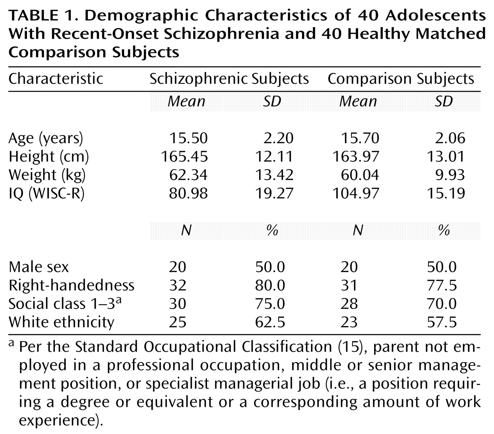
TABLE 1
TABLE 2

Footnote

Received June 1, 2000; revision received Nov. 8, 2000; accepted Dec. 28, 2000. From the Division of Psychological Medicine, Section of Neurobiology of Psychosis, Institute of Psychiatry. Address reprint requests to Dr. Frangou, Division of Psychological Medicine, Section of Neurobiology of Psychosis, Institute of Psychiatry, De Crespigny Park, London, SE5 8AF, U.K.; [email protected] (e-mail). Supported by a research award to Dr. Frangou from the Stanley Foundation.

References

1.
Barta PE, Pearlson GD, Powers RE, Richards SS, Tune LE: Auditory hallucinations and smaller superior temporal gyral volume in schizophrenia. Am J Psychiatry 1990; 147:1457-1462
2.
Shenton ME, Kikinis R, Jolesz FA, Pollak SD, LeMay M, Wible CG, Hokama H, Martin J, Metcalf D, Coleman M: Abnormalities of the left temporal lobe and thought disorder in schizophrenia: a quantitative magnetic resonance imaging study. N Engl J Med 1992; 327:604-612
3.
Woodruff PWR, Wright IC, Bullmore ET, Brammer M, Howard RJ, Williams SCR, Shapleske J, Rossell S, David AS, McGuire PK, Murray RM: Auditory hallucinations and the temporal cortical response to speech in schizophrenia: a functional magnetic resonance imaging study. Am J Psychiatry 1997; 154:1676-1682
4.
Levitan C, Ward PB, Catts SV: Superior temporal gyral volumes and laterality correlates of auditory hallucinations in schizophrenia. Biol Psychiatry 1999; 46:955-962
5.
Rajarethinam RP, DeQuardo JR, Nalepa R, Tandon R: Superior temporal gyrus in schizophrenia: a volumetric magnetic resonance imaging study. Schizophr Res 2000; 41:303-312
6.
Dierks T, Linden DE, Jandl M, Formisano E, Goebel R, Lanfermann H, Singer W: Activation of Heschl’s gyrus during auditory hallucinations. Neuron 1999; 22:615-621
7.
Lennox BR, Park SB, Jones PB, Morris PG: Spatial and temporal mapping of neural activity associated with auditory hallucinations (letter). Lancet 1999; 353:644; correction, 354:518
8.
Jones P, Rodgers B, Murray R, Marmot M: Child development risk factors for adult schizophrenia in the British 1946 birth cohort. Lancet 1994; 344:1398-1402
9.
Hollis C: Child and adolescent (juvenile onset) schizophrenia, a case control study of premorbid developmental impairments. Br J Psychiatry 1995; 166:489-495
10.
Flaum M, O’Leary DS, Swayze VW II, Miller DD, Arndt S, Andreasen NC: Symptom dimensions and brain morphology in schizophrenia and related psychotic disorders. J Psychiatr Res 1995; 29:261-276
11.
Marsh L, Harris D, Lim KO, Beal M, Hoff AL, Minn K, Csernansky JG, DeMent S, Faustman WO, Sullivan EV, Pfefferbaum A: Structural magnetic resonance imaging abnormalities in men with severe chronic schizophrenia and an early age at clinical onset. Arch Gen Psychiatry 1997; 54:1104-1112
12.
Hirayasu Y, Shenton ME, Salisbury DF, Dickey CC, Fischer IA, Mazzoni P, Kisler T, Arakaki H, Kwon JS, Anderson JE, Yurgelun-Todd D, Tohen M, McCarley RW: Lower left temporal lobe MRI volumes in patients with first-episode schizophrenia compared with psychotic patients with first-episode affective disorder and normal subjects. Am J Psychiatry 1998; 155:1384-1391
13.
Jacobsen LK, Giedd JN, Vaituzis AC, Hamburger SD, Rajapakse JC, Frazier JA, Kaysen D, Lenane MC, McKenna K, Gordon CT, Rapoport JL: Temporal lobe morphology in childhood-onset schizophrenia. Am J Psychiatry 1996; 153:355-361; correction: 153:851
14.
Jacobsen LK, Giedd JN, Castellanos FX, Vaituzis AC, Hamburger SD, Kumra S, Lenane MC, Rapoport JL: Progressive reduction of temporal lobe structures in childhood-onset schizophrenia. Am J Psychiatry 1998; 155:678-685
15.
Office of Population Censuses and Surveys: Standard Occupational Classification. London, Her Majesty’s Stationery Office, 1991
16.
Kay SR, Opler LA, Lindenmayer JP: The Positive and Negative Syndrome Scale (PANSS): rationale and standardisation. Br J Psychiatry 1989; 155:59-67
17.
First MB, Spitzer RL, Gibbon M, Williams JBW: Structured Clinical Interview for DSM-IV Axis I Disorders, Research Version, Patient Edition (SCID-P). New York, New York State Psychiatric Institute, Biometrics Research, 1997
18.
Matzner F, Silva R, Silvan M, Chowdhury M, Nastasi L: Preliminary test-retest reliability of the KID-SCID, in Scientific Proceedings of the 44th Meeting of the American Academy of Child and Adolescent Psychiatry. Washington, DC, AACAP, 1997, p 172-173
19.
Annett M: A classification of hand preference by association analysis. Br J Psychol 1970; 61:303-321
20.
Frangou S, Sharma T, Sigmudsson T, Barta P, Pearlson G, Murray RM: The Maudsley Family Study, 4: normal planum temporale asymmetry in familial schizophrenia: a volumetric MRI study. Br J Psychiatry 1997; 170:328-333
21.
Highley JR, McDonald B, Walker MA, Esiri MM, Crow TJ: Schizophrenia and temporal lobe asymmetry: a post-mortem stereological study of tissue volume. Br J Psychiatry 1999; 175:127-134
22.
Kulynych JJ, Vladar K, Jones DW, Weinberger DR: Superior temporal gyrus volume in schizophrenia: a study using MRI morphometry assisted by surface rendering. Am J Psychiatry 1996; 153:50-56
23.
Zipursky RB, Marsh L, Lim KO, DeMent S, Shear PK, Sullivan EV, Murphy GM, Csernansky JG, Pfefferbaum A: Volumetric MRI assessment of temporal lobe structures in schizophrenia. Biol Psychiatry 1994; 35:501-516
24.
Vita A, Dieci M, Giobbio GM, Caputo A, Ghiringhelli L, Comazzi M, Garbarini M, Mendini AP, Morganti C, Tenconi F: Language and thought disorder in schizophrenia: brain morphological correlates. Schizophr Res 1995; 15:243-251
25.
Holinger DP, Shenton ME, Wible CG, Donnino R, Kikinis R, Jolesz FA, McCarley RW: Superior temporal gyrus volume abnormalities and thought disorder in left-handed schizophrenic men. Am J Psychiatry 1999; 156:1730-1735
26.
Geschwind N, Galaburda AM: Cerebral lateralization: biological mechanisms, associations, and pathology; I: a hypothesis and a program for research. Arch Neurol 1985; 42:428-459
27.
Cannon M, Jones P, Huttunen MO, Tanskanen A, Huttunen T, Rabe-Hesketh S, Murray RM: School performance in Finnish children and later development of schizophrenia: a population-based longitudinal study. Arch Gen Psychiatry 1999; 56:457-463
28.
Eggers C, Bunk D, Volberg G, Ropcke B: The ESSEN study of childhood-onset schizophrenia: selected results. Eur Child Adolesc Psychiatry 1999; 8(suppl 1):121-128
29.
Eggers C, Bunk D: The long-term course of childhood-onset schizophrenia: a 42-year followup. Schizophr Bull 1997; 23:105-117
30.
Corson PW, Nopoulos P, Miller DD, Arndt S, Andreasen NC: Change in basal ganglia volume over 2 years in patients with schizophrenia: typical versus atypical neuroleptics. Am J Psychiatry 1999; 156:1200-1204
31.
Gur RE, Maany V, Mozley PD, Swanson C, Bilker W, Gur RC: Subcortical MRI volumes in neuroleptic-naive and treated patients with schizophrenia. Am J Psychiatry 1998; 155:1711-1717
32.
Chakos MH, Lieberman JA, Bilder RM, Borenstein M, Lerner G, Bogerts B, Wu H, Kinon B, Ashtari M: Increase in caudate nuclei volumes of first-episode schizophrenic patients taking antipsychotic drugs. Am J Psychiatry 1994; 151:1430-1436
33.
Selemon LD, Lidow MS, Goldman-Rakic PS: Increased volume and glial density in primate prefrontal cortex associated with chronic antipsychotic drug exposure. Biol Psychiatry 1999; 46:161-172
34.
Goldsmith SK, Joyce JN: Dopamine D2 receptors are organized in bands in normal human temporal cortex. Neuroscience 1996; 74:435-451
35.
Kessler RM, Whetsell WO, Ansari MS, Votaw JR, de Paulis T, Clanton JA, Schmidt DE, Mason NS, Manning RG: Identification of extrastriatal dopamine D2 receptors in post mortem human brain with [125I]epidepride. Brain Res 1993; 609:237-243
36.
Joyce JN, Janowsky A, Neve KA: Characterization and distribution of [125I]epidepride binding to dopamine D2 receptors in basal ganglia and cortex of human brain. J Pharmacol Exp Ther 1991; 257:1253-1263
37.
Bigliani V, Mulligan RS, Acton PD, Visvikis D, Ell PJ, Stephenson C, Kerwin RW, Pilowsky LS: In vivo occupancy of striatal and temporal cortical D2/D3 dopamine receptors by typical antipsychotic drugs: [123I]epidepride single photon emission tomography study. Br J Psychiatry 1999; 175:231-238
38.
Keshavan MS, Haas GL, Kahn CE, Aguilar E, Dick EL, Schooler NR, Sweeney JA, Pettegrew JW: Superior temporal gyrus and the course of early schizophrenia: progressive, static, or reversible? J Psychiatr Res 1998; 32:161-167
39.
Gur RE, Turetsky BI, Cowell PE, Finkelman C, Maany V, Grossman RI, Arnold SE, Bilker WB, Gur RC: Temporolimbic volume reductions in schizophrenia. Arch Gen Psychiatry 2000; 57:769-775

Information & Authors

Information

Published In

Go to American Journal of Psychiatry
American Journal of Psychiatry
Pages: 1299 - 1304
PubMed: 11481166

History

Published online: 1 August 2001
Published in print: August 2001

Authors

Details

Steven Williams, Ph.D.
Michael Hadjulis, M.D.

Metrics & Citations

Metrics

Citations

Export Citations

If you have the appropriate software installed, you can download article citation data to the citation manager of your choice. Simply select your manager software from the list below and click Download.

For more information or tips please see 'Downloading to a citation manager' in the Help menu.

Format
Citation style
Style
Copy to clipboard

View Options

View options

PDF/EPUB

View PDF/EPUB

Get Access

Login options

Already a subscriber? Access your subscription through your login credentials or your institution for full access to this article.

Personal login Institutional Login Open Athens login
Purchase Options

Purchase this article to access the full text.

PPV Articles - American Journal of Psychiatry

PPV Articles - American Journal of Psychiatry

Not a subscriber?

Subscribe Now / Learn More

PsychiatryOnline subscription options offer access to the DSM-5-TR® library, books, journals, CME, and patient resources. This all-in-one virtual library provides psychiatrists and mental health professionals with key resources for diagnosis, treatment, research, and professional development.

Need more help? PsychiatryOnline Customer Service may be reached by emailing [email protected] or by calling 800-368-5777 (in the U.S.) or 703-907-7322 (outside the U.S.).

Media

Figures

Other

Tables

Share

Share

Share article link

Share