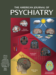
The present issue of the Journal features a series of articles on functional neuroimaging that reflect advances in the field and the contributions of neuroimaging to important clinical questions with relevance to treatment. The studies apply diverse methods—positron emission tomography (PET) to measure cerebral glucose metabolism (Mentis et al., Mayberg et al., Alexander et al.) and dopamine D1 receptor binding (Karlsson et al.) and fluorine magnetic resonance spectroscopy (19F MRS) to examine brain biochemistry (Strauss et al.).
The disorders examined in vivo across the life span illustrate the richness of the paradigms and findings. The methodological sophistication in neuroimaging data acquistion, processing, and statistical analysis is matched by clinical rigor in diagnosis, patient care, and ethical conduct of research. The study of children with pervasive developmental disorders by Strauss et al. addresses the gap in dosing guidelines for psychotropic agents in the treatment of children in view of increased use of selective serotonin reuptake inhibitors. 19F MRS is especially suitable for pediatric subjects as the technique is noninvasive, entails no ionizing radiation, and is less motion sensitive. The majority of participants (16 of 21) completed the study successfully, and the data yielded important pharmacokinetic information, indicating similar brain levels of fluvoxamine and fluoxetine when indexed for dose/body mass in adults and in children. This establishes a basis for scaling dose in relation to body mass. We can now go beyond “an educated guess” and have a scientific yardstick for therapeutic interventions.
Investigations in schizophrenia, commonly manifested in late adolescence and early adulthood, have contributed to a neurodevelopmental perspective of the disorder. Findings in neuroleptic-naive patients indicate that aberrations in brain structure and functioning are evident at first clinical presentation, before the introduction of neuroleptics. The value of examining first-episode, neuroleptic-naive patients is especially important in assessing receptor function. Painstaking efforts are required to enroll this unique population in studies that are technology based and take place in academic centers. Earlier postmortem and PET studies have suggested that D
1 binding is lower than normal in schizophrenia. In particular, the finding by Okubo et al.
(1) of low [
11C]SCH 23390 binding in the prefrontal cortex in schizophrenia supports findings from animal and human studies implicating a role of the D
1 receptor type in working memory, which is impaired in schizophrenia. However, the report by Karlsson and colleagues does not confirm these results. The discrepancy calls attention to variability among studies in study group and methods. The duration of illness before initial presentation might vary considerably among neuroleptic-naive patients, with likely concomitant changes in the brain. Differences in course may contribute to variability, especially in small groups. Furthermore, as the properties of PET ligands improve, stronger signals become available. The study by Karlsson et al. also illustrates the importance of negative findings in guiding future research and emphasizes the translational nature of PET receptor work, bridging clinical, human postmortem, and animal research.
Three of the studies reported in this issue used measures of cerebral glucose metabolism in innovative ways. Mayberg et al. dissected treatment response in middle-aged men hospitalized for unipolar depression who underwent 6-week double-blind administration of fluoxetine or placebo. Symptoms of depression and measures of metabolic activity were assessed at baseline and at 1 week and 6 weeks after treatment initiation. Clinical improvement was comparable in four responders treated with fluoxetine and four treated with placebo. Correspondingly, the patterns of metabolic change in cortical and paralimbic regions were similar in the two groups. However, specific additional changes in brainstem, striatum, and hippocampus activity were evident in the actively treated patients. Significant placebo effects have been noted in medicine, and their association with specific changes in brain activity in this small study group is intriguing, as is the unique effect of fluoxetine. The application of PET in treatment research is timely and can advance understanding of brain circuitry.
The clinical utility of PET is buttressed by the report of Mentis et al., who studied Parkinson’s disease, in which depression is common. The application of multivariate voxel-based analysis identified two topographic patterns of glucose metabolism: parieto-occipito-temporal activity correlated with visuospatial and mnemonic functioning, whereas lateral frontal and anterior limbic activity correlated with dysphoria. The relation between cognition and emotion in healthy people and in those with neuropsychiatric disorders has received increased attention. Functional imaging applying advanced statistical methods elucidates brain circuitry underlying pathophysiological processes that may have distinct effects. Such approaches can be useful in addressing questions regarding diagnostic dichotomies or continuum models of schizoaffective disorders and other psychotic disorders with cognitive and affective manifestations.
Alexander et al. extend PET methods to another neurodegenerative disorder affecting older adults, Alzheimer’s disease. Applying a brain mapping algorithm, they demonstrate significantly lower glucose metabolism in multiple brain regions among patients than among healthy elderly participants, with further reductions in Alzheimer’s disease documented at 1-year follow-up. The greatest decline was evident in the frontal association regions, indicating the sensitivity and the feasibility of using this method to detect response to interventions for Alzheimer’s disease. Indeed, the sensitivity of PET in detecting changes over 1 year was much greater than that of cognitive measures. The power analysis provided will yield a useful guide for future investigators designing studies of treatments for Alzheimer’s disease. At a time when the number of patients with Alzheimer’s disease is increasing as the population is aging, and when efforts are underway to slow disease progression, the application of PET methods to assess brain response to therapy can ultimately become a helpful predictor of outcome.
As someone who has participated in the initial efforts to apply neuroimaging in neuropsychiatric disorders, I am gratified by the opportunity to welcome five powerful studies into the Journal. For a long time, advances in neuroimaging methods have not been viewed as clinically relevant to psychiatry. Maturation of imaging technology has been accompanied by advances in statistical methods and in the sophistication of clinical assessment and incorporation of neurobehavioral system approaches. This combination is yielding integrative studies that are moving the field in new directions. Perhaps as important as the specific discoveries reported in these five articles are the span of the disorders and the recognition that all are brain disorders. It is time for clinicians to view the progress made in academic centers as relevant to patients’ assessment and care. Progress will be vastly enhanced if more patients take part in imaging studies. Imaging technology has come of age; it is harvest time.

