Full access
Article
Published Online: 1 April 2008

Regional White Matter Hyperintensity Burden in Automated Segmentation Distinguishes Late-Life Depressed Subjects From Comparison Subjects Matched for Vascular Risk Factors

Abstract

Objective: Segmented brain white matter hyperintensities were compared between subjects with late-life depression and age-matched subjects with similar vascular risk factor scores. Correlations between neuropsychological performance and whole brain-segmented white matter hyperintensities and white and gray matter volumes were also examined. Method: Eighty-three subjects with late-life depression and 32 comparison subjects underwent physical examination, psychiatric evaluation, neuropsychological testing, vascular risk factor assessment, and brain magnetic resonance imaging (MRI). Automated segmentation methods were used to compare the total brain and regional white matter hyperintensity burden between depressed patients and comparison subjects. Results: Depressed patients and comparison subjects did not differ in demographic variables, including vascular risk factor, or whole brain-segmented volumes. However, depressed subjects had seven regions of greater white matter hyperintensities located in the following white matter tracts: the superior longitudinal fasciculus, fronto-occipital fasciculus, uncinate fasciculus, extreme capsule, and inferior longitudinal fasciculus. These white matter tracts underlie brain regions associated with cognitive and emotional function. In depressed patients but not comparison subjects, volumes of three of these regions correlated with executive function; whole brain white matter hyperintensities correlated with executive function; whole brain white matter correlated with episodic memory, processing speed, and executive function; and whole brain gray matter correlated with processing speed. Conclusions: These findings support the hypothesis that the strategic location of white matter hyperintensities may be critical in late-life depression. Further, the correlation of neuropsychological deficits with the volumes of whole brain white matter hyperintensities and gray and white matter in depressed subjects but not comparison subjects supports the hypothesis of an interaction between these structural brain components and depressed status.
White matter hyperintensities on T2-weighted magnetic resonance imaging (MRI) have been associated with late-life depression. It has been proposed that vascular disease and concomitant white matter hyperintensities may contribute to the development or worsening of late-life depression by affecting frontal white matter pathways and subcortical structures involved in mood regulation. In the present study, we used the term white matter hyperintensity to denote both subcortical gray and white matter hyperintensities. A large body of literature has suggested higher rates and severity of white matter hyperintensities in elderly depressed patients relative to age-matched comparison subjects (14) .
Several factors are important contributors to the pathogenesis of white matter hyperintensities, particularly age (5) and medical comorbidity. Comorbid medical conditions such as hypertension (6), diabetes mellitus (7), cardiovascular disease (8), and higher Framingham Study risk factor scores (8, 9) are especially significant. While white matter hyperintensities are more prevalent in patients with vascular risk factors, they are also noted to occur at rates of up to 60% in healthy elderly patients (10) . Although studies examining the occurrence of white matter hyperintensities in depressed subjects have matched subjects for age, most studies have used either healthy comparison subjects or a comparison sample of convenience, rather than matching for vascular risk factors.
Another limitation of most studies examining white matter hyperintensities in late-life depression is the reliance on visual rating scales (11) for lesion severity and the use of relatively small sample sizes. Automated segmentation of white matter, gray matter, and CSF has become relatively common, but few existing paradigms have segmented white matter hyperintensity lesions. Current automated segmentation techniques remain unable to accurately classify deep cortical white matter hyperintensities and rely upon expert manual correction of misclassified hyperintensities (12) or manual grading of hyperintensities using ordinal rating scales for basal ganglia and deep cortical lesions (2) .
We previously described the development of an automated segmentation technique and demonstrated the reliability of this method to segment brain gray matter, white matter, and white matter hyperintensities and to localize white matter hyperintensities in atlas space (13) . In the present study, we applied this method to a large sample of subjects with late-life depression and a comparison sample of subjects matched for vascular risk factors. We further examined the functional significance of white matter hyperintensity lesions by correlating the severity with measures of neuropsychological functioning; we hypothesized that greater white matter hyperintensity lesion burden would be correlated with poorer performance on measures of executive functioning and processing speed.

Method

Subjects

Depressed patients (N=83) and nondepressed comparison subjects (N=32) age 59 years and older with a wide range of cerebrovascular risk factors were recruited for a National Institute of Mental Health (NIMH) study: Treatment Outcome in Vascular Depression. Patients and comparison subjects were evaluated by board-certified psychiatrists using the Diagnostic Interview for Genetic Studies criteria, a structured clinical interview that incorporates the Structured Clinical Interview for DSM-IV (SCID) criteria (14) . Depressed patients were evaluated for the presence of DSM-IV major depression, and nondepressed comparison subjects were evaluated for lifetime absence of psychiatric illness. All subjects were screened to rule out severe or unstable medical disorders, history of other axis I disorders prior to depression, and current suicide risk. Written informed consent approved by the institutional review board was obtained for all subjects.

Measures

Demographic variables were age, education, gender, race, depression symptom severity (scored on the Montgomery-Åsberg Depression Rating Scale), and vascular risk factors as defined by the Framingham Study (15) . The Framingham Study includes the following risk factors to predict 10-year stroke risk in both men and women: age, systolic blood pressure, use of antihypertensive therapy, diabetes mellitus, cigarette smoking, cardiovascular disease (coronary heart disease, cardiac failure, or intermittent claudication), atrial fibrillation, and left ventricular hypertrophy by electrocardiogram.
The neuropsychological testing was performed by a trained examiner who was closely supervised by a Ph.D.-level psychologist. Patients were tested prior to the initiation of antidepressant medication and were free of all psychotropic medication.

MRI

MRI was performed using a Siemens (Erlangen, Germany) Sonata 1.5T scanner. The following structural images were acquired: T1-weighted magnetization-prepared rapidly acquired gradient echo (MPRAGE) (sagittal TR=1900 msec, TE=4 msec, TI=1100 msec, matrix=222×256×128, resolution=1×1×1.25 mm); T2-weighted two-dimensional Turbospin echo (transverse TR=4000 msec, TE=97 msec, 17 echoes, 2 mm thickness, 10 mm gap, 6 interleaves, matrix=256×256×108, resolution=1×1×2 mm); and fluid-weighted inversion recovery scans (transverse TR=10,000 msec, TE=104 msec, TI=2310 msec, 5 mm thickness, 0 mm gap, matrix=224×256×30, resolution=1×1×5 mm). Four T1-weighted scans were acquired and coregistered using 9 parameter affine transform. The six T2-weighted images were collated and coregistered with the T1-weighted scans. The averaged T1-weighted and coregistered T2-weighted data were resampled to 1 mm 3 voxels in Talairach stereotaxic atlas space (T88) (16) . To correct for artifactual image intensity inhomogeneities in both T1- and T2-weighted data, a parametric bias field correction was used and all subsequent operations were performed using the parametric bias field corrected images in atlas space (13, 17) .

Segmentation Algorithm

Bispectral (T1- and T2-weighted) fuzzy class means (18, 19) was used to segment the whole brain into tissue classes (gray matter, white matter, and CSF) and white matter hyperintensities. Loci in the T1-/T2-weighted intensity plane corresponding to each tissue class were determined by a semiautomatic peak search of the two-dimensional T1-/T2-weighted intensity histogram (13) . A separate set of loci was used to segment deep (basal ganglia, thalamus, and ventricles) versus more superficial structures (cerebral cortex and underlying white matter), and the assembled segmentations were continuous at the boundary between deep and superficial structures. To segment the deep zone, gray matter, white matter, and CSF centroids were determined by sampling gray matter (caudate), white matter (corpus callosum), and CSF (lateral ventricles) using manually selected regions of interest. Fuzzy class means then generated a segmentation of the deep zone into four intracranial tissue types, including white matter hyperintensities. Post-fuzzy class mean corrections based on prior anatomical knowledge (20) were automatically performed as follows: small groups of white matter hyperintensity pixels located between gray matter and CSF (which clearly could not be white matter hyperintensities) were reassigned as gray matter; all “gray matter” entirely surrounded by white matter hyperintensities were assigned as white matter hyperintensities; and a single pixel of white matter hyperintensity adjacent to CSF was reassigned as white matter.

Validation of Segmentation Algorithm

Automated segmentation of white matter hyperintensities was validated in a subset of 20 subjects by comparison with manual threshold-based segmentation of fluid-weighted inversion recovery images. White matter hyperintensity on fluid-weighted inversion recovery imaging effectively constitutes a neuroradiological gold standard for white matter hyperintensity detection (21) . A fluid-weighted inversion recovery pixel intensity histogram was generated, and pixels with intensities that were two standard deviations greater than the white/gray matter mean were classified as white matter hyperintensities. Threshold-based segmentation of white matter hyperintensities was highly correlated with our semiautomated technique (r=0.979 overall) for the entire brain and for the subcortical volume only (r=0.925). In this same sample, the automated segmentation was further validated by comparison of the total brain-segmented white matter hyperintensity to expert manual ratings (Drs. Sheline and McKinstry) using the modified Fazekas Rating Scale (r=0.71, p=0.001) (13) .

Statistical Comparison of White Matter Hyperintensity Regions in Depressed Patients and Comparison Subjects

The segmentation results were converted to binary images (white matter hyperintensities or non-white matter hyperintensities), and the total number of white matter hyperintensity pixels was identified for each subject. To identify specific regions with group differences, the binary images of white matter hyperintensities were first smoothed (10 mm full-width-half-maximum Gaussian profile). We then conducted a pixel-by-pixel comparison using two-tailed unpaired t tests, with statistical threshold (p<0.01) and minimum cluster size (≥150 mm 3 ). The resultant image was reviewed to determine the areas in which significant lesion clusters occurred.

Neuropsychological Test Performance in Late-Life Depression

All participants were administered a large battery of neuropsychological tests covering cognitive domains relevant to late-life depression. We grouped cognitive tasks into domains based on our previous study (22) . To combine tasks, we created z scores for the primary dependent measure of interest across all participants and then summed the z scores. We created factor scores for the following assessments:

Language processing (Cronbach alpha=0.62)

The Shipley Vocabulary Test (number correct), Boston Naming Test (number correct), and word reading condition of the Stroop task (number completed) were used.

Processing speed (Cronbach alpha=0.81)

Symbol-digit modality (number completed), color naming condition of the Stroop task (number completed), and Trails A (reverse scored time to completion) were used.

Working memory (Cronbach alpha=0.69)

Digit span forward (number of trials correctly completed), digit span backward (number of trials correctly completed), and ascending digits (number of trials correctly completed) were used.

Episodic memory (Cronbach alpha=0.73)

Word list learning (total correct), logical memory (total correct immediate), constructional praxis (memory performance), and the Benton Visual Retention Test (total correct) were used.

Executive function (Cronbach alpha=0.75)

Verbal fluency (total phonological and semantic), Trails B (reverse scored time to completion), color-word interference condition of the Stroop task (number completed), initiation-perseveration subscales of the Mattis Dementia Rating Scale, and categories completed from the Wisconsin Card Sorting Test were used.

Correlation Between Brain Volumetric Measures and Neuropsychological Measures

In a multiple regression after accounting for age, gender, and education, there was a significant effect of cardiovascular disease risk factors on whole brain white matter hyperintensity measures (p=0.05). Since we found several regions with significant group differences in white matter hyperintensity burden (described in the Results section), we conducted Pearson correlations between the neuropsychological variables (cognitive domain z scores) and the white matter hyperintensity measure in each of the regions. These correlations were conducted separately in depressed patients and comparison subjects. We also conducted Pearson correlations between neuropsychological variables and whole brain-segmented volumes of white matter hyperintensities as well as gray and white matter. We followed up any significant simple correlations with partial correlations that removed shared variance with age and education to ensure that common variance with such demographic characteristics did not lead to spurious correlations between the anatomical and neuropsychological measures. To protect against false positives, we used a Bonferroni correction across the five cognitive domains for the correlations with each region of interest and whole brain measures and only considered a correlation significant at p<0.01 (0.05/5=0.01).

Results

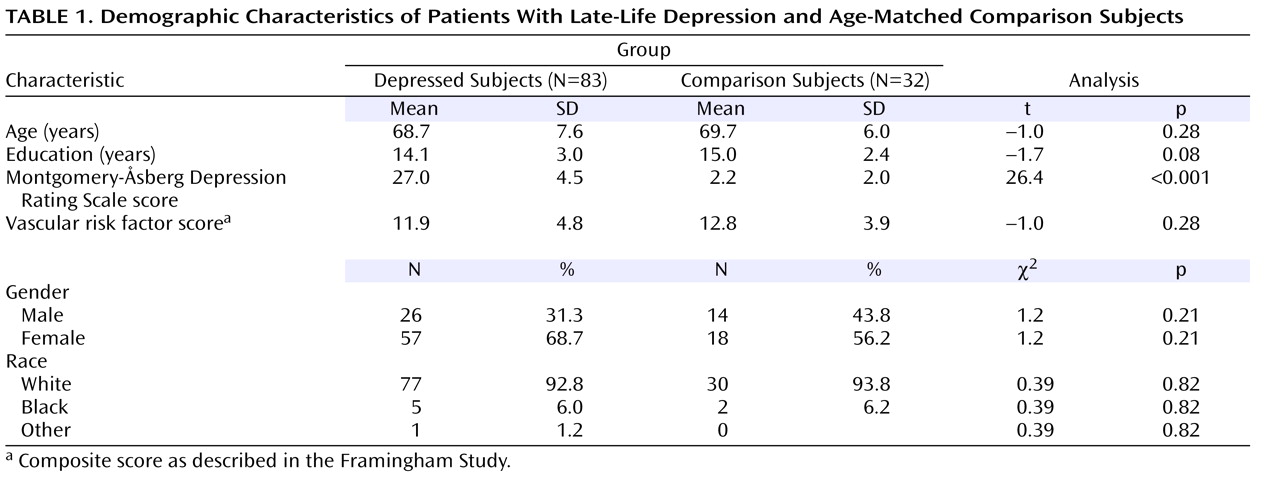
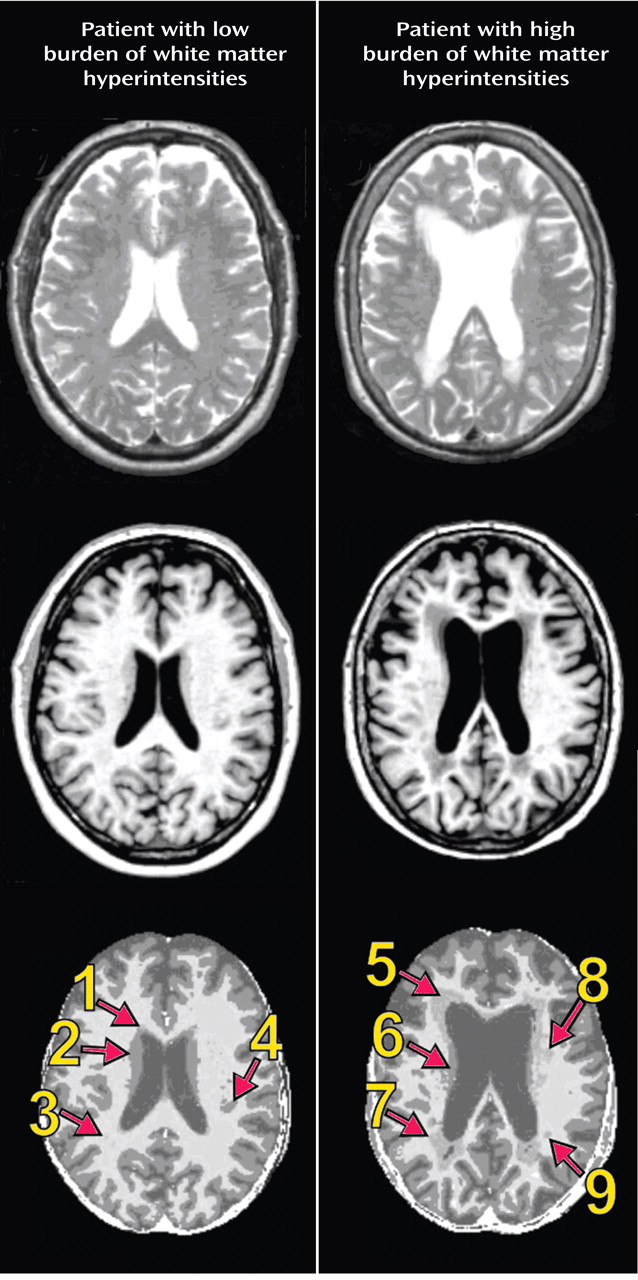
Table 1 shows demographic variables for depressed patients and comparison subjects, including age, education, gender, race, depression symptom severity on the Montgomery-Åsberg Depression Rating Scale, and vascular risk factor score as defined by the Framingham Study (15) . As shown in Table 1, depressed subjects had higher Montgomery-Åsberg Depression Rating Scale scores than comparison subjects, but the groups did not differ significantly on other variables. We compared whole brain volumes of white matter, gray matter, and white matter hyperintensities between depressed and comparison subjects. Figure 1 shows T1-weighted, T2-weighted, and segmented images for a subject with minimal white matter hyperintensities and a subject with significant white matter hyperintensities. A multivariate analysis of variance (MANOVA) was conducted to examine differences between the depressed and comparison groups in the three segmented measures of whole brain volume. The omnibus MANOVA was not significant (p>0.05), suggesting that whole brain volumes of white matter, gray matter, and white matter hyperintensities did not differ between groups for any of these three volume measurements.
Figure 1. Automated Segmentation of White Matter Hyperintensities a
a The first row shows T2-weighted images. The second row shows T1-weighted images. The third row shows images following automated segmentation. Of note are the following findings indicated by arrows in row 3: 1) minimal periventricular hyperintensity; 2) the caudate appears similar to white matter hyperintensity on the T2-weighted image but is correctly segmented as gray matter by the segmentation algorithm; 3) small white matter hyperintensity is not clearly identifiable on a T2-weighted image; 4) area of gray matter, from gyral folding in the temporal lobe that appears as white matter hyperintensity on T1- and T2-weighted images but is correctly segmented as gray matter; 5) large periventricular hyperintensity; 6) area potentially appearing as white matter hyperintensity that is actually correctly segmented as gray matter; 7) large periventricular hyperintensity; 8) white matter hyperintensity; and 9) white matter hyperintensity.
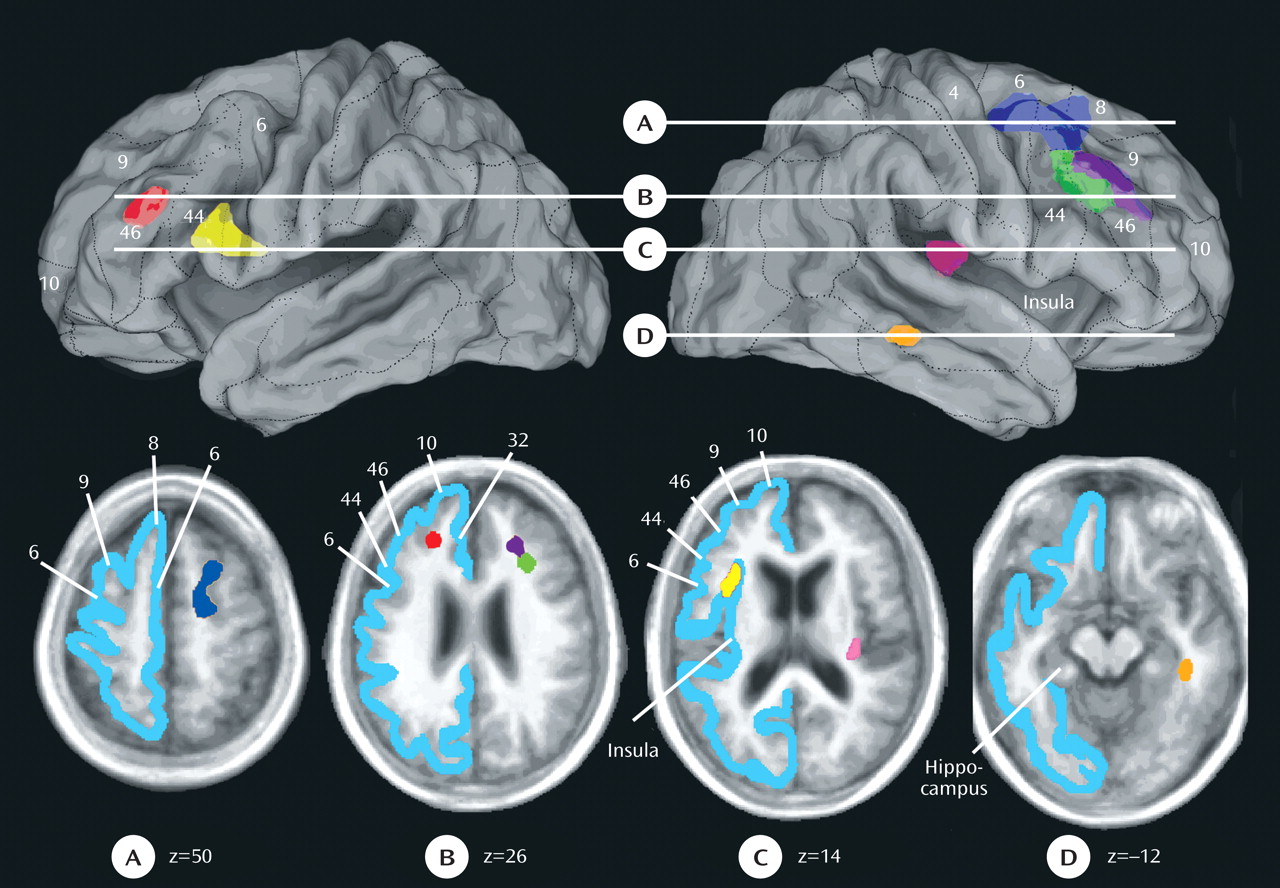
We then examined differences between the comparison and depressed groups in the prevalence of regional white matter hyperintensities. This analysis identified seven regions that met criteria and differed significantly between the two groups in white matter hyperintensity volumes ( Figure 2 ). The following regions were identified: 1) right superior longitudinal fasciculus division 1/fronto-occipital fasciculus; 2) right superior longitudinal fasciculus divisions 2 and 3; 3) a second region in the right superior longitudinal fasciculus divisions 2 and 3; 4) left superior longitudinal fasciculus divisions 2 and 3; 5) left uncinate fasciculus/frontal operculum; 6) right extreme capsule; and 7) right inferior longitudinal fasciculus. For each of these lesions, the white matter fasciculus involved and the relevant overlying cortical region are described in Figure 2 .
Figure 2. Differences Between Depressed Patients and Comparison Subjects in Regional White Matter Hyperintensity Volumes a
a The two top panels display the averaged difference maps between depressed and comparison subjects in oblique right and left views, respectively. The semitransparent display allows the lesions to be seen three-dimensionally, underlying the surface Brodmann area. Selected Brodmann areas are indicated in the upper three-dimensional brain representations as well as the bottom individual panels (A–D), which are four horizontal slices through the brain that indicate the z axis level of the panels. They are as follows: A) right superior longitudinal fasciculus 1/fronto-occipital fasciculus (blue) as defined by Petrides and Pandya (45) and Schmahmann and Pandya (28), underlying right Brodmann’s areas 6, 8, and 9 (extending from z=42–58); B) right superior longitudinal fasciculus 2 and 3 (purple) (26, 43), underlying the right dorsolateral prefrontal cortex (Brodmann’s area 9/46) (extending from z=26–38), a second lesion in the right superior longitudinal fasciculus 2 and 3 (green), underlying right Brodmann’s area 9/32 (extending from z=26–38), and the left superior longitudinal fasciculus 2 and 3 (red) in white matter underlying Brodmann’s area 46/32 (extending from z=18–26); C) left uncinate fasciculus/frontal operculum (yellow) in white matter underlying Brodmann’s areas 44/6 and 9 (extending from z=10–22) and the right extreme capsule (pink) in white matter underlying the insula (extending from z=6–14); and D) inferior longitudinal fasciculus (orange) in white matter adjacent to the right parahippocampus (extending from z=–14 to –6).
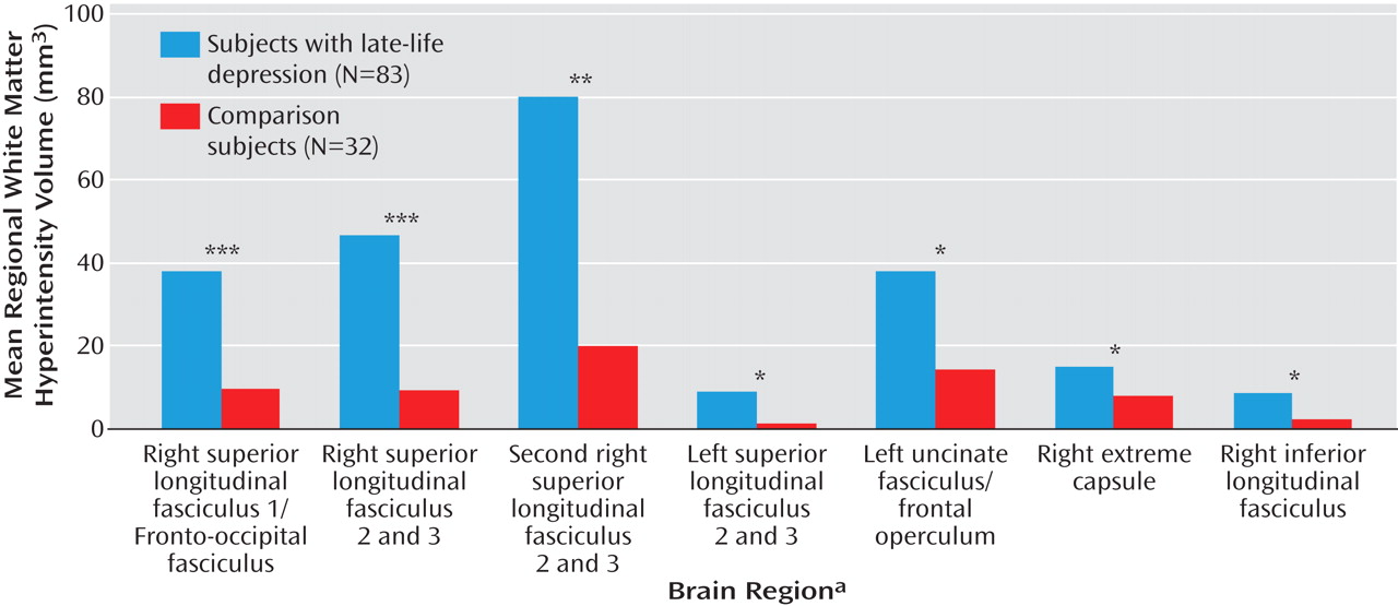
The mean white matter hyperintensity volume within each region in both depressed and comparison subjects was plotted and is shown in Figure 3 . The effect sizes for these differences were fairly similar across the seven regions and were as follows: 1) right superior longitudinal fasciculus 1/fronto-occipital fasciculus: Cohen’s d=0.43; 2) right superior longitudinal fasciculus divisions 2 and 3: Cohen’s d=0.39 (first region); 3) right superior longitudinal fasciculus divisions 2 and 3: Cohen’s d=0.37 (second region); 4) left superior longitudinal fasciculus divisions 2 and 3: Cohen’s d=0.33; 5) left uncinate fasciculus: Cohen’s d=0.37; 6) right extreme capsule: Cohen’s d=0.40; and 7) right inferior longitudinal fasciculus: Cohen’s d=0.43.
Figure 3. White Matter Hyperintensity Comparison by Region in Depressed Patients and Comparison Subjects a
a Regions correspond with the lesion locations shown in Figure 2: right superior longitudinal fasciculus division 1/fronto-occipital fasciculus [blue]; right superior longitudinal fasciculus divisions 2 and 3 [purple]; second region of right superior longitudinal fasciculus divisions 2 and 3 [green]; left superior longitudinal fasciculus divisions 2 and 3 [yellow]; left uncinate fasciculus/frontal operculum [red]; right extreme capsule [pink]; and right inferior longitudinal fasciculus [orange]).
*p<0.05. **p<0.01. ***p<0.005.
We then examined the relationship between the volumes of white matter hyperintensity within each of the seven regions and performance in the five different a priori domains of cognitive function (20) in the depressed and comparison subjects separately. Among the depressed patients, only three correlations were significant, and all three were with executive function as follows: 1) right superior longitudinal fasciculus 1/fronto-occipital fasciculus (r=–0.31, p=0.005); 2) left superior longitudinal fasciculus divisions 2 and 3 (r=–0.30, p=0.008); and 3) left uncinate fasciculus (r=–0.30, p=0.008). The magnitude of these correlations changed only minimally (with one going up) when age and education were partialled out. These changes were as follows: 1) right superior longitudinal fasciculus 1/fronto-occipital fasciculus (r=–0.35, p=0.002); 2) left superior longitudinal fasciculus divisions 2 and 3 (r= –0.28, p=0.02); and 3) left uncinate fasciculus (r=–0.26, p=0.02). Among comparison subjects, there were no significant correlations. Fisher’s r-to-z transformations indicated that there was a tendency toward the correlation between the right superior longitudinal fasciculus 1/fronto-occipital fasciculus and executive function being significantly more negative in patients relative to comparison subjects (z=–1.6, p=0.06).
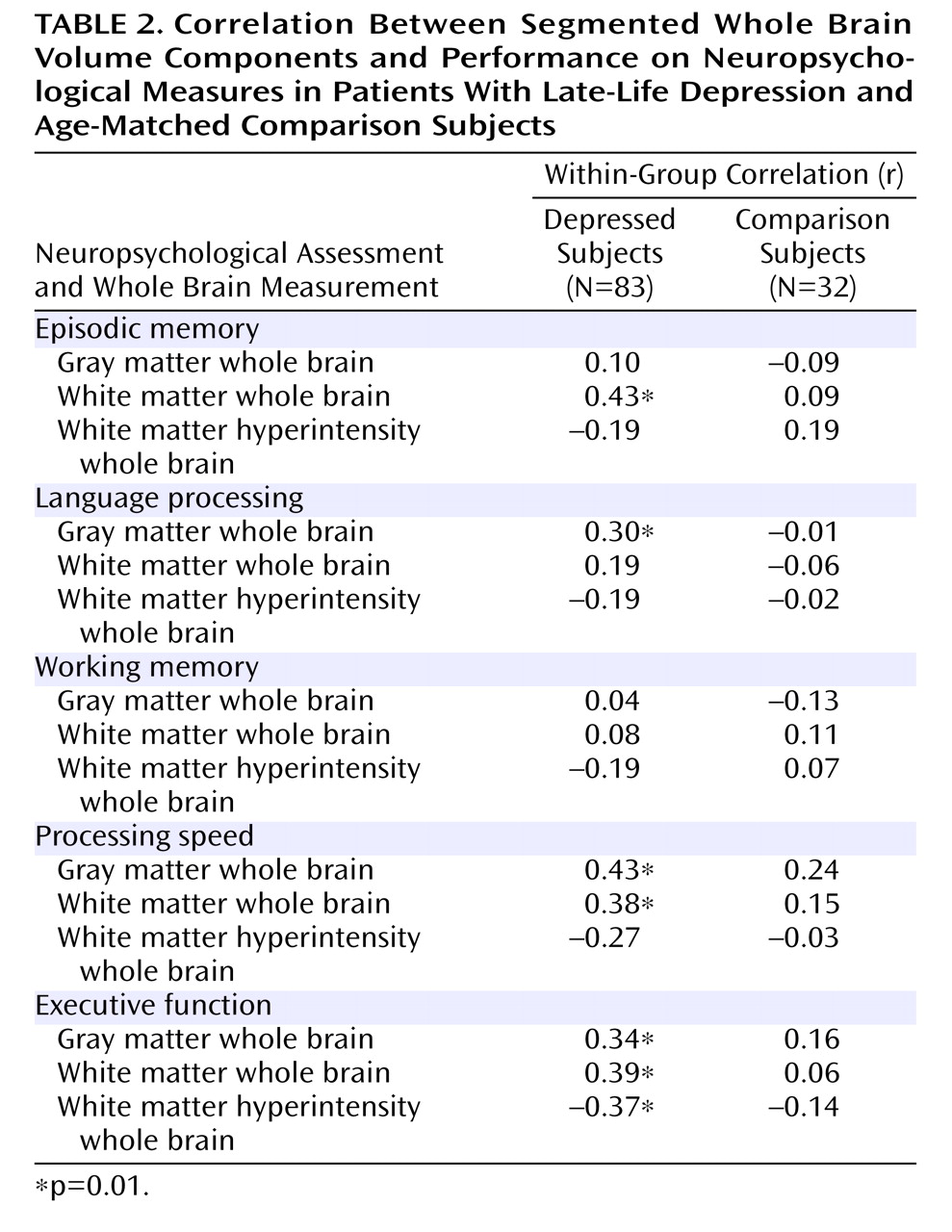
In addition, we examined the association between segmented whole brain volumetric measures and neuropsychological function. As shown in Table 2, there were no significant correlations between any of the whole brain measures and neuropsychological function in comparison subjects. Among depressed patients, there were significant correlations of white matter hyperintensities and gray and white matter with neuropsychological function. All of these correlations maintained their significance when age and education were partialled out. Fisher’s r-to-z transformations indicated that the correlation between executive function and white matter hyperintensities was significantly stronger in depressed patients relative to comparison subjects (z=1.65, p=0.05).

Discussion

This study adds to the understanding of the pathophysiology of late-life depression in several ways. We found increased white matter hyperintensity burden in patients with late-life depression relative to comparison subjects in specific brain regions, despite no differences in demographic variables, vascular risk factor scores, or whole brain gray matter, white matter, or white matter hyperintensity volumes. This supports the regional specificity of hyperintensities in depression. The finding (14) of increased overall volumes of white matter hyperintensities in late-life depressed subjects relative to comparison subjects in studies that did not match depressed and comparison subjects on vascular risk factors may indicate that more vascular events occur in depression. Depression has been associated with increased rates of cardiovascular illness (23), diabetes mellitus (24), smoking, and hypertension (25) . Thus, given that depressed subjects have increased rates of multiple factors comprising the overall vascular risk profile, it is important to control for vascular risk factors in comparing depressed and comparison subjects for white matter hyperintensities.
Second, we suggest that in order to optimize anatomical localization, it is important to utilize segmentation methodology that allows localization of white matter hyperintensities in atlas space. Current scoring techniques have variable interrater reliability (26) and homogenize white matter hyperintensity characteristics by assigning a general score for size and appearance in order to yield a global index of lesion load without spatial localization. In this study, we used an automated segmentation procedure that was reproducible, quantitatively validated (13), and precisely localized lesions in atlas space.
Third, using this technique we identified significant differences in white matter hyperintensity burden in specific brain regions. These lesions were located in deep white matter underlying cortical regions critical for executive function and emotional processing, and lesion volumes correlated with executive function in three regions. A potential interpretation of these findings is that regionally specific hyperintensities may increase susceptibility to depression and may be expressed functionally as cognitive impairment. An extensive literature has described the importance of cognitive control and disturbances thereof in late-life depression (27) . Further, of particular importance to emotional disturbances in depression are the white matter connections of the cingulate cortex, insula, and amygdala that were interrupted in the present study.
Lesions in the majority of fasciculi interrupted by hyperintensities in this study have been associated with specific cognitive impairment syndromes. The superior longitudinal fasciculus and fronto-occipital fasciculus are critical for cognitive control. A recent comprehensive compendium of white matter tracts in nonhuman primates and humans described these pathways in detail (28) . The primary long association fibers connecting the frontal cortex are the three subdivisions of the superior longitudinal fasciculus: components I, II, and III (28) . In the frontal lobe, superior longitudinal fasciculus 1 fibers project to the supplementary motor area and dorsal Brodmann’s areas 6 and 9 and convey information from these areas back to the parietal lobe cortices and probably into the precuneus. Superior longitudinal fasciculus 2 connections terminate in Brodmann’s areas 6, 8, 9/46, and 44, and superior longitudinal fasciculus 3 fibers terminate in ventral Brodmann’s areas 6 and 44.
Four regions of white matter hyperintensity lesions (right superior longitudinal fasciculus 1/fronto-occipital fasciculus; right superior longitudinal fasciculus 2/3; second region of right superior longitudinal fasciculus 2/3; left superior longitudinal fasciculus 2/3) were in white matter underling the dorsolateral prefrontal cortex (Brodmann’s area 9/46), and another region was located in the right extreme capsule of the basal ganglia, all interrupting the dorsolateral prefrontal circuit, namely connections between the basal ganglia and dorsolateral prefrontal cortex gray matter that are important for performance on tests of executive function (27, 29) . This circuit projects from Brodmann’s area 9/46 to the dorsolateral caudate, then to the lateral mediodorsal globus pallidus and rostrolateral substantia nigra. The basal ganglia projects to the ventral anterior and mediodorsal thalamus and back to the dorsolateral prefrontal cortex, completing the loop (30) . A large literature supports the association of the dorsolateral prefrontal cortex lesions with impairments in executive function (31, 32) . Such lesions are found with greater frequency in major depression (26) and in some studies were associated with worse treatment outcome (33) . Consistent with this prior work, in our study, white matter hyperintensity lesion burden in the superior longitudinal fasciculus 1 and superior longitudinal fasciculus divisions 2 and 3 regions and right extreme capsule was correlated with executive function in depressed subjects, although only the left superior longitudinal fasciculus divisions 2 and 3 correlation survived Bonferroni correction.
Many of the long fasciculi such as the superior longitudinal fasciculus 1, 2, and 3 have connections to Brodmann’s area 6. While traditionally thought of as a motor region, more recent work has shown Brodmann’s area 6 to be involved in cognitive tasks (34), including verbal operations (35) . Intriguingly, there were significant correlations between white matter hyperintensities in the left superior longitudinal fasciculus divisions 2 and 3 and executive function in depressed but not comparison subjects in our study. The extreme capsule is important for language function linking Wernicke’s area with Broca’s area (Brodmann’s areas 44 and 45), and stimulation of the cortical areas to which it links results in speech arrest (36) . Our depressed subjects had a significant relationship between lesions in this area and language function.
Several of the lesions in the present study interrupt pathways that are important in emotional circuitry. The cingulate gyrus has been implicated in many studies of emotional function. The major long association fibers that have been demonstrated emerging from the caudal cingulate gyrus include fibers in the dorsal part of the superior longitudinal fasciculus 1; fibers directed to the frontal lobe in the fronto-occipital fasciculus; and fibers in the cingulum bundle, uncinate fasciculus, and extreme capsule (28) . The uncinate fasciculus is a ventral limbic pathway connecting temporal regions with medial and orbital frontal cortices. A contingent of fibers within the uncinate fasciculus leads from the parahippocampal region to the orbitofrontal cortex. Some fibers terminate in medial forebrain areas, and others terminate in Brodmann’s areas 13, 47/12, 11, and 10, all areas involved in emotional circuitry. Some fibers from the posterior parahippocampal gyrus also terminate in the rostral cingulate cortex (Brodmann’s area 24).
The amygdala connections interrupted by lesions of the uncinate fasciculus have important implications in disorders of emotion processing (29, 37, 38) . White matter hyperintensity lesions in the inferior longitudinal fasciculus adjacent to the parahippocampal gyrus interrupt connections between V4 in the occipital cortex to the medial temporal structures, amygdala, hippocampus, and parahippocampus (39) . The amygdala is known to have neuromodulatory effects on the extrastriate visual cortex (40) . Thus, in depression it may be that the affective valence signals are not normally transmitted.
In addition to the impact of specific lesions, there were also significant correlations between segmented whole brain volumes and neuropsychological function in depressed subjects that were not found in comparison subjects, in particular, a significant correlation of whole brain white matter hyperintensities with executive function. Other studies have also found associations between white matter hyperintensity volumes and function, including (41) an association between white matter hyperintensities and global reductions in cortical metabolic rates. Whole brain white matter was correlated with executive function, episodic memory, and processing speed in depressed subjects but not comparison subjects. Whole brain gray matter was correlated with processing speed and showed tendencies toward correlation with executive function and language in depressed patients but not comparison subjects. These patterns suggest stronger relationships between whole brain white and gray matter volumes and neuropsychological performance in depressed patients relative to comparison subjects. However, this interpretation should be moderated by the greater power to detect relationships in depressed patients relative to comparison subjects because of their larger numbers. Further, it is somewhat surprising that we did not find a relationship between neuropsychological performance and whole brain measures of white matter hyperintensity burden in comparison subjects, since several previous studies (42, 43), but not all (44), have found such relationships. In the seven regions showing group differences in white matter hyperintensities, we did find significantly less white matter hyperintensity variance in comparison subjects relative to depressed subjects, potentially contributing to an absence of correlations with neuropsychological variables in comparison subjects. However, we found no group differences in variance for any of the neuropsychological measures themselves, or for any of the whole brain measures. Thus, we argue that in addition to the interruption of specific neuroanatomical pathways by white matter hyperintensities contributing to the development of late-life depression there is an interaction between overall white matter hyperintensity burden and gray and white matter loss in depressed subjects producing increased neuropsychological deficits. The mechanism for this interaction is unclear and worthy of further investigation.
Footnotes

A preliminary version of this study was presented as an abstract at the 2005 annual meeting of the American College of Neuropsychopharmacology. Received Jan. 25, 2007; revisions received Aug. 3 and Oct. 4, 2007; accepted Nov. 9, 2007 (doi: 10.1176/appi.ajp.2007.07010175). From the Departments of Psychiatry, Radiology, Neurology, Anatomy and Neurobiology, Psychology, Medicine, and Biostatistics, Washington University School of Medicine, St. Louis. Address correspondence and reprint requests to Dr. Sheline, Department of Psychiatry, Washington University School of Medicine, 660 South Euclid Ave., Campus Box 8134, St. Louis, MO 63110; [email protected] (e-mail).
Dr. Sheline serves as a consultant for Wyeth Pharmaceuticals, Cyberonics, and Eli Lilly; she also serves on the speaker’s bureau of Eli Lilly. Dr. Mintun serves as a consultant for Elan Pharmaceuticals, Hoffman-LaRoche, Avid Radiopharmaceuticals, and Eisai. Dr. McKinstry serves as a speaker for BioMarin Pharmaceutical. Drs. Price, Barch, Epstein, Wilkins, and Snyder and Mr. Vaishnavi, Mr. Couture, and Dr. Schechtman report no competing interests.
Supported by NIMH grants R01 MH60697 (Dr. Sheline) and K24 RR-18192 (Dr. Sheline) and a grant to cover sertraline pill costs by Pfizer.
The authors thank Brigitte Mittler for help in conducting this study and Asif Moinuddin for technical assistance and quality control on the MRI scanning.
The granting agencies had no role in any of the following aspects of this study: design and conduct of the study; collection, management, analysis and interpretation of the data or preparation, review, or approval of the article. Dr. Sheline is independent of any commercial provider, had full access to all of the data in this study, and takes responsibility for the integrity of the data and the accuracy of the data analysis.

References

1.
Greenwald BS, Kramer-Ginsberg E, Krishnan RR, Ashtari M, Aupperle PM, Patel M: MRI signal hyperintensities in geriatric depression. Am J Psychiatry 1996; 153:1212–1215
2.
Steffens DC, Bosworth HB, Provenzale JM, MacFall JR: Subcortical white matter lesions and functional impairment in geriatric depression. Depress Anxiety 2002; 15:23–28
3.
Taylor WD, MacFall JR, Payne ME, McQuoid DR, Steffens DC, Provenzale JM, Krishnan KR: Greater MRI lesion volumes in elderly depressed subjects than in control subjects. Psychiatry Res 2005; 139:1–7
4.
O’Brien JT, Firbank MJ, Krishnan MS, van Straaten EC, van der Flier WM, Petrovic K, Pantoni L, Simoni M, Erkinjuntti T, Wallin A, Wahlund LO, Inzitari D; LADIS Group: White matter hyperintensities rather than lacunar infarcts are associated with depressive symptoms in older people: the LADIS study. Am J Geriatr Psychiatry; 14:834–841
5.
Guttmann CR, Jolesz FA, Kikinis R, Killiany RJ, Moss MB, Sandor T, Albert MS: White matter changes with normal aging. Neurology 1998; 50:972–978
6.
Dufouil C, de Kersaint-Gilly A, Besancon V, Levy C, Auffray E, Brunnereau L, Alperovitch A, Tzourio C: Longitudinal study of blood pressure and white matter hyperintensities: the EVA MRI cohort. Neurology 2001; 56:921–926
7.
Novak V, Last D, Alsop DC, Abduljalil AM, Hu K, Lepicovsky L, Cavallerano J, Lipsitz LA: Cerebral blood flow velocity and periventricular white matter hyperintensities in type 2 diabetes. Diabetes Care 2006; 29:1529–1534
8.
Jeerakathil T, Wolf PA, Beiser A, Massaro J, Seshadri S, D’Agostino RB, DeCarli C: Stroke risk profile predicts white matter hyperintensity volume. the Framingham Study. Stroke 2004; 35:1857–1861
9.
DeCarli C, Massaro J, Harvey D, Hald J, Tullberg M, Au R, Beiser A, D’Agostino R, Wolf PA: Measures of brain morphology and infarction in the Framingham Heart Study: establishing what is normal. Neurobiol Aging 2005; 26:491–510
10.
Mirsen TR, Lee DH, Wong CJ, Diaz JF, Fox AJ, Hachinski VC, Merskey H: Clinical correlates of white-matter changes on magnetic resonance imaging scans of the brain. Arch Neurol 1991; 48:1015–1021
11.
Fazekas F, Chawluk JB, Alavi A, Hurtig HI, Zimmerman RA: MR signal abnormalities at 1.5. T in Alzheimer’s dementia and normal aging. AJR Am J Roentgenol 1987; 149:351–356
12.
Payne ME, Fetzer DL, MacFall JR, Provenzale JM, Byrum CE, Krishnan KR: Development of a semi-automated method for quantification of MRI gray and white matter lesions in geriatric subjects. Psychiatry Res 2002; 115:63–77
13.
McKinstry RC, Sheline YI, Snyder AZ, Moinuddin A, Christensen JJ, Mintun MA: Bispectral analysis of white matter lesions (WML) using T2W and 3D T1W MRI. Abstr  Soc Neurosci 2003; 33:640.7
14.
Nurnberger J, Blehar M, Kaufmann C, York-Cooler C, Simpson S, Harkavy-Friedman J, Severe J, Malaspina D, Reich T: Diagnostic Interview for Genetic Studies: rationale, unique features and training. NIMH Genetics Initiative. Arch Gen Psychiatry 1994; 51:849–859
15.
Wolf PA, D’Agostino RB, Belanger AJ, Kannel WB: Probability of stroke: a risk profile from the Framingham Study. Stroke 1991; 22:312–318
16.
Buckner RL, Head D, Parker J, Fotenos AF, Marcus D, Morris JC, Snyder AZ: A unified approach for morphometric and functional data analysis in young, old, and demented adults using automated atlas-based head size normalization: reliability and validation against manual measurement of total intracranial volume. Neuroimage 2004; 23:724–738
17.
Styner M, Brechbuhler C, Szekely G, Gerig G: Parametric estimate of intensity inhomogeneities applied to MRI. IEEE Trans Med Imaging 2000; 19:153–165
18.
Pham DL, Prince JL: Adaptive fuzzy segmentation of magnetic resonance images. IEEE Trans Med Imaging 1999; 18:737–752
19.
Bezdek JC, Hall LO, Clark MC, Goldgof DB, Clarke LP: Medical image analysis with fuzzy models. Stat Meth Med Res 1997; 6:191–214
20.
Admiraal-Behloul F, van den Heuvel DM, Olofsen H, van Osch MJ, van der Grond J, van Buchem MA, Reiber JH: Fully automatic segmentation of white matter hyperintensities in MR images of the elderly. Neuroimage 2005; 28:607–617
21.
Barkhof F, Scheltens P: Imaging of white matter lesions. Cerebrovasc Dis 2002; 13(suppl 2):21–30
22.
Sheline YI, Barch DM, Garcia K, Gersing K, Piper C, Welsh-Bohmer KA, Steffens DC, Doraiswamy PM: Cognitive function in late life depression: relationships to depression severity, cerebrovascular risk factors and processing speed. Biol Psychiatry 2006; 60:58–65
23.
Carney RM, Blumenthal JA, Stein PK, Watkins L, Catellier D, Berkman LF, Czajkowski SM, O’Connor C, Stone PH, Freedland KE: Depression, heart rate variability and acute myocardial infarction. Circulation 2001; 104:2024–2028
24.
Lustman PJ, Griffith LS, Gavard JA, Clouse RE: Depression in adults with diabetes. Diabetes Care 1992; 15:1631–1639
25.
Rutledge T, Hogan BE: A quantitative review of prospective evidence linking psychological factors with hypertension development. Psychosom Med 2002; 64:758–766
26.
Mantyla R, Erkinjuntti T, Salonen O, Aronen HJ, Peltonen T, Pohjasvaara T, Standertskjold-Nordenstam CG: Variable agreement between visual rating scales for white matter hyperintensities on MRI: comparison of 13 rating scales in a poststroke cohort. Stroke 1997; 28:1614–1623
27.
Rogers MA, Kasai K, Koji M, Fukuda R, Iwanami A, Nakagome K, Fukuda M, Kato N: Executive and prefrontal dysfunction in unipolar depression: a review of neuropsychological and imaging evidence. Neurosci Res 2004; 50:1–11
28.
Schmahmann JD, Pandya DN: Fiber pathways of the brain. Oxford, United Kingdom, Oxford University Press, 2006
29.
Drevets WC: Neuroimaging and neuropathological studies of depression: implications for the cognitive-emotional features of mood disorders. Curr Opin Neurobiol 2001; 11:240–249
30.
Alexander GE, DeLong MR, Strick PL: Parallel organization of functionally segregated circuits linking basal ganglia and cortex. Annu Rev Neurosci 1986; 9:357–381
31.
Tekin S, Cummings JL: Frontal-subcortical neuronal circuits and clinical neuropsychiatry: an update. J Psychosom Res 2002; 53:647–654
32.
Miller EK, Cohen JD: An integrative theory of prefrontal cortex function. Annu Rev Neurosci 2001; 24:167–202
33.
Alexopoulos GS, Kiosses DN, Heo M, Murphy CF, Shanmugham B, Gunning-Dixon F: Executive dysfunction and the course of geriatric depression. Biol Psychiatry. 2005; 58:204–210
34.
Hanakawa T, Honda M, Sawamoto N, Okada T, Yonekura Y, Fukuyama H, Shibasaki H: The role of rostral Brodmann area 6 in mental-operation tasks: an integrative neuroimaging approach. Cereb Cortex 2002; 112:1157–1170
35.
Tanaka S, Honda M, Sadato N: Modality-specific cognitive function of medial and lateral human Brodmann area 6. J Neurosci 2005; 25:496–501
36.
Rasmussen T, Milner B: Clinical and surgical studies of the cerebral speech areas in man, in Cerebral Localization. Edited by Zulch KJ, Creutzfeldt O, Galbraith GC. Berlin, Springer-Verlag, 1975, pp 238–257
37.
Sheline YI, Barch DM, Donnelly JM, Ollinger JM, Snyder AZ, Mintun MA: Increased amygdalar response to masked emotional faces in depressed subjects resolves with antidepressant treatment: an fMRI study. Biol Psychiatry 2001; 50:651–659
38.
Price JL: Comparative aspects of amygdala connectivity. Ann N Y Acad Sci 2003; 985:50–58
39.
Catani M, Jones DK, Donato R, Ffytche DH: Occipito-temporal connections in the human brain. Brain 2003; 126:2093–2107
40.
Pessoa L, McKenna M, Gutierrez E, Ungerleider LG: Neural processing of emotional faces requires attention. Proc Natl Acad Sci USA 2002; 99:11458–11463
41.
Reed BR, Eberling JL, Mungas D, Weiner M, Kramer JH, Jagust WJ: Effects of white matter lesions and lacunes on cortical function. Arch Neurol 2004; 61:1545–1550
42.
Gunning-Dixon FM, Raz N: The cognitive correlates of white matter abnormalities in normal aging: a quantitative review. Neuropsychology 2000; 14:224–232
43.
Tullberg M, Fletcher E, DeCarli C, Mungas D, Reed BR, Harvey DJ, Weiner MW, Chui HC, Jagust WJ: White matter lesions impair frontal lobe function regardless of their location. Neurology 2004; 63:246–253
44.
Bunce D, Anstey KJ, Christensen H, Dear K, Wen W, Sachdev P: White matter hyperintensities and within-person variability in community-dwelling adults aged 60–64 years. Neuropsychologia 2007; 45:2009–2015
45.
Petrides M, Pandya D: Comparative architectonic analysis of the human and the macaque frontal cortex, in Handbook of Neuropsychology, Volume 9. Edited by Boller F, Grafman J. Amsterdam, Elsevier, 1994, pp 17–58

Information & Authors

Information

Published In

Go to American Journal of Psychiatry
American Journal of Psychiatry
Pages: 524 - 532
PubMed: 18281408

History

Published online: 1 April 2008
Published in print: April, 2008

Authors

Details

Yvette I. Sheline, M.D.
Joseph L. Price, Ph.D.
S. Neil Vaishnavi, B.S.
Deanna M. Barch, Ph.D.
Adrian A. Epstein, B.A.
Consuelo H. Wilkins, M.D.
Abraham Z. Snyder, M.D.
Kenneth Schechtman, Ph.D.
Robert C. McKinstry, M.D., Ph.D.

Metrics & Citations

Metrics

Citations

Export Citations

If you have the appropriate software installed, you can download article citation data to the citation manager of your choice. Simply select your manager software from the list below and click Download.

For more information or tips please see 'Downloading to a citation manager' in the Help menu.

Format
Citation style
Style
Copy to clipboard

View Options

View options

PDF/EPUB

View PDF/EPUB

Login options

Already a subscriber? Access your subscription through your login credentials or your institution for full access to this article.

Personal login Institutional Login Open Athens login
Purchase Options

Purchase this article to access the full text.

PPV Articles - American Journal of Psychiatry

PPV Articles - American Journal of Psychiatry

Not a subscriber?

Subscribe Now / Learn More

PsychiatryOnline subscription options offer access to the DSM-5-TR® library, books, journals, CME, and patient resources. This all-in-one virtual library provides psychiatrists and mental health professionals with key resources for diagnosis, treatment, research, and professional development.

Need more help? PsychiatryOnline Customer Service may be reached by emailing [email protected] or by calling 800-368-5777 (in the U.S.) or 703-907-7322 (outside the U.S.).

Media

Figures

Other

Tables

Share

Share

Share article link

Share