Abstract

Objective

The role of the thalamus in the genesis of attention deficit hyperactivity disorder (ADHD) remains poorly understood. The authors used anatomical MRI to examine the morphology of the thalamus in youths with ADHD and healthy comparison youths.

Method

The authors examined 46 youths with ADHD and 59 comparison youths 8–18 years of age in a cross-sectional case-control study. Conventional volumes and measures of surface morphology of the thalamus served as the main outcome measures.

Results

A mixed-effects model comparing whole thalamic volumes revealed no significant differences between groups. Maps of the thalamic surface revealed significantly smaller regional volumes bilaterally in the pulvinar in youths with ADHD relative to comparison subjects. Post hoc analyses showed that ADHD patients who received stimulants (N=31) had larger conventional thalamic volumes than untreated youths with ADHD, and maps of the thalamic surface showed enlargement over the pulvinar in those receiving stimulants. Smaller regional volumes in the right lateral and left posterior thalamic surfaces were associated with more severe hyperactivity symptoms, whereas larger regional volumes in the right medial thalamic surfaces were associated with more severe symptoms of inattention.

Conclusion

These findings demonstrate reduced pulvinar volumes in youths with ADHD and indicate that this same area is relatively enlarged in patients treated with stimulants compared to those untreated. Associations of hyperactivity scores with smaller regional volumes on the lateral thalamic surface and inattention scores with larger regional volumes on the medial thalamic surface suggest the differential involvement of thalamic subcircuits in the pathogenesis of differing ADHD symptoms.
Contemporary theories of the pathogenesis of attention deficit hyperactivity disorder (ADHD) conceptualize symptoms of inattention, hyperactivity, and impulsivity as consequences of dysfunction in the cortico-striato-thalamo-cortical (CSTC) loops that subserve, among other processes, executive function and effortful control (13). Abnormal activation in these networks is thought to produce hypoarousal and inadequate suppression of both irrelevant sensory inputs and premature behavioral responses, which in turn manifest as the cardinal symptoms of inattention, hyperactivity, and impulsivity that define ADHD (36). Animal studies have shown that CSTC loops convey information from the cortex to the basal ganglia, then to thalamic nuclei, and then back to the cortex (79). In principle, disturbances in any of the structures along these CSTC pathways could produce abnormal information processing and ultimately the symptoms of ADHD (6, 10). Most anatomical imaging studies in youths with ADHD, however, have focused on the basal ganglia and cortex (1117) and have largely neglected the thalamus, a key component of the larger CSTC network that relays basal ganglia output to the cortex and mediates information flow between cortical circuits (4, 1822).
Neuropsychological and neuroanatomical imaging studies generally implicate the thalamus in the pathogenesis of ADHD. Trauma to the thalamus and basal ganglia, for example, can produce new-onset ADHD symptoms in children and adolescents (23). Functional imaging studies have shown that activation of distributed portions of CSTC circuits, including the thalamus, is abnormal in individuals with ADHD (6). Stimulant medications administered to adolescents with ADHD have been reported to alter portions of the EEG that originate in the thalamic reticular nucleus (24). These reports together provide compelling preliminary evidence for the presence of anatomical and functional abnormalities of the thalamus in persons with ADHD. To our knowledge, however, no studies have examined thalamic morphology in youths with ADHD. We present findings from the first high-resolution anatomical MRI study that tests the hypothesis that morphological features of the thalamus in youths with ADHD differ significantly from those of healthy comparison subjects.

Method

Participants

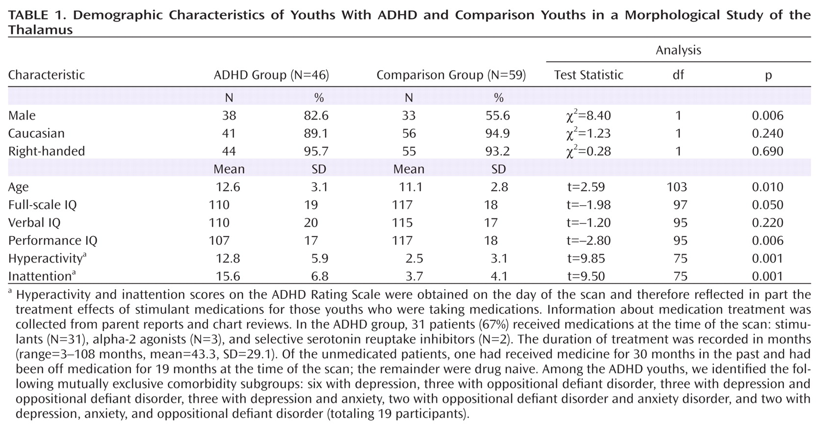
We studied a cohort of 105 children and adolescents, 8–18 years of age, including 46 youths who met DSM-IV criteria (25) for ADHD, combined type, and 59 comparison youths who were recruited randomly from a telemarketing list and matched to the patient group by postal code. Written informed consent was obtained from all parents, and all participants provided written assent. Participants' demographic characteristics are summarized in Table 1.
Table 1. Demographic Characteristics of Youths With ADHD and Comparison Youths in a Morphological Study of the Thalamus
Diagnoses of ADHD were established using the Schedule for Affective Disorders and Schizophrenia for School-Age Children–Present and Lifetime Version (26) and a best-estimate consensus procedure that considered all available clinical and diagnostic information (27). Additional instruments used included the ADHD Rating Scale (28), the Revised Children's Manifest Anxiety Scale (29), and the Children's Depression Inventory (30). Exclusion criteria for participants with ADHD were premature birth (gestation ≤36 weeks) and lifetime diagnosis of any bipolar, psychotic, obsessive-compulsive, or tic disorder. Exclusion criteria for comparison subjects were a lifetime or a current DSM-IV axis I disorder. Additional exclusion criteria for both groups were epilepsy, head trauma with loss of consciousness, lifetime substance abuse, developmental delay, or IQ below 80, as measured by the WISC-III (31), WAIS (32), or Kaufmann Brief Intelligence Test (33). Socioeconomic status was estimated using the Hollingshead Four-Factor Index of Social Status (34).

MRI Scanning and Image Analysis

High-resolution MR images were obtained using a single 1.5-T scanner (GE Signa, Milwaukee). Head position was standardized using canthomeatal landmarks. T1-weighted sagittal three-dimensional volume images were acquired using a spoiled gradient echo pulse sequence (repetition time=24 msec, echo time=5 msec, flip angle=45º, matrix=256×192, field of view=30 cm, excitations=2, slice thickness=1.2 mm, contiguous slices=124).

Preprocessing

Image processing was performed on SunUltra10 workstations with the ANALYZE 7.5 software program (Biomedical Imaging Resource, Mayo Foundation, Rochester, Minn.). Operators were blind to participant characteristics and hemisphere (images were randomly flipped left to right prior to analysis). Large-scale variations in image intensity were removed (35), and images were reformatted to standardize head positioning prior to region definition (36). Axial slices were oriented parallel to both the anterior and posterior commissures, and sagittal slices were oriented parallel to standard midline landmarks (36).

Whole-brain volume

An isointensity contour function was used in conjunction with manual editing to isolate the cerebrum. This whole-brain volume measure included gray and white matter, ventricular CSF, cisterns, fissures, and cortical sulci. CSF was included using a connected components analysis. Whole-brain volume was used as a covariate in statistical analyses to control for scaling effects (37).

Thalamus definition

After excluding non-brain tissue, an anisotropic diffusion filter was applied to the remaining brain tissue to improve the discrimination of the lateral surface of the thalamus from the white matter of the internal capsule. The procedures for defining the thalamus were identical to those previously published (38, 39). The thalamus was segmented by sampling gray-scale values of the thalamus and internal capsule throughout the entire three-dimensional extent of each structure and then averaging the peaks for white and gray matter. An isointensity contour function applied at the calculated threshold and grown from a seed within the thalamus provided an initial definition of the structure that was then edited manually. The intra- and interrater intraclass correlation coefficients were 0.95. The thalamus was distinguished from the hypothalamus by a line defining the hypothalamic sulcus on sagittal views, which excluded portions of the geniculate nuclei from the analysis.

Surface morphometry

We calculated the distance from a voxel-sized point on the surface of each participant's thalamus to the corresponding point on the surface of the thalamus in a template brain. This previously validated method of surface analysis (40) was customized to accommodate independent analysis of right and left hemi-thalami. Briefly, a rigid-body similarity transformation with global scaling was used to register the entire brain of each participant with the template brain, thereby eliminating the need to further adjust for differences in overall brain volume. The thalamus was then rigidly coregistered to the template thalamus. This second transformation created a "refined registration" in which isolated thalami could be compared. Each hemi-thalamus was warped to the corresponding anatomy of the template thalamus using a high-dimensional nonrigid warping based on fluid-flow dynamics that permitted point-to-point matching of homologous tissue between the two thalami. The warped images were then unwarped to the refined registration while maintaining these point-to-point correspondences, which permitted calculation of the distance of each point on the surface of the test thalamus from the corresponding point on the surface of the template thalamus.
We applied a rigorous two-step procedure to select a thalamic template that was as close as possible (in the sense of least-squares mean) to the average shape of the thalamus for all our healthy comparison subjects. First, a preliminary reference was selected as the thalamus of the comparison subject who demographically was as representative as possible of all healthy comparison subjects. The thalami for all other comparison subjects were then normalized to this preliminary reference. The point correspondences on the thalamic surfaces were determined, and we computed the distance from the template surface for each of the corresponding points on the surfaces of the thalami for all other participants. Second, the thalamus for which all points across its surface were closest, in terms of least-squares mean, to the average of the computed distances was selected as the final template. As we have done previously, we used a single representative brain as a template rather than an averaged brain from many youths, because use of a single brain, which has sharp borders at the CSF-gray matter or gray matter-white matter interface, improves the accuracy of registration (36, 41, 42). Averaging images for a template blurs these boundaries and increases registration errors that are subtle but important when distinguishing subtle effects across populations. In addition, precise surface morphometry requires a brain with smooth gray and white matter surfaces that are devoid of topological defects, which cannot be reconstructed by averaging brains from many youths.

Statistical Analyses

Conventional volumes

Statistical analyses were performed in SAS, version 9.0 (SAS Institute, Inc., Cary, N.C.). We tested our a priori hypothesis that conventional measures of overall thalamic volume would differ across diagnostic groups by assessing the main effect of diagnosis in a mixed-models analysis with repeated measures over a spatial domain (volumes of each hemi-thalamus). The model included the within-subjects factor "hemisphere" with two levels (left and right), the between-subjects factor of diagnosis (ADHD=1 and comparison subjects=0), and the covariates of age, sex, and whole-brain volume. In addition to these independent variables, we considered all two- and three-way interactions of diagnosis, sex, hemisphere, and age, as well as the two-way interactions of whole-brain volume with hemisphere. Other variables included medication, IQ, and lifetime diagnoses of depression, oppositional-defiant disorder, and anxiety disorders. Statistically nonsignificant terms were eliminated via backward stepwise regression, with the constraint that the model at each step had to be hierarchically well formulated (i.e., all possible lower-order component terms of an interaction were included in the model, regardless of statistical significance). We considered p values <0.05 statistically significant. All p values were two-sided.

Surface morphometry

The distances between points on the surface of the thalamus for each participant and the corresponding points on the template thalamus were compared statistically between groups using linear regression while covarying for age and sex. We color-coded p values at each voxel and displayed them across the surface of the template thalamus. We used the theory of Gaussian random fields (GRFs) to correct p values appropriately for the multiple comparisons performed across the thalamic surface (42).

Cytoarchitectonically Defined Thalamic Atlas

We superimposed a cytoarchitectonic atlas for the thalamus (43, 44) on our thalamic template to identify precisely the regions of significant differences in regional volumes between groups of subjects. The cytoarchitectonic atlas was coregistered to our thalamic template (40) using the same registration and warping procedures as employed for surface morphometry (above).

Results

Conventional Volumes

Hypothesis testing

The main effect of diagnosis indicated no significant differences across groups in overall conventional volumes of the thalamus.

Post hoc analyses

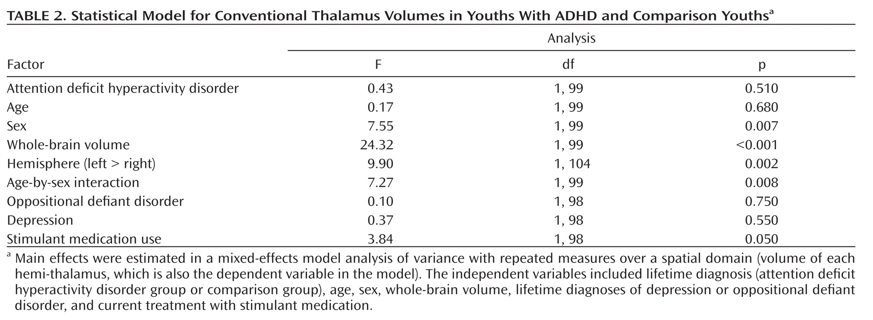
Significant covariates in the model included whole-brain volume (F=24.32; df=1, 99, p<0.001), indicating the presence of significant scaling effects in the thalamus; hemisphere (F=9.90; df=1, 104, p<0.002), reflecting significantly larger volumes in the left hemisphere for all participants; and sex (F=7.55; df=1, 99, p<0.007), demonstrating sex-specific differences in thalamic volumes across the whole sample. The main effect for age was not significant, and the absence of a diagnosis-by-age interaction indicated the stability of findings across the age range of the youths studied (Table 2). The effects of stimulant medication, evaluated by including stimulant treatment as an independent variable in the linear regression, revealed that patients who were receiving stimulants at the time of scanning had larger thalamic volumes than did untreated patients, although this difference fell short of statistical significance (F=3.84, df=1, 98, p=0.053).
Table 2. Statistical Model for Conventional Thalamus Volumes in Youths With ADHD and Comparison Youthsa

Morphological Features of the Thalamic Surface

Hypotheses testing
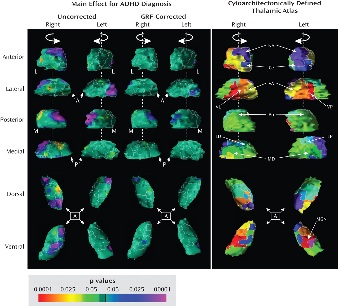
Relative to comparison subjects, youths with ADHD exhibited significantly smaller regional volumes corresponding to the anterior, posterior, and ventral lateral surfaces of the right hemi-thalamus and the posterior surface of the left hemi-thalamus. Using the thalamic atlas template, these morphological differences between the groups were localized in the right anterior and lateral posterior nuclei and the lateral portion of the pulvinar bilaterally. GRF-corrected analyses showed that the differences in regional morphology were concentrated predominantly in the lateral pulvinar bilaterally (Figure 1).
Figure 1. Main Effect for ADHD Diagnosis and Cytoarchitectonically Defined Thalamic Atlasa
aIn the left panel, each row of images shows the statistical maps and thalamic atlas in varying rotational views. The color bar indicates the color coding for p values associated with the main effect of ADHD diagnosis, ranging from p<0.0001 in red (increased regional volumes) to p<0.0001 in purple (reduced regional volumes). The theory of Gaussian random fields (GRFs) was used to correct the maps for the multiple statistical comparisons performed. The maps show significantly smaller regional thalamic volumes predominantly in the pulvinar bilaterally in youths with ADHD relative to comparison subjects. A=anterior; L=lateral; M=medial; P=posterior. The right panel shows the cytoarchitectonically defined thalamic atlas. Ce=central medial nucleus; LD=lateral dorsal nucleus; LP=lateral posterior nucleus; MD=medial dorsal nucleus; MGN=medial geniculate nucleus; NA=nucleus anterior; Pu=pulvinar nucleus; VA=ventral anterior nucleus; VL=ventral lateral nucleus; VP=ventral posterior nucleus.

Post Hoc Analyses

Medication effects
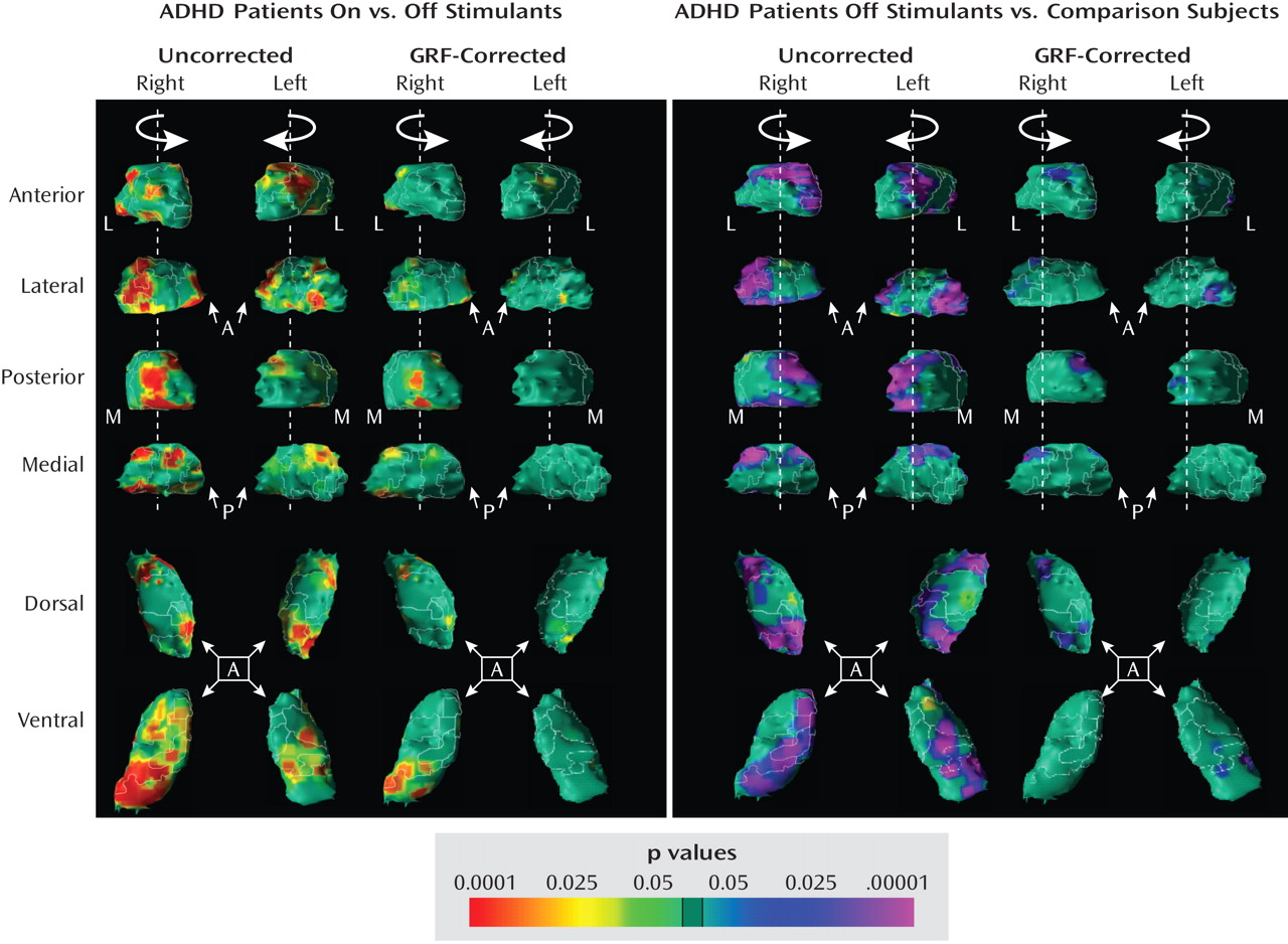
Youths with ADHD who were receiving stimulant medications at the time of scanning had significantly larger regional volumes over the posterior dorsal, ventral, and anterior dorsal thalamic surfaces bilaterally compared to untreated patients. The thalamic atlas template further localized these regions to the anterior nucleus and pulvinar bilaterally. GRF-corrected maps revealed larger regional volumes in stimulant-treated youths predominantly in the pulvinar, closely overlapping previously identified locations of smaller regional volumes in the ADHD group relative to the comparison group. These findings did not change when we compared youths with ADHD on and off stimulants after excluding the five participants with ADHD who were taking both stimulant and nonstimulant medications and one participant who had taken stimulants in the past but had been off them for 19 months before the scan. Additionally, GRF-corrected analyses demonstrated smaller regional volumes of the right anterior nucleus and the pulvinar bilaterally in untreated patients with ADHD relative to comparison subjects (Figure 2). Thus, we obtained the finding of smaller regional volumes in both the posterior and anterior thalamic surfaces in the ADHD group relative to the comparison group when we compared healthy youths either to all patients with ADHD or to untreated patients only.
Figure 2. Comparison of Local Morphological Features of the Thalamus in Youths With ADHD On and Off Stimulant Medications and Comparison Subjectsa
aThe left panel shows regional thalamic volumes in stimulant-treated (N=31) relative to untreated (N=15) youths with ADHD. The right panel shows regional thalamic volumes in untreated youths with ADHD (N=15) relative to healthy comparison subjects (N=59). These maps demonstrate that youths with ADHD receiving stimulants exhibit significantly larger regional volumes predominantly in the right pulvinar relative to their untreated counterparts, whereas untreated youths with ADHD exhibit significantly smaller regional volumes in the pulvinar bilaterally relative to comparison subjects. The color bar indicates the color coding for p values associated with the main effect of stimulant treatment at the time of the scan, ranging from p<0.0001 in red (increased regional volume) to p<0.0001 in purple (reduced regional volume). GRF=Gaussian random field; A=anterior; L=lateral; M=medial; P=posterior.
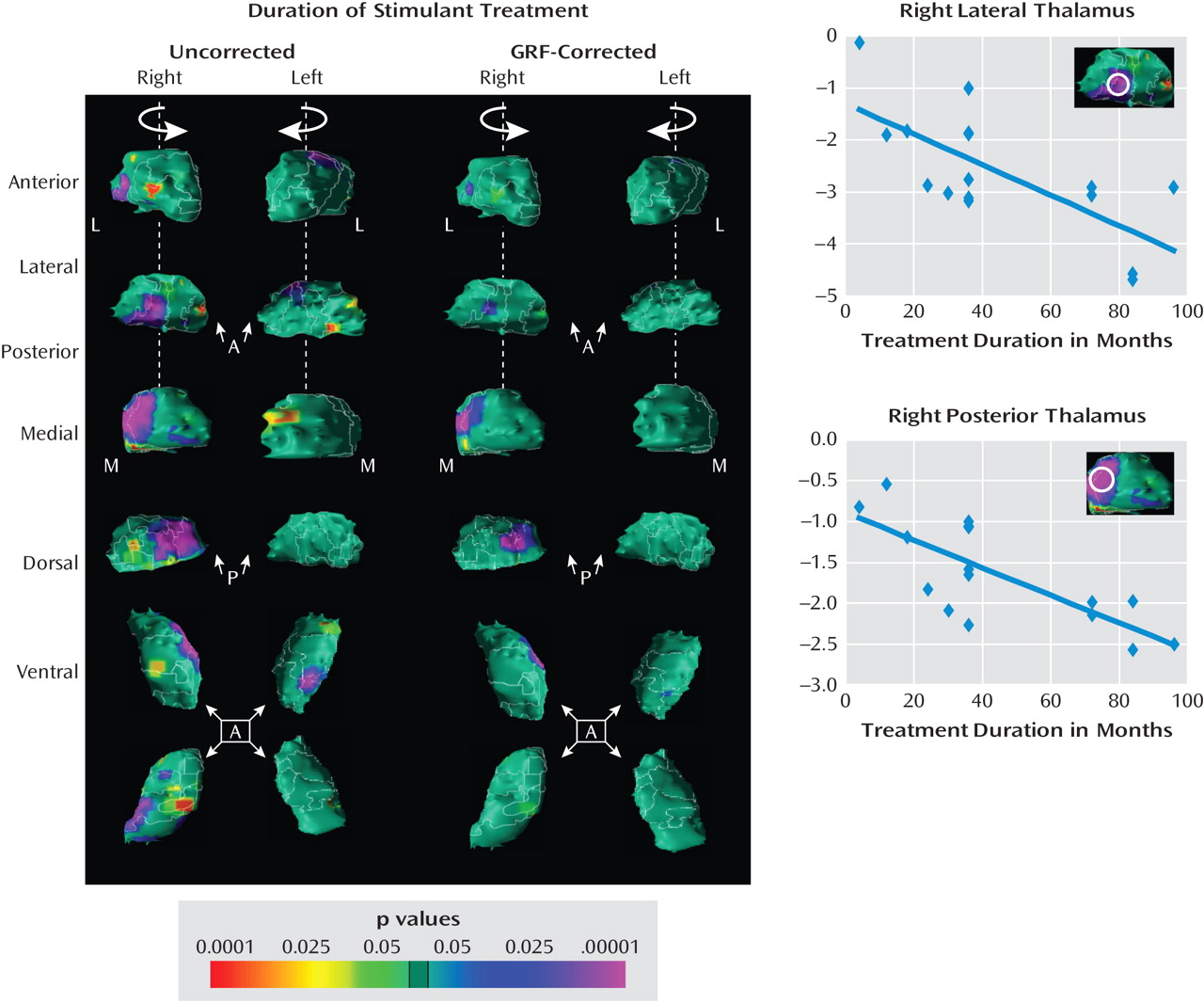
Surface maps for the subgroup of youths with ADHD for whom we collected sufficient data on duration of treatment with stimulants (N=17) revealed that a longer duration of treatment was associated with smaller regional volumes in the right lateral and medial posterior thalamic surfaces (Figure 3). GRF-corrected analyses localized these correlations predominantly to the right lateral and medial pulvinar, a location that did not overlap with the main effects of diagnosis or stimulant treatment.
Figure 3. Correlation of Local Morphological Features of the Thalamus With the Duration of Stimulant Treatmenta
aIn the left panel, surface maps demonstrate correlations of regional morphological features of the thalamus with the duration of stimulant treatment in the ADHD subgroup (N=17). The scatterplot at upper right demonstrates that the duration of stimulant treatment was inversely correlated with regional volumes on the right lateral thalamic surfaces (r=–0.49, p<0.05) in youths with ADHD (N=17). The scatterplot at lower right demonstrates that duration of stimulant treatment was inversely correlated with regional volumes on the right posterior thalamic surfaces (r=–0. 57, p<0.05) in youths with ADHD (N=17). The surface maps indicate that youths with ADHD who were treated longer with stimulants had smaller regional volumes. The color bar depicts the statistical significance of the correlation coefficients, ranging from p<0.0001 in red (positive correlations) to p<0.0001 in purple (inverse correlations). Regions where these correlations were most pronounced and survived Gaussian random field (GRF) correction are probed (white circles). Distance is calculated in millimeters from the surface of the template thalamus. A=anterior; L=lateral; M=medial; P=posterior.

Correlations with symptom severity

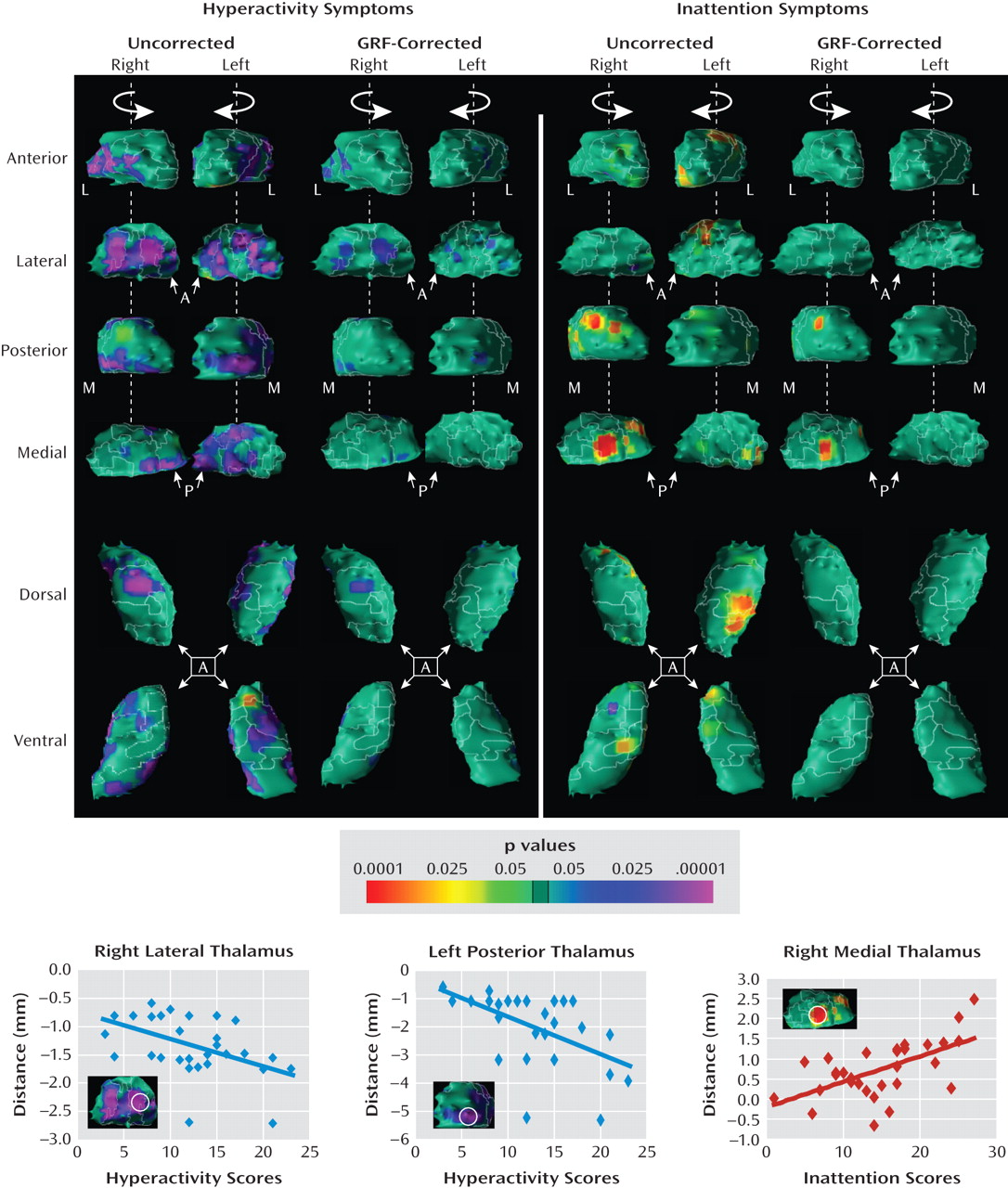
More severe hyperactivity accompanied smaller regional volumes in the lateral and posterior thalamic regions bilaterally, which were localized in the ventral anterior, ventral lateral, ventral posterior, lateral posterior, and pulvinar nuclei. GRF-corrected maps showed that these differences were concentrated on the ventral lateral nucleus bilaterally and the left lateral posterior nucleus. In contrast, greater inattention scores accompanied larger regional volumes in the right posterior and medial thalamus and in the left anterior and lateral thalamic regions, corresponding to the right pulvinar, medial dorsal, left central medial, ventral lateral, and lateral posterior nuclei. GRF-corrected maps localized these differences to the right pulvinar and the medial dorsal nuclei (Figure 4), extending beyond the locations of the main effects near the pulvinar, where regional volumes were smaller in the ADHD group.
Figure 4. Correlation of Local Morphological Features of the Thalamus With Severity of ADHD Symptomsa
aIn the upper left panel, surface maps demonstrate correlations of regional morphological features of the thalamus with severity of hyperactivity symptoms in the ADHD group (N=46). In the upper right panel, surface maps demonstrate correlations of regional morphological features of the thalamus with severity of inattention symptoms in the ADHD group (N=46). The scatterplot at lower left demonstrates that current hyperactivity scores on the ADHD Rating Scale are inversely correlated with regional volumes on the right lateral thalamic surfaces (r=–0.23, p<0.05) in youths with ADHD (N=46). The lower center scatterplot demonstrates that current hyperactivity scores on the ADHD Rating Scale are inversely correlated with regional volumes on the left posterior thalamic surfaces (r=–0.31, p<0.05) in youths with ADHD (N=46). The lower right scatterplot demonstrates that current inattention scores on the ADHD Rating Scale are positively correlated with the regional volumes on the right medial thalamic surface (r=0.35, p<0.05) in youths with ADHD (N=46). The surface maps indicate that youths with ADHD with higher hyperactivity scores have smaller regional volumes, whereas youths with ADHD with higher inattention scores have larger regional volumes. The color bar depicts the statistical significance of the correlation coefficients, ranging from p<0.0001 in red (positive correlations) to p<0.0001 in purple (inverse correlations). Regions where these correlations were most pronounced and survived Gaussian random field (GRF) correction are probed (white circles). Distance is calculated in millimeters from the surface of the template thalamus. A=anterior; L=lateral; M=medial; P=posterior.

Possible confounders

In analyses of conventional volumes and surface morphology, we did not discern appreciable effects of age, comorbid disorders (see Table 2, as well as Figures S1 and S2 in the data supplement that accompanies the online edition of this article), duration of stimulant treatment (not shown), or IQ (not shown), which suggests that these variables did not unduly influence our findings.

Discussion

Regional volumes were significantly reduced over the pulvinar bilaterally in youths with ADHD, despite the presence of normal overall thalamic volumes. These morphological abnormalities were not associated with the effects of naturalistic stimulant medication treatment or the presence of comorbid conditions in the ADHD group. In addition, conventional thalamic volumes in youths with ADHD treated naturalistically with stimulants tended to be larger than in untreated individuals (p=0.053), and regional volumes in those treated with stimulants were significantly larger in the pulvinar bilaterally. Finally, smaller regional thalamic volumes in the ventral lateral nucleus bilaterally and the left lateral posterior nucleus were associated with more hyperactivity, and larger regional volumes in the right pulvinar and medial dorsal nuclei were associated with more inattention.
The absence of a significant main effect for diagnosis in analyses of conventional thalamic volumes likely derives from the insensitivity of our ability to detect abnormalities in conventional volumes of a heterogeneous brain region such as the thalamus when the anatomical abnormalities in that region are restricted to a small number of substructures. Several studies, however, have reported significant group differences in conventional volumes of the hippocampus (36), the putamen (45, 46), the globus pallidus (47), the frontal cortex (47, 48), and the cerebellum (47, 49) in youths with ADHD relative to comparison subjects. Thus, examination of both conventional thalamic volumes and morphological features of the thalamic surface is appropriate in the study of ADHD.

The Role of the Thalamus in the Pathogenesis of ADHD

We used the cytoarchitectonically defined thalamic atlas (43) to determine precisely the regional morphological features on the surface of the thalamus overlying specific thalamic nuclei and to interpret morphological alterations in the context of the known thalamic connectivity derived from studies in humans. This atlas allowed us to map anatomical findings in youths with ADHD onto larger anatomical circuits that subserve specific cognitive processes. These morphological findings suggest that the pulvinar and ventral lateral nuclei are involved in the pathophysiology of ADHD.

The pulvinar nucleus

The pulvinar projects to the frontal and parietal association cortices (50), two thalamocortical pathways that support attentional processes. These circuits are important for stimulus seeking and for distinguishing between contextually relevant and irrelevant somatosensory stimuli (51). Within this overall thalamocortical network, the pulvinar integrates and coordinates responses to auditory and visual stimuli (5254). Morphological disturbances in the pulvinar in youths with ADHD therefore may contribute to difficulties in allocating and directing attentional resources toward salient stimuli.
The pulvinar also connects with limbic structures, including the amygdala (5559). We previously reported morphological disturbances in the basolateral complex of the amygdala (36), and others have reported abnormal amygdala activation (60, 61) in individuals with ADHD. The basolateral complex of the amygdala supports emotional regulation, fear conditioning, and the attribution of affective valence to sensory stimuli (6264). Therefore, disturbances of the thalamolimbic network could interfere with emotional learning and the regulation of affect, thereby contributing to the emotional dysregulation and affective illnesses that affect many children with ADHD (65, 66).

The ventral lateral nucleus

The ventral lateral nucleus receives projections from the cerebellum and connects reciprocally with the basal ganglia, motor cortex, and premotor cortices to form a sensorimotor network that drives the acquisition and implementation of learned motor behaviors (6769). Smaller regional volumes in the ventral lateral nucleus could therefore contribute to the gross and fine motor disturbances and the slower processing speed that have been documented in children with ADHD (7074).

Effects of Stimulant Medications on Thalamic Morphology

Surface maps show that the pulvinar was significantly larger bilaterally in treated compared with untreated youths with ADHD. These areas closely overlapped regions where volumes were smaller in youths with ADHD, which suggests that stimulant treatment may help attenuate morphological abnormalities in the thalamus in individuals with ADHD. If that is true, then the absence of statistically significant differences in conventional volumes of the thalamus in youths with ADHD relative to comparison subjects may be partially explained by these hypothesized effects of stimulant treatment. Alternatively, youths with ADHD who had larger thalami may for unknown reasons have been preferentially treated with stimulants. The effects of stimulants on local morphology should be more definitively determined in a longitudinal randomized controlled trial that would obviate any possible selection biases that may have confounded treatment effects in this cross-sectional, naturalistic study.
The duration of stimulant treatment was inversely correlated with regional volumes in the right lateral and medial posterior hemi-thalamus. These regions did not overlap those where the main effects of an ADHD diagnosis or where the main effects of stimulants were located. These findings suggest that stimulants may have differing short- and long-term morphological effects on differing thalamic nuclei. Treatment with stimulants for any duration was associated with larger volumes in the dorsal anterior, posterior, and ventral posterior surfaces bilaterally, whereas progressively longer treatment was associated with progressively smaller regional volumes in the right lateral and medial posterior surfaces. Alternatively, youths with ADHD who received stimulants for longer durations may have differed systematically and in unknown ways from those who received stimulants for shorter durations, producing only an apparent association of regional volumes with duration that was not actually caused by longer exposure to stimulant medication. Moreover, we must emphasize that the association of treatment duration with thalamic morphology should be interpreted with caution, as treatment duration is exceedingly difficult to estimate reliably. Treatment is often interrupted by medication holidays and lapses in compliance that are not documented or recalled, and pharmacological agents and their dosing often vary widely over time.
We do not know what neurobiological mechanisms produced the local morphological differences we observed between the ADHD and comparison groups and that seemed to have attenuated those differences in the stimulant-treated youths with ADHD. Previous reports of depleted levels of dopamine and norepinephrine suggest that disturbances in catecholamine transmission may contribute to the pathogenesis of ADHD (75). Stimulants are thought to attenuate ADHD symptoms by inhibiting presynaptic dopamine and norepinephrine transporters and thereby potentiating dopamine and norepinephrine neurotransmission. Both dopamine and norepinephrine have powerful influences on the physiology and cellular morphology of the prefrontal cortex, basal ganglia, and thalamic portions of CSTC circuits (7679). In humans, multiple thalamic nuclei, including the pulvinar, receive dopamine and norepinephrine inputs (8083), so theoretically stimulants should increase cathecholaminergic transmission within the thalamus, although this has not been shown definitively. Nevertheless, numerous preclinical studies in rodents have shown that repeated exposure to stimulants increases the length of dendrites and the density of dendritic spines within the basal ganglia, limbic system, and frontal cortices (8486). We speculate that stimulants may have a similar effect in the thalamus. Alternatively, the stimulant-induced cellular and functional changes in the prefrontal cortex (79) or basal ganglia could in turn alter cellular activity and dendritic architecture within the thalamus. The effects of stimulants on catecholamines have also been shown to alter the number of astrocytes in the striatum (87), cells that regulate the energetics of neuronal excitation (88). Stimulants could have similar effects on thalamic astrocytes.

The Differential Association of Thalamic Nuclei With ADHD Symptom Domains

Smaller regional volumes in the ventral lateral nucleus bilaterally and the left lateral posterior nucleus were associated with more severe hyperactivity, whereas larger regional volumes in the right pulvinar and the medial dorsal nucleus were associated with more severe inattention. The localization of these differences is consistent with reports that the projections of the ventral lateral nucleus to the striatum participate in the control of motor actions (4, 89) and that the pulvinar, through its cortical connections, may be involved in alerting and in the allocation of attention (5254). Smaller regional volumes in the lateral thalamus suggest the presence of disturbances in networks related to motor functions, possibly producing more severe symptoms of hyperactivity. Larger regional volumes in the pulvinar may represent alterations in systems that subserve attention, either exacerbating the symptoms of inattention or compensating for functional disturbances in attention-related networks located outside the thalamus.

Limitations

Several limitations of this study should be mentioned. First, our study design did not allow us to determine the direction of causality in the associations of regional thalamic volumes with diagnosis or stimulant treatment. Second, the use of the thalamic atlas has inherent inaccuracies in the exact boundaries that it ascribes to thalamic subnuclei, and our analyses of the thalamic surface assign the source of morphological differences to the most superficial nucleus, when in fact it may derive instead from abnormalities in deeper underlying nuclei. Third, the imaging methods we used cannot alone identify the cellular and molecular bases for our morphological findings, which would require studies using either different imaging modalities or postmortem histological analyses. Finally, the multiple statistical tests in our exploratory analyses increased the likelihood of type I error, although we minimized false positive findings through the use of conservative statistical thresholds and GRF-based corrections of p values for the multiple comparisons performed (90).

Conclusions

Our findings provide new evidence that abnormalities at the level of the thalamus are involved in the pathogenesis of ADHD. Our findings additionally suggest that the therapeutic effects of stimulants may attenuate these morphological abnormalities, possibly via the local changes that they produce in dendritic architecture and cellular composition within CSTC circuits. Finally, we hypothesize that the hyperactivity and inattention symptoms of ADHD derive from abnormalities in different thalamic nuclei that may reflect the differential involvement of both pathogenic and compensatory processes.

Supplementary Material

File (ajp_167_04_397_01.pdf)

References

1.
Kimura M, Minamimoto T, Matsumoto N, Hori Y: Monitoring and switching of cortico-basal ganglia loop functions by the thalamo-striatal system. Neurosci Res 2004; 48:355–360
2.
Schiff ND: Central thalamic contributions to arousal regulation and neurological disorders of consciousness. Ann NY Acad Sci 2008; 1129:105–118
3.
Rowe DL, Robinson PA, Lazzaro IL, Powles RC, Gordon E, Williams LM: Biophysical modeling of tonic cortical electrical activity in attention deficit hyperactivity disorder. Int J Neurosci 2005; 115:1273–1305
4.
McFarland NR, Haber SN: Thalamic relay nuclei of the basal ganglia form both reciprocal and nonreciprocal cortical connections, linking multiple frontal cortical areas. J Neurosci 2002; 22:8117–8132
5.
Smith Y, Raju DV, Pare JF, Sidibe M: The thalamostriatal system: a highly specific network of the basal ganglia circuitry. Trends Neurosci 2004; 27:520–527
6.
Dickstein SG, Bannon K, Castellanos FX, Milham MP: The neural correlates of attention deficit hyperactivity disorder: an ALE meta-analysis. J Child Psychol Psychiatry 2006; 47:1051–1062
7.
Alexander GE, DeLong MR, Strick PL: Parallel organization of functionally segregated circuits linking basal ganglia and cortex. Annu Rev Neurosci 1986; 9:357–381
8.
Parent A, Hazrati LN: Functional anatomy of the basal ganglia, I: the cortico-basal ganglia-thalamo-cortical loop. Brain Res Brain Res Rev 1995; 20:91–127
9.
Parent A, Hazrati LN: Functional anatomy of the basal ganglia, II: the place of subthalamic nucleus and external pallidum in basal ganglia circuitry. Brain Res Brain Res Rev 1995; 20:128–154
10.
Perlov E, Philipsen A, Matthies S, Drieling T, Maier S, Bubl E, Hesslinger B, Buechert M, Henning J, Ebert D, van Elst LT: Spectroscopic findings in attention-deficit/hyperactivity disorder: review and meta-analysis. World J Biol Psychiatry 2008; 12:1–11
11.
Kates WR, Frederikse M, Mostofsky SH, Folley BS, Cooper K, Mazur-Hopkins P, Kofman O, Singer HS, Denckla MB, Pearlson GD, Kaufmann WE: MRI parcellation of the frontal lobe in boys with attention deficit hyperactivity disorder or Tourette syndrome. Psychiatry Res 2002; 116:63–81
12.
Kelly AM, Margulies DS, Castellanos FX: Recent advances in structural and functional brain imaging studies of attention-deficit/hyperactivity disorder. Curr Psychiatry Rep 2007; 9:401–407
13.
Castellanos FX, Lee PP, Sharp W, Jeffries NO, Greenstein DK, Clasen LS, Blumenthal JD, James RS, Ebens CL, Walter JM, Zij­denbos A, Evans AC, Giedd JN, Rapoport JL: Developmental trajectories of brain volume abnormalities in children and adolescents with attention-deficit/hyperactivity disorder. JAMA 2002; 288:1740–1748
14.
Seidman LJ, Valera EM, Makris N: Structural brain imaging of attention-deficit/hyperactivity disorder. Biol Psychiatry 2005; 57:1263–1272
15.
Durston S, Fossella JA, Mulder ML, Casey BJ, Ziermans TB, Vessaz MN, Van Engeland H: Dopamine transporter genotype conveys familial risk of attention-deficit/hyperactivity disorder through striatal activation. J Am Acad Child Adolesc Psychiatry 2008; 47:61–67
16.
Durston S: A review of the biological bases of ADHD: what have we learned from imaging studies? Ment Retard Dev Disabil Res Rev 2003; 9:184–195
17.
Bush G, Valera EM, Seidman LJ: Functional neuroimaging of attention-deficit/hyperactivity disorder: a review and suggested future directions. Biol Psychiatry 2005; 57:1273–1284
18.
Kunzle H: Thalamic projections from the precentral motor cortex in Macaca fascicularis. Brain Res 1976; 105:253–267
19.
Kunzle H, Akert K: Efferent connections of cortical, area 8 (frontal eye field) in Macaca fascicularis: a reinvestigation using the autoradiographic technique. J Comp Neurol 1977; 173:147–164
20.
Kunzle H: Thalamo-striatal projections in the hedgehog tenrec. Brain Res 2006; 1100:78–92
21.
Russchen FT, Amaral DG, Price JL: The afferent input to the magnocellular division of the mediodorsal thalamic nucleus in the monkey, Macaca fascicularis. J Comp Neurol 1987; 256:175–210
22.
Siwek DF, Pandya DN: Prefrontal projections to the mediodorsal nucleus of the thalamus in the rhesus monkey. J Comp Neurol 1991; 312:509–524
23.
Gerring J, Brady K, Chen A, Quinn C, Herskovits E, Bandeen-Roche K, Denckla MB, Bryan RN: Neuroimaging variables related to development of secondary attention deficit hyperactivity disorder after closed head injury in children and adolescents. Brain Inj 2000; 14:205–218
24.
Rowe DL, Robinson PA, Gordon E: Stimulant drug action in attention deficit hyperactivity disorder (ADHD): inference of neurophysiological mechanisms via quantitative modelling. Clin Neurophysiol 2005; 116:324–335
25.
American Psychiatric Association: Diagnostic and Statistical Manual of Mental Disorders, 4th ed (DSM-IV). Washington, DC, American Psychiatric Association, 1994
26.
Kaufman J, Birmaher B, Brent D, Rao U, Flynn C, Moreci P, Williamson D, Ryan N: Schedule for Affective Disorders and Schizophrenia for School-Age Children–Present and Lifetime Version (K-SADS-PL): initial reliability and validity data. J Am Acad Child Adolesc Psychiatry 1997; 36:980–988
27.
Leckman JF, Sholomskas D, Thompson WD, Belanger A, Weissman MM: Best estimate of lifetime psychiatric diagnosis: a methodological study. Arch Gen Psychiatry 1982; 39:879–883
28.
DuPaul GJ, Power TJ, Anastopoulos AD, Reid R: ADHD Rating Scale–IV: Checklists, Norms, and Clinical Interpretations. New York, Guilford, 1998
29.
Reynolds CR, Richmond BO: Revised Children's Manifest Anxiety Scale, 3rd ed, revised ed (RCMAS). Washington, DC, Western Psychological Services, 1985
30.
Kovacs M: The Children's Depression Inventory (CDI). Psychopharmacol Bull 1985; 21:995–998
31.
Wechsler D: WISC-III Manual, Canadian Supplement. Toronto, Psychological Corporation, 1996
32.
Wechsler D: Wechsler Adult Intelligence Scale–III. New York, Psychological Corporation, 1991
33.
Grados JJ, Russo-Garcia KA: Comparison of the Kaufman Brief Intelligence Test and the Wechsler Intelligence Scale for Children–Third Edition in economically disadvantaged African American youth. J Clin Psychol 1999; 55:1063–1071
34.
Hollingshead AB: Four-Factor Index of Social Status. New Haven, Conn, Yale University, Department of Sociology, 1975
35.
Sled JG, Zijdenbos AP, Evans AC: A nonparametric method for automatic correction of intensity nonuniformity in MRI data. IEEE Trans Med Imaging 1998; 17:87–97
36.
Plessen KJ, Bansal R, Zhu H, Whiteman R, Amat J, Quackenbush GA, Martin L, Durkin K, Blair C, Royal J, Hugdahl K, Peterson BS: Hippocampus and amygdala morphology in attention-deficit/hyperactivity disorder. Arch Gen Psychiatry 2006; 63:795–807
Arndt S, Cohen G, Alliger RJ, Swayze VW, Andreasen NC: Problems with ratio and proportion measures of imaged cerebral structures. Psychiatry Res 1991; 40:79–89
38.
Frazier JA, Chiu S, Breeze JL, Makris N, Lange N, Kennedy NDN, Herbert MR, Bent EK, Koneru VK, Dieterich ME, Hodge SM, Rauch SL, Grant PE, Cohen BM, Seidman LJ, Caviness VS, Biederman J: Structural brain magnetic resonance imaging of limbic and thalamic volumes in pediatric bipolar disorder. Am J Psychiatry 2005; 162:1256–1265
39.
Seidman LJ, Faraone SV, Goldstein JM, Goodman JM, Kremen WS, Toomey R, Tourville J, Kennedy D, Makris N, Caviness VS, Tsuang MT: Thalamic and amygdala-hippocampal volume reductions in first-degree relatives of patients with schizophrenia: an MRI-based morphometric analysis. Biol Psychiatry 1999; 46:941–954
40.
Bansal R, Staib LH, Whiteman R, Wang YM, Peterson BS: ROC-based assessments of 3D cortical surface-matching algorithms. Neuroimage 2005; 24:150–162
41.
Amat JA, Whiteman R, Bansal R, Davies M, Haggerty R, Peterson BS: The cognitive correlates of amygdala and hippocampus volumes in healthy adults. Brain Cognit 2008; 66:105–114
42.
Bansal AS, Staib LH, Xu D, Zhu H, Peterson BS: Statistical analyses of brain surfaces using Gaussian random fields on 2-D manifolds. IEEE Trans Med Imaging 2007; 26:46–57
43.
Chakravarty MM, Bertrand G, Hodge CP, Sadikot AF, Collins DL: The creation of a brain atlas for image guided neurosurgery using serial histological data. Neuroimage 2006; 30:359–376
44.
Chakravarty MM, Sadikot AF, Germann J, Bertrand G, Collins DL: Towards a validation of atlas warping techniques. Med Image Anal 2008; 12:713–726
45.
Overmeyer S, Bullmore ET, Suckling J, Simmons A, Williams SC, Santosh PJ, Taylor E: Distributed grey and white matter deficits in hyperkinetic disorder: MRI evidence for anatomical abnormality in an attentional network. Psychol Med 2001; 31:1425–1435
46.
Wang J, Jiang T, Cao Q, Wang Y: Characterizing anatomic differences in boys with attention-deficit/hyperactivity disorder with the use of deformation-based morphometry. AJNR Am J Neuroradiol 2007; 28:543–547
47.
Castellanos FX, Giedd JN, Marsh WL, Hamburger SD, Vaituzis AC, Dickstein DP, Sarfatti SE, Vauss YC, Snell JW, Lange N, Kaysen D, Krain AL, Ritchie JF, Rajapakse JC, Rapoport JL: Quantitative brain magnetic resonance imaging in attention-deficit hyperactivity disorder. Arch Gen Psychiatry 1996; 53:607–616
48.
Sowell ER, Thompson PM, Welcome SE, Henkenius AL, Toga AW, Peterson BS: Cortical abnormalities in children and adolescents with attention-deficit hyperactivity disorder. Lancet 2003; 362:1699–1707
49.
Mackie S, Shaw P, Lenroot R, Pierson R, Greenstein DK, Nugent TF, Sharp WS, Giedd JN, Rapoport JL: Cerebellar development and clinical outcome in attention deficit hyperactivity disorder. Am J Psychiatry 2007; 164:647–655
50.
Romanski LM, Giguere M, Bates JF, Goldman-Rakic PS: Topographic organization of medial pulvinar connections with the prefrontal cortex in the rhesus monkey. J Comp Neurol 1997; 379:313–332
51.
Wester K, Irvine DR, Hugdahl K: Auditory laterality and attentional deficits after thalamic haemorrhage. J Neurol 2001; 248:676–683
52.
McAlonan K, Cavanaugh J, Wurtz RH: Guarding the gateway to cortex with attention in visual thalamus. Nature 2008; 456:391–394
53.
Arend I, Rafal R, Ward R: Spatial and temporal deficits are regionally dissociable in patients with pulvinar lesions. Brain 2008; 131:2140–2152
54.
Kastner S, O'Connor DH, Fukui MM, Fehd HM, Herwig U, Pinsk MA: Functional imaging of the human lateral geniculate nucleus and pulvinar. J Neurophysiol 2004; 91:438–448
55.
Jones EG, Burton H: A projection from the medial pulvinar to the amygdala in primates. Brain Res 1976; 104:142–147
56.
Morel A, Magnin M, Jeanmonod D: Multiarchitectonic and stereotactic atlas of the human thalamus. J Comp Neurol 1997; 387:588–630
57.
Ohman A: The role of the amygdala in human fear: automatic detection of threat. Psychoneuroendocrinology 2005; 30:953–958
58.
Das P, Kemp AH, Liddell BJ, Brown KJ, Olivieri G, Peduto A, Gordon E, Williams LM: Pathways for fear perception: modulation of amygdala activity by thalamo-cortical systems. Neuroimage 2005; 26:141–148
59.
Liddell BJ, Brown KJ, Kemp AH, Barton MJ, Das P, Peduto A, Gordon E, Williams LM: A direct brainstem-amygdala-cortical "alarm" system for subliminal signals of fear. Neuroimage 2005; 24:235–243
60.
Volkow ND, Wang G-J, Newcorn J, Telang F, Solanto MV, Fowler JS, Logan J, Ma Y, Schulz K, Pradhan K, Wong C, Swanson JM: Depressed dopamine activity in caudate and preliminary evidence of limbic involvement in adults with attention-deficit/hyperactivity disorder. Arch Gen Psychiatry 2007; 64:932–940
61.
Marsh AA, Finger EC, Mitchell DG, Reid ME, Sims C, Kosson DS, Towbin KE, Leibenluft E, Pine DS, Blair RJ: Reduced amygdala response to fearful expressions in children and adolescents with callous-unemotional traits and disruptive behavior disorders. Am J Psychiatry 2008; 165:712–720
62.
Bechara A, Damasio H, Damasio AR: Role of the amygdala in decision-making. Ann NY Acad Sci 2003; 985:356–369
63.
Cardinal RN, Parkinson JA, Hall J, Everitt BJ: Emotion and motivation: the role of the amygdala, ventral striatum, and prefrontal cortex. Neurosci Biobehav Rev 2002; 26:321–352
64.
Winstanley CA, Theobald DE, Cardinal RN, Robbins TW: Contrasting roles of basolateral amygdala and orbitofrontal cortex in impulsive choice. J Neurosci 2004; 24:4718–4722
65.
Levy F: Synaptic gating and ADHD: a biological theory of comorbidity of ADHD and anxiety. Neuropsychopharmacology 2004; 29:1589–1596
66.
Pliszka SR: Patterns of psychiatric comorbidity with attention-deficit/hyperactivity disorder. Child Adolesc Psychiatr Clin N Am 2000; 9:525–540
67.
Stepniewska I, Preuss TM, Kaas JH: Thalamic connections of the dorsal and ventral premotor areas in New World owl monkeys. Neuroscience 2007; 147:727–745
68.
Jeljeli M, Strazielle C, Caston J, Lalonde R: Effects of ventrolateral-ventromedial thalamic lesions on motor coordination and spatial orientation in rats. Neurosci Res 2003; 47:309–316
69.
Fabre-Thorpe M, Levesque F: Visuomotor relearning after brain damage crucially depends on the integrity of the ventrolateral thalamic nucleus. Behav Neurosci 1991; 105:176–192
70.
Pitcher TM, Piek JP, Hay DA: Fine and gross motor ability in males with ADHD. Dev Med Child Neurol 2003; 45:525–535
71.
Whitmont S, Clark C: Kinaesthetic acuity and fine motor skills in children with attention deficit hyperactivity disorder: a preliminary report. Dev Med Child Neurol 1996; 38:1091–1098
72.
Barkley RA: Behavioral inhibition, sustained attention, and executive functions: constructing a unifying theory of ADHD. Psychol Bull 1997; 121:65–94
73.
Piek JP, Dyck MJ, Francis M, Conwell A: Working memory, processing speed, and set-shifting in children with developmental coordination disorder and attention-deficit-hyperactivity disorder. Dev Med Child Neurol 2007; 49:678–683
74.
Mayes SD, Calhoun SL: Learning, attention, writing, and processing speed in typical children and children with ADHD, autism, anxiety, depression, and oppositional-defiant disorder. Child Neuropsychol 2007; 13:469–493
75.
Prince J: Catecholamine dysfunction in attention-deficit/hyperactivity disorder: an update. J Clin Psychopharmacol 2008; 28(3 suppl 2):S39–S45
76.
Arnsten AF, Scahill L, Findling RL: alpha2-Adrenergic receptor agonists for the treatment of attention-deficit/hyperactivity disorder: emerging concepts from new data. J Child Adolesc Psychopharmacol 2007; 17:393–406
77.
Madras BK, Miller GM, Fischman AJ: The dopamine transporter and attention-deficit/hyperactivity disorder. Biol Psychiatry 2005; 57:1397–1409
78.
Bymaster FP, Katner JS, Nelson DL, Hemrick-Luecke SK, Threlkeld PG, Heiligenstein JH, Morin SM, Gehlert DR, Perry KW: Atomoxetine increases extracellular levels of norepinephrine and dopamine in prefrontal cortex of rat: a potential mechanism for efficacy in attention deficit/hyperactivity disorder. Neuropsychopharmacology 2002; 27:699–711
79.
Robinson TE, Kolb B: Structural plasticity associated with exposure to drugs of abuse. Neuropharmacology 2004; 47(suppl 1):33–46
80.
García-Cabezas M, Rico B, Sánchez-González M, Cavada C: Distribution of the dopamine innervation in the macaque and human thalamus. Neuroimage 2007; 34:965–984
81.
Aston-Jones G, Cohen JD: An integrative theory of locus coeruleus-norepinephrine function: adaptive gain and optimal performance. Annu Rev Neurosci 2005; 28:403–450
82.
Berridge CW, Waterhouse BD: The locus coeruleus-noradrenergic system: modulation of behavioral state and state-dependent cognitive processes. Brain Res Brain Res Rev 2003; 42:33–84
83.
Aston-Jones G, Rajkowski J, Cohen J: Locus coeruleus and regulation of behavioral flexibility and attention. Prog Brain Res 2000; 126:165–182
84.
Ujike H, Takaki M, Kodama M, Kuroda S: Gene expression related to synaptogenesis, neuritogenesis, and MAP kinase in behavioral sensitization to psychostimulants. Ann NY Acad Sci 2002; 965:55–67
85.
Lee KW, Kim Y, Kim AM, Helmin K, Nairn AC, Greengard P: Cocaine-induced dendritic spine formation in D1 and D2 dopamine receptor-containing medium spiny neurons in nucleus accumbens. Proc Natl Acad Sci USA 2006; 103:3399–3404
86.
Robinson TE, Kolb B: Persistent structural modifications in nucleus accumbens and prefrontal cortex neurons produced by previous experience with amphetamine. J Neurosci 1997; 17:8491–8497
87.
Magistretti PJ: Neuron-glia metabolic coupling and plasticity. J Exp Biol 2006; 209:2304–2311
88.
Todd RD, Botteron KN: Is attention-deficit/hyperactivity disorder an energy deficiency syndrome? Biol Psychiatry 2001; 50:151–158
89.
Graybiel AM: The basal ganglia: learning new tricks and loving it. Curr Opin Neurobiol 2005; 15:638–644
90.
Friston KJ, Penny WD, Glaser DE: Conjunction revisited. Neuroimage 2005; 25:661–667

Information & Authors

Information

Published In

Go to American Journal of Psychiatry
American Journal of Psychiatry
Pages: 397 - 408
PubMed: 20123910

History

Received: 20 March 2009
Accepted: 2 November 2009
Published online: 1 April 2010
Published in print: April 2010

Authors

Details

Cristoph Kellendonk, Ph.D.
Juan Sanchez-Pena, M.S.
Ann M. Miller, M.D., Ph.D.
M. Mallar Chakravarty, Ph.D.
Laurence L. Greenhill, M.D.
Bradley S. Peterson, M.D.

Notes

Received March 20, 2009; revisions received Aug. 3 and Oct. 22, 2009; accepted Nov. 2, 2009. From Mount Sinai School of Medicine, New York; Columbia College of Physicians and Surgeons, New York; New York State Psychiatric Institute, New York; University of North Carolina, Chapel Hill; University of Chicago, Pritzker School of Medicine; and the Allen Institute for Brain Science, Seattle. Address correspondence and reprint requests to Dr. Ivanov, Division of Child and Adolescent Psychiatry, Mount Sinai School of Medicine, One Gustave L. Levy Pl., Box 1228, New York, NY 10029; [email protected] (e-mail).

Competing Interests

r. Greenhill has received research support from NIMH, Johnson & Johnson, and Otsuka and travel support and honoraria from the American Academy of Child and Adolescent Psychiatry. The other authors report no financial relationships with commercial interests.

Funding Information

Supported in part by a grant from the Tourette Syndrome Association, grant K23 PA-00-003 from the National Institute on Drug Abuse and the American Academy of Child and Adolescent Psychiatry, and NIMH grants MH068318, MH59139, and K02 74677.

Metrics & Citations

Metrics

Citations

Export Citations

If you have the appropriate software installed, you can download article citation data to the citation manager of your choice. Simply select your manager software from the list below and click Download.

For more information or tips please see 'Downloading to a citation manager' in the Help menu.

Format
Citation style
Style
Copy to clipboard

View Options

View options

PDF/EPUB

View PDF/EPUB

Login options

Already a subscriber? Access your subscription through your login credentials or your institution for full access to this article.

Personal login Institutional Login Open Athens login
Purchase Options

Purchase this article to access the full text.

PPV Articles - American Journal of Psychiatry

PPV Articles - American Journal of Psychiatry

Not a subscriber?

Subscribe Now / Learn More

PsychiatryOnline subscription options offer access to the DSM-5-TR® library, books, journals, CME, and patient resources. This all-in-one virtual library provides psychiatrists and mental health professionals with key resources for diagnosis, treatment, research, and professional development.

Need more help? PsychiatryOnline Customer Service may be reached by emailing [email protected] or by calling 800-368-5777 (in the U.S.) or 703-907-7322 (outside the U.S.).

Media

Figures

Other

Tables

Share

Share

Share article link

Share