T
o the Editor: We incidentally detected complete agenesis of the corpus callosum (
1,
2) in a 34-year-old married African American man with schizophrenia who was referred for a resting-state functional MRI (fMRI) and diffusion tensor imaging (DTI) research study. The patient had a 5-year history of schizophrenia that was treated with risperidone, 4 mg/day, and he was mildly ill at the time of the study (Positive and Negative Syndrome Scale subscores of 7 [positive], 10 [negative], and 20 [general psychopathology]). We compared his images from the 3-T scanner with those of an age-, gender-, and ethnicity-matched comparison subject.
The images in
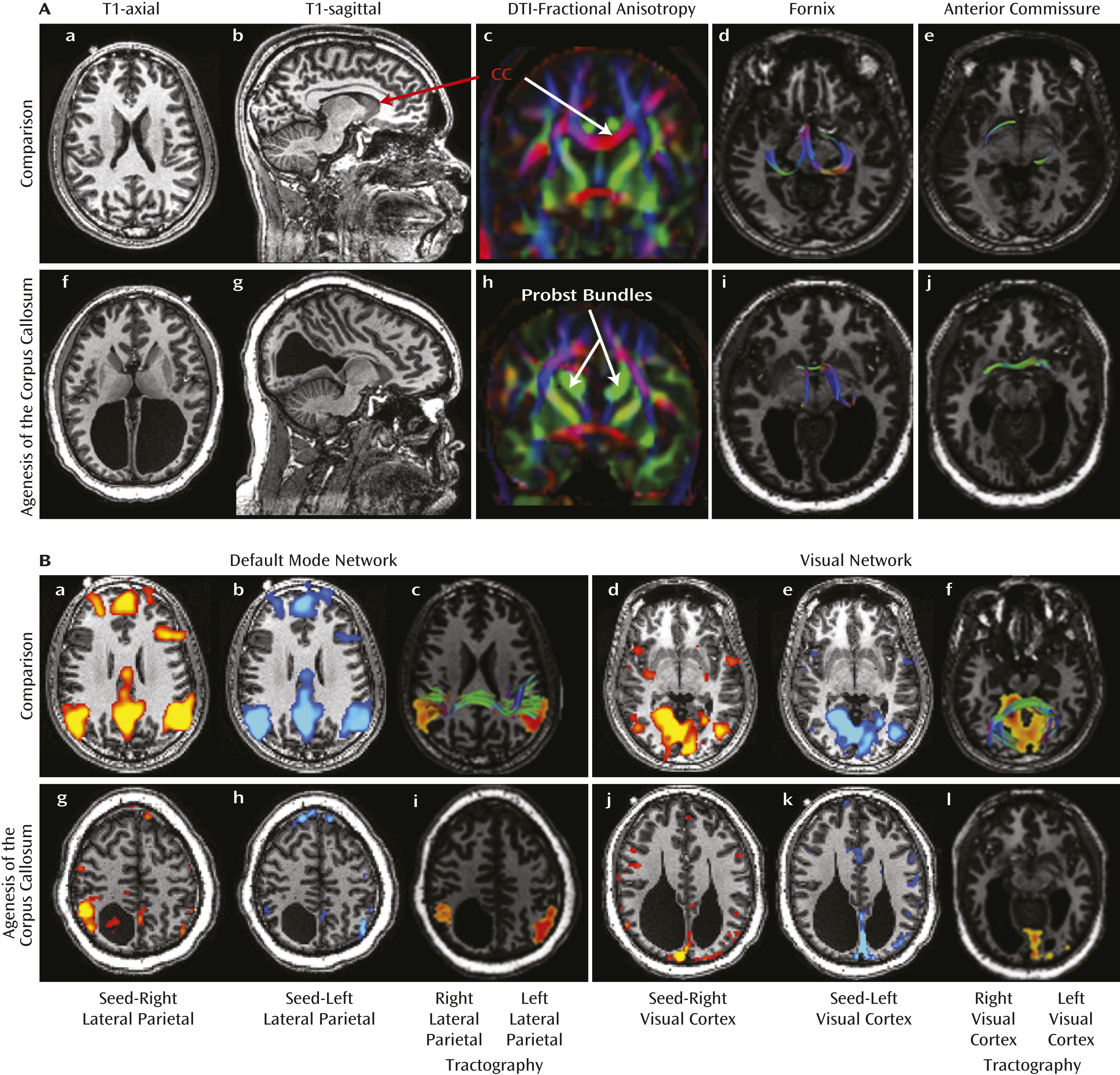
Figure 1A show the absent corpus callosum in the patient and prominent Probst bundles running parallel to the interhemispheric fissure. The fornix, although skewed in orientation, was intact, and the anterior commissure was more prominent in our patient than in the comparison subject.
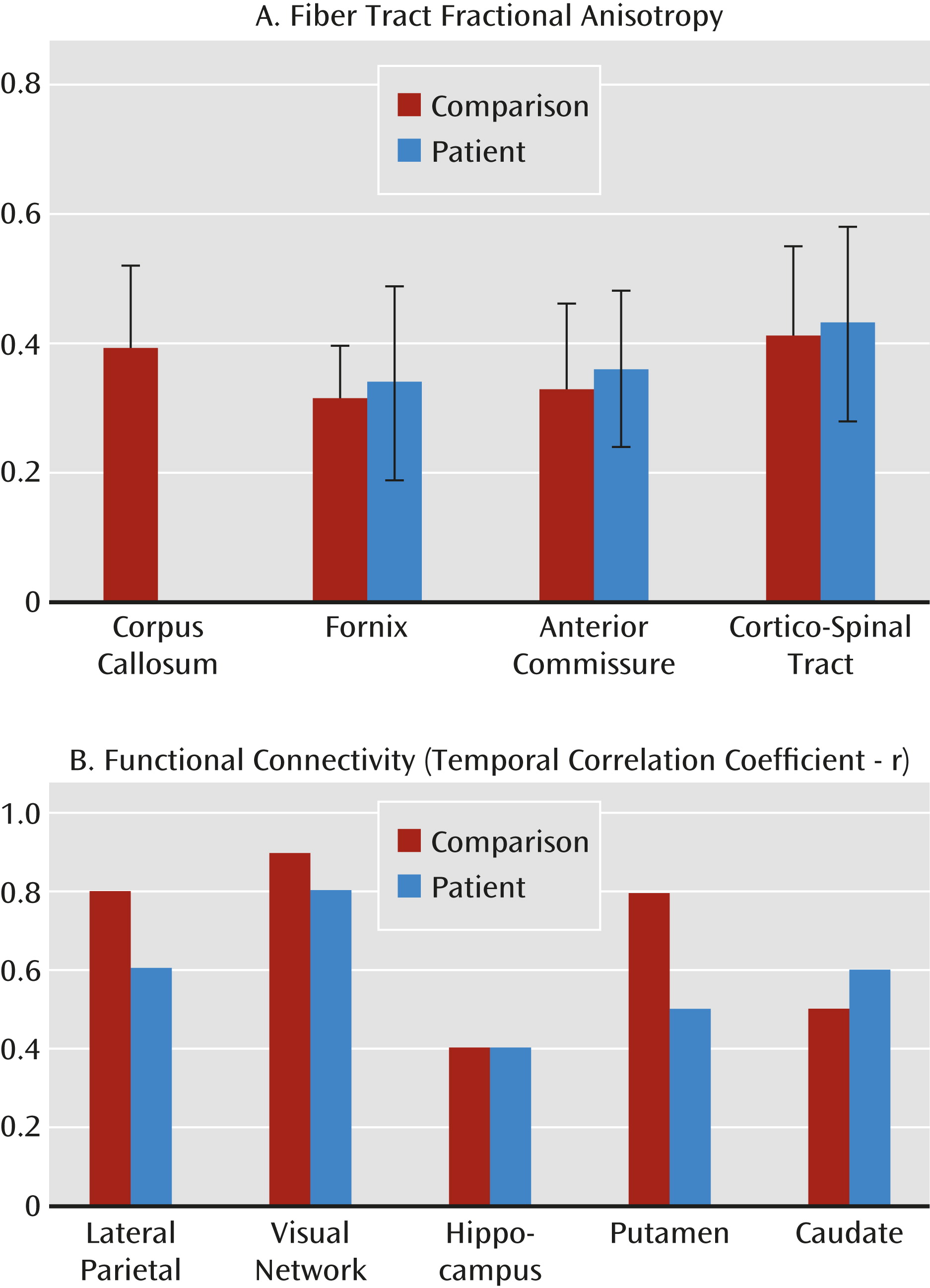
Figure 2A shows that the patient had slightly higher fractional anisotropy values in the fornix and anterior commissure.
We used independent component analysis in FMRIB’s Software Library to identify resting-state activity involving the lateral parietal region in the default mode network and involving the primary visual cortex in the visual network. These functionally defined regions of interest in the lateral parietal and primary visual cortex were then used for two-region DTI tractography using MedINRIA (
3) to evaluate interhemispheric connections via the corpus callosum as well as noncallosal connections via the cortico-spinal tract, fornix, and anterior commissure.
The patient showed dramatically reduced interhemispheric but strong ipsilateral functional connectivity in the default mode network and the primary visual cortex (
Figure 1B). DTI tractography showed that connections between the left and right lateral parietal and primary visual cortex regions, mediated via the corpus callosum, were absent in the patient (panels i and l of
Figure 1B). We calculated correlation coefficients between the left and right brain regions (
Figure 2B) and found reduced temporal synchrony in the lateral parietal and primary visual cortex (connected via the corpus callosum) but normal synchrony in the hippocampus (connected via the fornix and anterior commissure) and the caudate nuclei (possibly from long-term compensatory or robustly developed indirect pathways).
Consistent with the view that functional connectivity demonstrated by resting-state blood-oxygen-level-dependent fMRI is related to anatomical connectivity, we observed highly lateralized and reduced interhemispheric connectivity in regions dependent on an intact corpus callosum in our patient. Nonetheless, functional and structural connectivity across the two brain hemispheres via other interhemispheric fiber tracts were preserved. This case report provides a broad understanding of the morphological and functional modifications that occur in agenesis of the corpus callosum. Altered brain function in a patient with complete agenesis of the corpus callosum was successfully illustrated using fMRI. Furthermore, these changes also conformed to the altered anatomical connectivity that was observed with DTI.


