Full access
New Research
Published Online: 1 December 2013

Drug-Induced Long QT in Adult Psychiatric Inpatients: The 5-Year Cross-Sectional ECG Screening Outcome in Psychiatry Study

Abstract

Routine electrocardiograms of 6,790 patients admitted to a Swiss public psychiatric hospital over 5 years showed the likelihood of a long QT interval increases if a patient taking certain medications has hypokalemia, hepatitis C, HIV, or abnormal T wave morphology.

Abstract

Objective

The authors aimed to determine the prevalence of drug-induced long QT at admission to a public psychiatric hospital and to document the associated factors using a cross-sectional approach.

Method

All ECG recordings over a 5-year period were reviewed for drug-induced long QT (heart-rate corrected QT ≥500 ms and certain or probable drug imputability) and associated conditions. Patients with drug-induced long QT (N=62) were compared with a sample of patients with normal ECG (N=143).

Results

Among 6,790 inpatients, 27.3% had abnormal ECG, 1.6% had long QT, and 0.9% qualified as drug-induced long QT case subjects. Sudden cardiac death was recorded in five patients, and torsade de pointes was recorded in seven other patients. Relative to comparison subjects, patients with drug-induced long QT had significantly higher frequencies of hypokalemia, hepatitis C virus (HCV) infection, HIV infection, and abnormal T wave morphology. Haloperidol, sertindole, clotiapine, phenothiazines, fluoxetine, citalopram (including escitalopram), and methadone were significantly more frequent in patients with drug-induced long QT. After adjustment for hypokalemia, HCV infection, HIV infection, and abnormal T wave morphology, the effects of haloperidol, clotiapine, phenothiazines, and citalopram (including escitalopram) remained statistically significant. Receiver operating characteristic curve analysis based on the number of endorsed factors per patient indicated that 85.5% of drug-induced long QT patients had two or more factors, whereas 81.1% of patients with normal ECG had fewer than two factors.

Conclusions

Drug-induced long QT and arrhythmia propensity substantially increase when specific psychotropic drugs are administered to patients with hypokalemia, abnormal T wave morphology, HCV infection, and HIV infection.
Electrocardiogram (ECG) is the primary tool for detecting long QT syndrome, the most common cause of marketed drug withdrawal (1) and a surrogate marker for the propensity to develop specific ventricular arrhythmias called torsade de pointes. The term was coined in 1966 to describe a malignant polymorphic ventricular tachycardia characterized by a twisting pattern of the QRS complex (2). The probability of torsade de pointes increases with the extent of heart rate-corrected QT (QTc) prolongation, but no threshold has been established below which the risk can be considered absent. Most torsade de pointes episodes are transient and self-limited. The transition probability to sudden death may be distributed between 1% and 17%, a wide range that reflects uncertainty around the estimate (3). In spite of methodological heterogeneity across studies, a recent meta-analysis reported consistent associations between prolonged QT interval and both increased total mortality and sudden cardiac death (4).
QT interval prolongation occurs with noncardiac medications, particularly antipsychotic drugs (5). Epidemiological studies indicate that the propensity of torsade de pointes and fatal outcome increases with the dosage of conventional and atypical antipsychotics (6), as well as antidepressants, mood stabilizers, and methadone (7). In a large study based on Medicaid data, users and nonusers of antipsychotic drugs were followed for the occurrence of sudden cardiac death; users of either conventional or atypical antipsychotics had higher rates of sudden cardiac death than nonusers, with incidence rate ratios of 1.99 and 2.26, respectively (8). Furthermore, administration of QT-prolonging drugs appears more likely to generate ventricular arrhythmias if associated with exacerbating conditions that are commonly observed in hospitalized patients (9). Well-documented risk factors for drug-induced torsade de pointes include older age, female sex, electrolyte abnormalities, renal or hepatic dysfunction, preexisting heart disease (e.g., infarction), bradycardia or rhythms with pauses, treatment with more than one QT-prolonging drug, and genetic predisposition (9, 10).
The ECG Screening Outcome in Psychiatry (ESOP) study reviewed all ECG recordings performed at hospital admission for psychiatric inpatients over a 5-year period. Its objectives were to determine the prevalence of drug-induced long QT and to identify associated factors based on pharmacovigilance data.

Method

Patients and Study Design

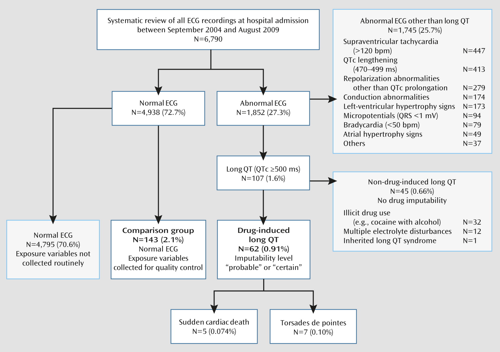
The survey included all patients who had ECG recordings at admission to the public Psychiatric Hospital of Geneva (Department of Mental Health and Psychiatry, University Hospitals of Geneva, Switzerland) between September 2004 and August 2009. Patient inclusion is depicted in Figure 1.
FIGURE 1. Patient Inclusion in a Study of Drug-Induced Long QT in Adult Psychiatric Inpatients
The study was based on data collected as part of a pharmacovigilance program. The characteristics of all patients with drug-induced long QT in the study period (N=62; see drug imputability) were compared with the characteristics of a sample of patients with normal ECG recordings at admission (N=143). The comparison group included patients sequentially admitted to the hospital within three periods (September 18−29, 2006; January 14−25, 2008; and July 6−31, 2009), during which quality control was implemented (i.e., ECG was examined by two psychopharmacologists and two internists in charge of the periodic review of somatic health care in adult psychiatric inpatients).
The study protocol was approved by the ethics committee of the Department of Mental Health and Psychiatry (protocol number 09–180R) and the General College of Physicians, University Hospitals of Geneva. The protocol was in agreement with the Declaration of Helsinki (most recent amendment, October 2008).

ECG Assessment

ECG recording is required at admission to the Geneva psychiatric hospital. All included ECGs were recorded within 24 hours of hospital admission and were screened by a senior physician trained in internal medicine as part of routine practice. Strict procedures were followed to ensure that individuals remained in the supine position for complete QT adaptation for 3 minutes before and throughout the recordings. QT morphology was analyzed on 12-lead electrocardiograms recorded at 25 mm/second speed, 10 mm/mV amplification, and 0.05–35 Hz filter settings with an AT−2 plus C 2.22 Cardiovit ECG recorder (Schiller AG, Baar, Switzerland). QT intervals and heart rate derived from R-R duration were measured electronically and confirmed manually from leads V5 and II. QT length was assessed as follows (11): the intersection of a tangent along the descending limb slope of the T wave with the isoelectric line was used. U-waves with at least 50% of the T wave amplitude were not counted into the QT interval. If a wave following the T wave merged with the end of the T-wave, the position of the notch between the waves was used for measurement. QT interval was corrected for heart rate according to the Bazett and Fridericia correction (QTc) (12). Patients were diagnosed with long QT if one of the two QTc values exceeded 500 ms. This cutoff value was selected because risk stratification indicates substantially higher risk of arrhythmias and sudden cardiac death beyond this value (10). ECG reviewers were blind to patient medication status. Each long QT syndrome was confirmed by an experienced cardiologist (DS).
Because previous studies suggested that morphology of the T wave may be an additional relevant parameter in the evaluation of cardiac safety, we derived a dichotomous variable to describe abnormal T wave morphology based on asymmetry, flatness, and notch (13).

Drug Imputability

Drug imputability of all long QT cases was assessed shortly after ECG recording by three experienced psychopharmacologists (FG, PB, PD). They had access to all data, in keeping with recommendations for pharmacovigilance notification. According to the World Health Organization–Uppsala Monitoring Centre system for standardized case causality assessment (14), all patients in whom long QT was detected were given ratings in one of four categories (unlikely, possible, probable, or certain). Only those patients who met criteria for probable or certain causality were considered as drug-induced long QT case subjects. Each individual case led to a pharmacovigilance report to the Swiss agency for therapeutic products (Swissmedic), which was forwarded to the database center in Uppsala, Sweden. This implies that the event was unlikely attributable to concurrent diseases or other drugs, that a temporal relationship was established between drug administration and effect, and that improvement occurred after drug de-challenge. These conditions were ascertained by two criteria: 1) long QT in the presence of the target drug was preceded by normal ECG in the absence of the drug or 2) long QT in the presence of the target drug was followed by QT normalization after drug withdrawal or dose reduction. All patients in whom drug-induced long QT was detected were followed for possible subsequent events, such as torsade de pointes and sudden death.

Exposure Variables

Sociodemographic and clinical information were extracted from medical records and complemented or confirmed by contacting the patients’ attending psychiatrists. According to routine practice, clinical pharmacologists collect such information immediately after long QT detection for pharmacovigilance purposes (not blind to ECG results). Thus, all data refer to the time of ECG screening at hospital admission. A similar procedure was implemented for patients in the comparison group, but it was not performed routinely for patients with normal ECG, abnormal ECG other than long QT, and non-drug-induced long QT. The following variables were considered: psychiatric diagnosis according to ICD-10 criteria, sex, age, obesity (body mass index >30), hypokalemia (potassium level <3.5 mEq/L), hepatic insufficiency (based on prothrombin level and factor V level), chronic hepatitis B virus (HBV) infection, chronic hepatitis C virus (HCV) infection, human immunodeficiency virus (HIV) infection, cardiovascular risk factors (i.e., hypertension, diabetes, hyperlipidemia, and family history), coronary heart disease, and tobacco smoking. Exposure to medication was carefully reviewed for all participants by two experienced pharmacologists (FG, PB). It should be emphasized that medication usually referred to treatment initiated before admission, as a majority of patients were followed in public outpatient services or in private practice before hospitalization.

Data Analysis

Patients with drug-induced long QT and with normal ECG were compared for binary variables with the Fisher-Boschloo’s exact unconditional test, with Berger-Boos correction (15). The Mann-Whitney U test was used for continuous variables. All drugs administered to at least 10 patients were first tested for a possible association with drug-induced long QT using cross-tabulation, unadjusted odds ratios, and 95% confidence intervals (CIs). Drugs that were significantly associated with drug-induced long QT in univariate analyses were then entered into a multivariable logistic regression model, adjusted for hypokalemia, HCV infection, HIV infection, and abnormal T wave morphology. Adjusted odds ratios and 95% confidence intervals were estimated. Considering the four clinical factors and seven drugs associated with drug-induced long QT (adjusted odds ratio >3), we counted the number of endorsed factors for each individual patient. A receiver operating characteristic curve was built and compared with the one that took only clinical conditions into account. For each possible cutoff, sensitivity (proportion of drug-induced long QT patients at or above the cutoff) and specificity (proportion of normal ECG patients below the cutoff) were calculated. Best cutoff was defined as the one that maximized the sum of sensitivity and specificity. Except for the Fisher-Boschloo test (available at http://www4.stat.ncsu.edu/~boos/exact/), statistics were computed using SPSS, version 17 (SPSS Inc., Chicago). All tests were two-tailed with the significance level set at 0.05.

Results

The study flowchart is illustrated in Figure 1. Of 6,790 ECG recordings, 27.3% were abnormal. Long QT was diagnosed in 107 patients (1.6%), of whom 62 (0.9%) met criteria for drug-induced long QT, with probable or certain drug imputability according to World Health Organization–Uppsala Monitoring Centre criteria.
Sudden death was recorded in five patients and torsade de pointes in seven patients within 72 hours following drug-induced long QT detection, indicating that a time relationship was plausible (Table 1). Although these events were rare (<0.2% of the entire sample with ECG recordings at admission), they represented 19.4% of patients identified with drug-induced long QT. Eleven of these 12 patients had concomitant T wave abnormalities, and seven had received methadone. All torsade de pointes patients recovered after drug de-challenge.
TABLE 1. Characteristics of Patients With Torsade de Pointes (N=7) and Sudden Death (N=5)
 GenderAge (Years)Clinical DataaT Wave MorphologyDrug Treatment
Torsade de pointes     
 Male42HIVAsymmetric, notchedFluvoxamine, 100 mg; clozapine, 300 mg; lopinavir, 800 mg; ritonavir, 200 mg; sertindole, 16 mg; tenofovir, 136 mg; zidovudine, 300 mg
 Male24Hypokalemia, HCVFlat, U wave presentMethadone overdose; clozapine, 100 mg; lorazepam, 1.5 mg; clotiapine, 15 mg
 Male55HypomagnesemiaU wave presentMethadone, 80 mg; escitalopram, 30 mg; olanzapine, 20 mg; ibuprofen, 1,200 mg; acetaminophen, 2 g
 Male56HCV, HIVAsymmetricMethadone, 80 mg; escitalopram, 30 mg; fluoxetine, 20 mg; zuclopenthixol, 40 mg; atazanavir, 400 mg; zidovudine, 600 mg; lamivudine, 300 mg; ritonavir, 200 mg
 Female69HypokalemiaAlternans with nine torsade de pointes episodesHaloperidol, 20 mg; procyclidine, 7.5 mg; clotiapine, 40 mg
 Female43HypocalcemiaAlternans with two torsade de pointes episodesClozapine, 100 mg; haloperidol, 3 mg
 Female36HCV, HIVAsymmetric, flatMethadone, 120 mg; venlafaxine, 225 mg; modafinil, 100 mg; topiramate, 100 mg; abacavir, 600 mg; lamivudine, 150 mg; zidovudine, 300 mg
Sudden death     
 Male42Obesity, cardiomegaly (autopsy)Asymmetric, flat, notchedAmisulpride, 200 mg; clozapine, 300 mg; fluvoxamine, 50 mg; clotiapine, 40 mg; esomeprazole, 20 mg; simvastatin, 20 mg; alprazolam, 0.5 mg; zolpidem, 10 mg
 Male38Cocaine and alcohol abuse Haloperidol, 15 mg; olanzapine, 20 mg; promazine, 100 mg
 Male20HypokalemiaU wave presentMethadone, 20 mg (first dose); olanzapine, 5 mg; clonazepam, 1.5 mg; zolpidem, 10 mg; mirtazapine, 30 mg
 Male55HCV, HIVAlternansMethadone abuse; escitalopram, 30 mg; atazanavir, 400 mg; levomepromazine, 50 mg; zidovudine, 600 mg; lamivudine, 300 mg
 Male25 AlternansMethadone abuse; clozapine, 100 mg (plasma concentration, 1,160 ng/ml, therapeutic range, 350–600 ng/ml); lorazepam, 15 mg
a
HCV=Hepatitis C virus.
Patients with drug-induced long QT did not differ significantly from patients with normal ECG with respect to age, sex, obesity, cardiovascular risk factors, and coronary heart disease (Table 2). In contrast, drug-induced long QT patients had significantly higher frequencies of hypokalemia (19.4% compared with 5.6%, p=0.004), HCV infection (41.9% compared with 9.8%, p=0.001), HIV infection (24.2% compared with 6.3%, p=0.001), and abnormal T wave morphology (35.5% compared with 15.4%, p=0.003).
TABLE 2. Characteristics of Patients With and Without Drug-Induced Long QT at Admission to the Psychiatric Hospital
CharacteristicDrug-Induced Long QT (N=62)Normal ECG (N=143)Analysisa
 N%N%p
Female3658.16948.30.22
Obesity (body mass index ≥30)b1626.22316.40.10
Hypokalemia (potassium <3.5 mEq/L)1219.485.60.004
Hepatitis C virus infection2641.9149.80.001
Hepatitis B virus infection00.021.41
HIV infection1524.296.30.001
Cardiovascular risk factors (>2)46.51711.90.27
Coronary heart disease23.274.90.63
Abnormal T wave morphology2235.52215.40.003
ICD–10 psychiatric diagnoses     
 Disorders due to psychoactive substance use2946.84229.40.023
 Schizophrenia, schizotypal, and delusional disorders1930.62819.60.09
 Mood disorders1930.65941.30.15
 MedianRangeMedianRangep
Age (years)4020–744118–660.96
RR interval (ms)791450–1,600739485–13840.026
QT interval (ms)463340–680360308–492not tested
QTc (ms, Bazett's formula)510474–647421357–499not tested
QTc (ms, Fridericia’s formula)496444–616399357–494not tested
a
Fisher-Boschloo’s exact unconditional test with Berger-Boos correction for binary variables; Mann-Whitney U test for other variables.
b
Drug-induced long QT, N=61; normal ECG, N=140.
The association between prolonged QTc interval and drugs prescribed to at least 10 patients was first investigated with univariate statistics (Table 3). The antipsychotics clotiapine (a dibenzothiazepine, not marketed in the United States), haloperidol, phenothiazines (promazine and levomepromazine), and sertindole were significantly more frequent among drug-induced long QT patients, but no difference was observed for aripiprazole, clozapine, olanzapine, quetiapine, and risperidone (including paliperidone). The serotonin-selective reuptake inhibitors citalopram (including escitalopram) and fluoxetine were significantly associated with drug-induced long QT, unlike trazodone and venlafaxine. Prolonged QTc was significantly associated with methadone, which was the most frequent drug among patients with drug-induced long QT (37.1%). A significant inverse association was found for flurazepam, which was less frequent in the prolonged QTc sample than in the comparison group. Other benzodiazepines and drugs such as zolpidem, lamotrigine, valproic acid, biperiden, and omeprazole were not associated with drug-induced long QT.
TABLE 3. Univariate and Multivariate Analysis of Drugs Associated With Long QT at Admission to the Psychiatric Hospital
DrugaDrug-Induced Long QT (N=62)Normal ECG (N=143)AnalysisRisk of Torsade de Pointesb
 N%N%Unadjusted Odds Ratio95% CIAdjusted Odds Ratioc95% CI 
Antipsychotics         
 Aripiprazole34.874.90.990.25–3.95   
 Clotiapine914.564.23.881.32–11.45.011.16–21.6 
 Clozapine711.32114.70.740.30–1.84  Possible
 Haloperidol1016.164.24.391.52–12.75.161.38–19.3Yes
 Olanzapine1016.11711.91.430.61–3.32   
 Promazine / levomepromazine1930.6128.44.822.17–10.76.742.34–19.4 
 Quetiapine1016.13323.10.640.29–1.40  Possible
 Risperidone / paliperidone1422.61913.31.900.88–4.10  Possible
 Sertindole711.321.48.971.81–44.55.240.88–31.4Possible
Antidepressants         
 Citalopram / escitalopram1524.21510.52.721.24–6.004.381.45–13.3Yes / possible
 Fluoxetine914.542.85.901.74–20.05.550.92–33.4Conditional
 Trazodone11.6128.40.180.02–1.41  Conditional
 Venlafaxine34.8139.10.510.14–1.85  Possible
Mood stabilizers         
 Lamotrigine11.6149.80.150.02–1.18   
 Valproic acid23.285.60.560.12–2.73   
Sedative drugs and tranquilizers         
 Clonazepam69.7128.41.170.42–3.27   
 Flurazepam11.61711.90.120.02–0.93   
 Lorazepam1219.42718.91.030.48–2.20   
 Oxazepam69.72920.30.420.17–1.07   
 Zolpidem69.71611.20.850.32–2.29   
Other drugs         
 Biperiden11.6117.70.200.03–1.56   
 Methadone2337.1128.46.442.94–14.13.110.98–9.85Yes
 Omeprazole914.52114.70.990.42–2.30   
a
All drugs administered to at least 10 patients are listed, except for sertindole (N=9).
b
Arizona Center for Education and Research on Therapeutics (http://www.azcert.org/).
c
Logistic regression model, adjusted for hypokalemia, hepatitis C virus infection, HIV infection, and abnormal T wave morphology.
In a multivariate model adjusted for hypokalemia (odds ratio=6.5, p=0.005), HCV infection (odds ratio=4.4, p=0.009), HIV infection (odds ratio=3.9, p=0.036), and abnormal T wave morphology (odds ratio=4.0, p=0.004), the drugs haloperidol, clotiapine, phenothiazines, and citalopram (including escitalopram) remained significantly associated with drug-induced long QT, whereas sertindole, fluoxetine, and methadone did not, despite adjusted odds ratios above 3 (p<0.10; Table 3). The correlation matrix of estimated coefficients did not reveal any multicollinearity problem, despite expected associations between some predictors (e.g., HCV infection, HIV, and methadone prescription). No other drug was significantly associated with long QT when added into the multivariate model.
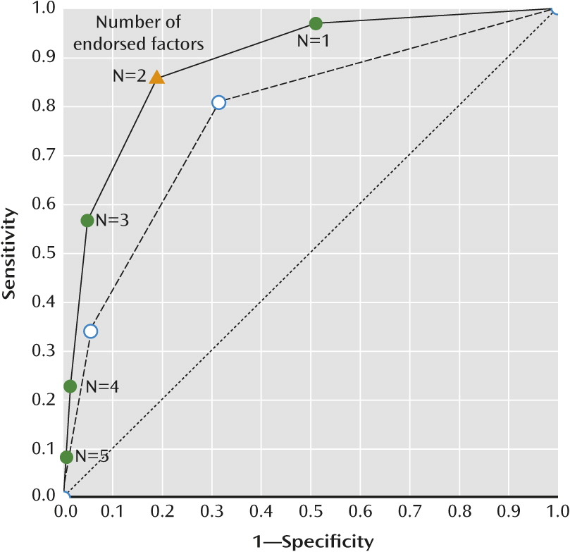
Considering the four clinical factors (hypokalemia, HCV infection, HIV, and abnormal T wave morphology) and the seven drugs associated with drug-induced long QT (clotiapine, haloperidol, phenothiazines, sertindole, citalopram, fluoxetine, and methadone), we counted the number of factors endorsed by each individual patient (median=1, range=0–5 in normal ECG patients; median=3, range=0–5 in long QT patients). As illustrated in Figure 2, the area under the receiver operating characteristic curve was 0.89 (95% CI=0.84–0.94) when taking into account clinical conditions and medication and 0.78 (95% CI=0.71–0.84) when only clinical conditions were counted (p<0.001). Whereas 81.1% of patients with normal ECG endorsed fewer than two factors (specificity), 85.5% of patients with drug-induced long QT endorsed two or more factors (sensitivity).
FIGURE 2. Receiver Operating Characteristic Curves for Distinguishing Between Patients With Drug-Induced Long QT and Normal ECGa
a Curves are built as a function of the number of endorsed factors among four clinical conditions (hypokalemia, hepatitis C virus infection, HIV, and abnormal T wave morphology) and seven drugs (clotiapine, haloperidol, phenothiazines, sertindole, citalopram, fluoxetine, and methadone). Area under the curve was 0.78 (95% CI=0.71–0.84) when only clinical conditions were counted (dashed line, open symbols) and 0.89 (95% CI=0.84–0.94) when taking into account clinical conditions and medication (solid line, closed symbols). The cutoff value (triangle) that maximized the sum of sensitivity and specificity was two factors, with 85.5% sensitivity (drug-induced long QT patients with two or more factors) and 81.1% specificity (normal ECG patients with fewer than two factors).

Discussion

In this 5-year study, 6,790 patients had a routine ECG at admission to the psychiatric hospital. Prevalence of drug-induced long QT (QTc >500 ms and probable or certain drug imputability) was 0.9%, in agreement with previous studies that reported prevalence rates between 1.2% and 2.6% (1618). Almost 20% of patients in whom drug-induced long QT was detected were subsequently documented for torsade de pointes (N=7) and sudden death (N=5). Because seven of these 12 patients were prescribed methadone for opioid substitution, a possible role of illicit drug use cannot be ruled out. Nevertheless, this observation emphasizes that the detection of a QTc interval >500 ms at admission to the psychiatric hospital should lead to prompt action, including discontinuation of the offending drug unless the benefits of treatment clearly outweigh the risk, correction of electrolyte disturbances, and careful cardiac monitoring (19).
The ESOP study indicated significant associations between drug-induced long QT and clinical conditions, such as hypokalemia, HIV, and HCV infection, and concomitant ECG features, such as morphological T wave abnormalities. Hypokalemia is the most common electrolyte abnormality in clinical practice, with potassium values <3.6 mmol/L in over 20% of inpatients (20). It is a well-known risk factor for torsade de pointes (9).
In HIV-infected individuals, high viral load and low CD4+ cell count (cutoff values, 17,900 copies/mL and 144 cells/mm3, respectively) were associated with QT lengthening (21). Autonomic neuropathy was reported as a possible explanation (22). Another mechanism might be through the HIV Tat protein, which inhibits human ether-a-go-go-related gene (hERG) potassium channels (23). A large cross-sectional study in HIV-infected patients revealed no significant effect of anti-HIV medication on QTc (24).
HCV infection frequently causes impaired liver function, which is a documented risk factor for long QT because of the increased likelihood of drug-drug interactions. HCV infection nearly doubled the propensity of QTc ≥470 ms in patients with concurrent HIV infection (29.6% compared with 15.8%) (25). Furthermore, HCV-triggered liver-kidney-microsomal type 1 antibodies (LKM−1) are associated with reduced CYP2D6 activity (26), a crucial metabolic pathway for many antipsychotics and antidepressants. The effects of HIV and HCV infections most likely encompass indirect effects as well, through uncontrolled variables such as injection drug use and risk behaviors.
In addition to QTc interval, T wave morphology descriptors are valuable covariates in cardiac safety trials. Basic (peak amplitude and area under the curve) and advanced descriptors (notch, asymmetry, and flatness) are sensitive to drug-induced alterations (27, 28). T-U wave distortion, T wave alternans, and new-onset ventricular ectopies are further ECG features promoting arrhythmias by increasing the heterogeneity of repolarization across the myocardial wall (13). A composite score of T wave morphology, either alone or in combination with the QT interval, allowed efficient discrimination between normal and abnormal repolarization (29).
The ESOP study examined 23 drugs for a possible association with drug-induced long QT. Seven drugs were associated with QTc >500 ms (odds ratio >3), of which five had been previously documented for conditional, possible, or actual risk of torsade de pointes and two showed structural and pharmacological similarities to QT-prolonging drugs (Table 3).
A previous study in methadone maintenance patients indicated that QT lengthening was dosage-dependent and increased with concomitant administration of CYP3A4 inhibitors, hypokalemia, and impaired liver function (11). A modeling approach using pooled data from five clinical trials supported the need for ECG monitoring and arrhythmia risk assessment in patients receiving methadone dosages greater than 120 mg/day (30). Substitution of racemic methadone with the R-methadone enantiomer, which mainly mediates the opioid effect, was associated with reduced QTc interval and thus safer cardiac profile (31).
The antipsychotic haloperidol is associated with an increased risk of torsade de pointes, particularly for intravenous administration and at high dosage (32). As far as we know, promazine and levomepromazine have not been associated with QT prolongation. However, these phenothiazines have properties similar to chlorpromazine and thioridazine, for which cases of ventricular arrhythmias and torsade de pointes have been reported (33). Thus, similar caution may apply for these different drugs. To our knowledge, QT prolongation had not been documented for clotiapine, an atypical antipsychotic agent with a dibenzothiapine structure and properties similar to those of phenothiazines. The present study suggests that risk of drug-induced long QT with clotiapine might be of the same order of magnitude as for phenothiazines and haloperidol. Sertindole is associated with a possible risk of torsade de pointes. However, a multicenter postmarketing surveillance study indicated that QT interval prolongation might not translate into increased overall mortality (34).
The antidepressants citalopram and escitalopram (its S-enantiomer) increase QTc in a dose-dependent manner at therapeutic dosages (35), leading to a safety announcement by the U.S. Food and Drug Administration, updated in March 2012 (http://www.fda.gov/Drugs/DrugSafety/ucm297391.htm). Fluoxetine has been associated with QTc prolongation and torsade de pointes in case reports (36). It is a potent inhibitor of CYP2D6, and pharmacokinetic interactions may result in a synergistic effect on QTc prolongation when fluoxetine is taken with QT-prolonging CYP2D6 substrates.
Exploratory multivariable analysis suggested that all four clinical conditions (hypokalemia, HCV infection, HIV, and abnormal T wave morphology) and seven drugs (clotiapine, haloperidol, phenothiazines, sertindole, citalopram, fluoxetine, and methadone) may independently contribute to drug-induced long QT. The analysis emphasized that drug-induced long QT may depend on the co-occurrence of several risk factors. Indeed, we observed that more than 85% of patients with drug-induced long QT endorsed two or more factors, as compared with less than 20% in the comparison group. This observation is in line with a review of 249 cases of torsade de pointes as a result of noncardiac medication (37).
Strengths of the ESOP study include a systematic review of a large number of ECG recordings at admission to the psychiatric hospital over a 5-year period. Long QT is a rare event and such a survey allowed us to identify a relatively large sample that had been thoroughly documented as part of an ongoing pharmacovigilance program.
Nonetheless, some limitations need to be acknowledged. First, cross-sectional design provides no information about the temporal relationship between exposure and outcome and does not allow risk estimation and further causality assessment. Second, the comparison sample may not have been representative of all patients admitted in the 2004–2009 period. A selection bias cannot be excluded, due to medication withdrawal from the market (thioridazine in 2005), drug reintroduction (sertindole in 2006), increased use of generic drugs (e.g., citalopram, clozapine, and lithium salts), and evolving prescription patterns. A third limitation is sample size. The lack of routinely collected reliable data did not allow us to include the entire population with ECG results at hospital admission or to select a larger random sample of patients with normal ECG. Furthermore, the small numbers of patients taking some drugs resulted in limited statistical power to detect their possible associations with long QT and uncertainty in estimating effect size, as reflected by wide confidence intervals in Table 3. Sample size also prevented investigating possible dosage-related effects and interactions between different factors. Fourth, associations based on data collected in hospital settings may not reflect associations in the general population because of admission rate bias, sometimes called Berkson’s fallacy (38). This bias would occur if exposure (e.g., psychotropic medication) was associated with hospitalization rate on its own right, which cannot be fully excluded. Using QTc >500 ms as a single outcome variable may be considered a fifth limitation. Although an absolute threshold has not been established, a cutoff at 500 ms is widely accepted as indicative of a high risk for arrhythmic events. Finally, it should be emphasized that the complex interplay between the different factors responsible for QT prolongation deserves further investigation in large samples. Whereas some degree of collinearity between the investigated factors was possible in the present study, additional risk factors (e.g., hypomagnesemia) or possible confounders (e.g., illicit drug use) were not taken into account. In addition, some psychotropic drugs were not investigated for QT prolongation because they were rarely prescribed (e.g., amisulpride) or unavailable in Switzerland (e.g., ziprasidone).
The American Heart Association and the American College of Cardiology Foundation published a scientific statement in order to raise awareness about risk factors, ECG monitoring, and managing drug-induced long QT in hospital settings (9). Incidence of sudden cardiac death in patients taking antipsychotic medication is about 2.9 events per 1,000 patient-years (39), as compared with 0.2 events per 1,000 patient-years for death from clozapine-induced agranulocytosis (40). Yet no formal risk-management program has been established to detect prolonged QT at the psychiatric hospital, in contrast with white blood cell count monitoring for clozapine prescription. While the ESOP study speaks in favor of ECG surveillance in psychiatric inpatients, in particular when several QT-prolonging drugs and clinical conditions are identified, the cost-utility of a formal detection program deserves further analysis.
Acknowledgments

The authors thank Dr. Pierre Schulz for his role in the initial implementation of the project, Anne-Lise Balsiger for the standardized ECG recordings, Victoria Rollason, Ph.D., for supervising the pharmacovigilance recording process, and Drs. Katalin Delessert and Anne Zaninetti for their medical assistance.

References

1.
Roden DM: Drug-induced prolongation of the QT interval. N Engl J Med 2004; 350:1013–1022
2.
Dessertenne F: [Ventricular tachycardia with 2 variable opposing foci]. Arch Mal Coeur Vaiss 1966; 59:263–272 (French)
3.
Salle P, Rey JL, Bernasconi P, Quiret JC, Lombaert M: [Torsades de pointe: apropos of 60 cases]. Ann Cardiol Angeiol (Paris) 1985; 34:381–388 (French)
4.
Zhang Y, Post WS, Blasco-Colmenares E, Dalal D, Tomaselli GF, Guallar E: Electrocardiographic QT interval and mortality: a meta-analysis. Epidemiology 2011; 22:660–670
5.
Glassman AH, Bigger JT: Antipsychotic drugs: prolonged QTc interval, torsade de pointes, and sudden death. Am J Psychiatry 2001; 158:1774–1782
6.
Wang PS, Schneeweiss S, Avorn J, Fischer MA, Mogun H, Solomon DH, Brookhart MA: Risk of death in elderly users of conventional vs atypical antipsychotic medications. N Engl J Med 2005; 353:2335–2341
7.
Piguet V, Desmeules J, Ehret G, Stoller R, Dayer P: QT interval prolongation in patients on methadone with concomitant drugs. J Clin Psychopharmacol 2004; 24:446–448
8.
Ray WA, Chung CP, Murray KT, Hall K, Stein CM: Atypical antipsychotic drugs and the risk of sudden cardiac death. N Engl J Med 2009; 360:225–235
9.
Drew BJ, Ackerman MJ, Funk M, Gibler WB, Kligfield P, Menon V, Philippides GJ, Roden DM, Zareba W; American Heart Association Acute Cardiac Care Committee of the Council on Clinical Cardiology, the Council on Cardiovascular Nursing, and the American College of Cardiology Foundation: Prevention of torsade de pointes in hospital settings: a scientific statement from the American Heart Association and the American College of Cardiology Foundation. Circulation 2010; 121:1047–1060
10.
Priori SG, Schwartz PJ, Napolitano C, Bloise R, Ronchetti E, Grillo M, Vicentini A, Spazzolini C, Nastoli J, Bottelli G, Folli R, Cappelletti D: Risk stratification in the long-QT syndrome. N Engl J Med 2003; 348:1866–1874
11.
Ehret GB, Voide C, Gex-Fabry M, Chabert J, Shah D, Broers B, Piguet V, Musset T, Gaspoz JM, Perrier A, Dayer P, Desmeules JA: Drug-induced long QT syndrome in injection drug users receiving methadone: high frequency in hospitalized patients and risk factors. Arch Intern Med 2006; 166:1280–1287
12.
Chiladakis J, Kalogeropoulos A, Arvanitis P, Koutsogiannis N, Zagli F, Alexopoulos D: Heart rate-dependence of QTc intervals assessed by different correction methods in patients with normal or prolonged repolarization. Pacing Clin Electrophysiol 2010; 33:553–560
13.
Graff C, Struijk JJ, Matz J, Kanters JK, Andersen MP, Nielsen J, Toft E: Covariate analysis of QTc and T-wave morphology: new possibilities in the evaluation of drugs that affect cardiac repolarization. Clin Pharmacol Ther 2010; 88:88–94
14.
Meyboom RH, Hekster YA, Egberts AC, Gribnau FW, Edwards IR: Causal or casual? the role of causality assessment in pharmacovigilance. Drug Saf 1997; 17:374–389
15.
Lydersen S, Fagerland MW, Laake P: Recommended tests for association in 2 x 2 tables. Stat Med 2009; 28:1159–1175
16.
Sadanaga T, Sadanaga F, Yao H, Fujishima M: Abnormal QT prolongation and psychotropic drug therapy in psychiatric patients: significance of bradycardia-dependent QT prolongation. J Electrocardiol 2004; 37:267–273
17.
Ramos-Ríos R, Arrojo-Romero M, Paz-Silva E, Carballal-Calvo F, Bouzón-Barreiro JL, Seoane-Prado J, Codesido-Barcala R, Crespí-Armenteros A, Fernández-Pérez R, López-Moríñigo JD, Tortajada-Bonaselt I, Diaz FJ, de Leon J: QTc interval in a sample of long-term schizophrenia inpatients. Schizophr Res 2010; 116:35–43
18.
Pasquier M, Pantet O, Hugli O, Pruvot E, Buclin T, Waeber G, Aujesky D: Prevalence and determinants of QT interval prolongation in medical inpatients. Intern Med J 2012; 42:933–940
19.
Nielsen J, Graff C, Kanters JK, Toft E, Taylor D, Meyer JM: Assessing QT interval prolongation and its associated risks with antipsychotics. CNS Drugs 2011; 25:473–490
20.
Paice BJ, Paterson KR, Onyanga-Omara F, Donnelly T, Gray JM, Lawson DH: Record linkage study of hypokalaemia in hospitalized patients. Postgrad Med J 1986; 62:187–191
21.
Qaqa AY, Shaaban H, DeBari VA, Phung S, Slim J, Costeas CA, Perez G, Shamoon FE: Viral load and CD4+ cell count as risk factors for prolonged QT interval in HIV-infected subjects: a cohort-nested case-control study in an outpatient population. Cardiology 2010; 117:105–111
22.
Villa A, Foresti V, Confalonieri F: Autonomic neuropathy and prolongation of QT interval in human immunodeficiency virus infection. Clin Auton Res 1995; 5:48–52
23.
Bai YL, Liu HB, Sun B, Zhang Y, Li Q, Hu CW, Zhu JX, Gong DM, Teng X, Zhang Q, Yang BF, Dong DL: HIV Tat protein inhibits hERG K+ channels: a potential mechanism of HIV infection induced LQTs. J Mol Cell Cardiol 2011; 51:876–880
24.
Reinsch N, Buhr C, Krings P, Kaelsch H, Neuhaus K, Wieneke H, Erbel R, Neumann T; German Heart Failure Network: Prevalence and risk factors of prolonged QTc interval in HIV-infected patients: results of the HIV-HEART study. HIV Clin Trials 2009; 10:261–268
25.
Nordin C, Kohli A, Beca S, Zaharia V, Grant T, Leider J, Marantz P: Importance of hepatitis C coinfection in the development of QT prolongation in HIV-infected patients. J Electrocardiol 2006; 39:199–205
26.
Girardin F, Daali Y, Gex-Fabry M, Rebsamen M, Roux-Lombard P, Bihl F, Cerny A, Binek J, Moradpour D, Negro F, Desmeules JA: Liver kidney microsomal type 1 antibodies reduce the CYP2D6 activity in patients with chronic hepatitis C virus infection. J Viral Hepat 2012; 19:568–573
27.
Couderc JP, Vaglio M, Xia X, McNitt S, Wicker P, Sarapa N, Moss AJ, Zareba W: Impaired T-amplitude adaptation to heart rate characterizes I(Kr) inhibition in the congenital and acquired forms of the long QT syndrome. J Cardiovasc Electrophysiol 2007; 18:1299–1305
28.
Andersen MP, Xue JQ, Graff C, Kanters JK, Toft E, Struijk JJ: New descriptors of T-wave morphology are independent of heart rate. J Electrocardiol 2008; 41:557–561
29.
Shakibfar S, Graff C, Ehlers LH, Toft E, Kanters JK, Struijk JJ: Assessing common classification methods for the identification of abnormal repolarization using indicators of T-wave morphology and QT interval. Comput Biol Med 2012; 42:485–491
30.
Florian J, Garnett CE, Nallani SC, Rappaport BA, Throckmorton DC: A modeling and simulation approach to characterize methadone QT prolongation using pooled data from five clinical trials in MMT patients. Clin Pharmacol Ther 2012; 91:666–672
31.
Ansermot N, Albayrak O, Schläpfer J, Crettol S, Croquette-Krokar M, Bourquin M, Déglon JJ, Faouzi M, Scherbaum N, Eap CB: Substitution of (R,S)-methadone by (R)-methadone: impact on QTc interval. Arch Intern Med 2010; 170:529–536
32.
Sharma ND, Rosman HS, Padhi ID, Tisdale JE: Torsades de pointes associated with intravenous haloperidol in critically ill patients. Am J Cardiol 1998; 81:238–240
33.
Hoehns JD, Stanford RH, Geraets DR, Skelly KS, Lee HC, Gaul BL: Torsades de pointes associated with chlorpromazine: case report and review of associated ventricular arrhythmias. Pharmacotherapy 2001; 21:871–883
34.
Kasper S, Möller HJ, Hale A: The European post-marketing observational sertindole study: an investigation of the safety of antipsychotic drug treatment. Eur Arch Psychiatry Clin Neurosci 2010; 260:59–68
35.
de Gregorio C, Morabito G, Cerrito M, Dattilo G, Oreto G: Citalopram-induced long QT syndrome and torsade de pointes: role for concomitant therapy and disease. Int J Cardiol 2011; 148:226–228
36.
Wilting I, Smals OM, Holwerda NJ, Meyboom RH, de Bruin ML, Egberts TC: QTc prolongation and torsades de pointes in an elderly woman taking fluoxetine. Am J Psychiatry 2006; 163:325
37.
Zeltser D, Justo D, Halkin A, Prokhorov V, Heller K, Viskin S: Torsade de pointes due to noncardiac drugs: most patients have easily identifiable risk factors. Medicine (Baltimore) 2003; 82:282–290
38.
Brown GW: Berkson fallacy revisited: spurious conclusions from patient surveys. Am J Dis Child 1976; 130:56–60
39.
Schneeweiss S, Avorn J: Antipsychotic agents and sudden cardiac death: how should we manage the risk? N Engl J Med 2009; 360:294–296
40.
Alvir JM, Lieberman JA, Safferman AZ, Schwimmer JL, Schaaf JA: Clozapine-induced agranulocytosis: incidence and risk factors in the United States. N Engl J Med 1993; 329:162–167

Information & Authors

Information

Published In

Go to American Journal of Psychiatry
American Journal of Psychiatry
Pages: 1468 - 1476
PubMed: 24306340

History

Received: 30 June 2012
Revision received: 30 November 2012
Revision received: 19 March 2013
Accepted: 17 May 2013
Published online: 1 December 2013
Published in print: December 2013

Authors

Details

François R. Girardin, M.D., M.Sc.
From the Division of Clinical Pharmacology and Toxicology, the Medical and Quality Directorate, the Department of Community Medicine, Primary Care and Emergency Medicine, the Department of Mental Health and Psychiatry, and the Division of Cardiology, University Hospitals of Geneva and Geneva University, Geneva, Switzerland.
Marianne Gex-Fabry, Ph.D.
From the Division of Clinical Pharmacology and Toxicology, the Medical and Quality Directorate, the Department of Community Medicine, Primary Care and Emergency Medicine, the Department of Mental Health and Psychiatry, and the Division of Cardiology, University Hospitals of Geneva and Geneva University, Geneva, Switzerland.
Patricia Berney, M.D.
From the Division of Clinical Pharmacology and Toxicology, the Medical and Quality Directorate, the Department of Community Medicine, Primary Care and Emergency Medicine, the Department of Mental Health and Psychiatry, and the Division of Cardiology, University Hospitals of Geneva and Geneva University, Geneva, Switzerland.
Dipen Shah, M.D.
From the Division of Clinical Pharmacology and Toxicology, the Medical and Quality Directorate, the Department of Community Medicine, Primary Care and Emergency Medicine, the Department of Mental Health and Psychiatry, and the Division of Cardiology, University Hospitals of Geneva and Geneva University, Geneva, Switzerland.
Jean-Michel Gaspoz, M.D., M.Sc.
From the Division of Clinical Pharmacology and Toxicology, the Medical and Quality Directorate, the Department of Community Medicine, Primary Care and Emergency Medicine, the Department of Mental Health and Psychiatry, and the Division of Cardiology, University Hospitals of Geneva and Geneva University, Geneva, Switzerland.
Pierre Dayer, M.D.
From the Division of Clinical Pharmacology and Toxicology, the Medical and Quality Directorate, the Department of Community Medicine, Primary Care and Emergency Medicine, the Department of Mental Health and Psychiatry, and the Division of Cardiology, University Hospitals of Geneva and Geneva University, Geneva, Switzerland.

Notes

Address correspondence to Dr. Girardin ([email protected]).
Presented in part at the Swiss-Western Annual Meeting of Psychiatry, Geneva, Switzerland, June 14th, 2012.

Funding Information

Dr. Shah has received research grants, speakers honoraria, consultant fees, stock options, or scientific advisory board honoraria from Biosense Webster, Biotronik, Endosense, and St. Jude. The other authors report no financial relationships with commercial interests.The ECG Screening Outcome in Psychiatry (ESOP) study was supported by the Department of Anesthesiology, Intensive Care, and Clinical Pharmacology and Toxicology (University Hospitals of Geneva). Some inpatients were included in the Swiss Hepatitis C Cohort Study funded by the Swiss National Science Foundation (3347C0-108782/1), the Swiss Federal Office for Education and Sciences (03.0599), and the European Commission (LSHM-CT-2004-503359; VIRGIL Network of Excellence on Antiviral Drug Resistance).

Metrics & Citations

Metrics

Citations

Export Citations

If you have the appropriate software installed, you can download article citation data to the citation manager of your choice. Simply select your manager software from the list below and click Download.

For more information or tips please see 'Downloading to a citation manager' in the Help menu.

Format
Citation style
Style
Copy to clipboard

View Options

View options

PDF/EPUB

View PDF/EPUB

Get Access

Login options

Already a subscriber? Access your subscription through your login credentials or your institution for full access to this article.

Personal login Institutional Login Open Athens login
Purchase Options

Purchase this article to access the full text.

PPV Articles - American Journal of Psychiatry

PPV Articles - American Journal of Psychiatry

Not a subscriber?

Subscribe Now / Learn More

PsychiatryOnline subscription options offer access to the DSM-5-TR® library, books, journals, CME, and patient resources. This all-in-one virtual library provides psychiatrists and mental health professionals with key resources for diagnosis, treatment, research, and professional development.

Need more help? PsychiatryOnline Customer Service may be reached by emailing [email protected] or by calling 800-368-5777 (in the U.S.) or 703-907-7322 (outside the U.S.).

Media

Figures

Other

Tables

Share

Share

Share article link

Share