Abstract

For severely and chronically irritable children, frustration during a feedback game revealed striking deactivation in the brain regions associated with spatial attention, reward processing, and emotion. Severe mood dysregulation in children reduces attention flexibility, which may contribute to insufficient emotional regulation.

Abstract

Objective

Irritability is common in children and adolescents and is the cardinal symptom of disruptive mood dysregulation disorder, a new DSM-5 disorder, yet its neural correlates remain largely unexplored. The authors conducted a functional MRI study to examine neural responses to frustration in children with severe mood dysregulation.

Method

The authors compared emotional responses, behavior, and neural activity between 19 severely irritable children (operationalized using criteria for severe mood dysregulation) and 23 healthy comparison children during a cued-attention task completed under nonfrustrating and frustrating conditions.

Results

Children in both the severe mood dysregulation and the healthy comparison groups reported increased frustration and exhibited decreased ability to shift spatial attention during the frustration condition relative to the nonfrustration condition. However, these effects of frustration were more marked in the severe mood dysregulation group than in the comparison group. During the frustration condition, participants in the severe mood dysregulation group exhibited deactivation of the left amygdala, the left and right striatum, the parietal cortex, and the posterior cingulate on negative feedback trials, relative to the comparison group (i.e., between-group effect) and to the severe mood dysregulation group’s responses on positive feedback trials (i.e., within-group effect). In contrast, neural response to positive feedback during the frustration condition did not differ between groups.

Conclusions

In response to negative feedback received in the context of frustration, children with severe, chronic irritability showed abnormally reduced activation in regions implicated in emotion, attention, and reward processing. Frustration appears to reduce attention flexibility, particularly in severely irritable children, which may contribute to emotion regulation deficits in this population. Further research is needed to relate these findings to irritability specifically, rather than to other clinical features of severe mood dysregulation.
Irritability can be defined as a low threshold for experiencing negative affect in response to frustration, with frustration being the emotional response to blocked goal attainment (1, 2). Irritability is both common and impairing in child and adult psychiatric disorders (3), and its importance is recognized in DSM-5 with the adoption of disruptive mood dysregulation disorder, a disorder whose defining feature is excessive and impairing irritability. Given the importance and pervasiveness of irritability, it is notable that few neuroimaging studies have been conducted to examine its pathophysiology. Since irritability can be viewed as a decreased threshold for experiencing frustration, one approach to studying its pathophysiology is by evoking frustration in the scanner. In this study, we compared the neural correlates of frustration in healthy children and children with chronic and impairing irritability.
Frustration is typically induced in the laboratory by using paradigms that increase task difficulty or deceive participants into believing that because their performance is substandard, they cannot earn a reward (410). The few functional MRI (fMRI) studies that have investigated the circuitry mediating frustration have implicated the amygdala, parietal attentional networks, and the dorsal and ventromedial prefrontal cortex. Healthy adults exhibit increased amygdala activation with increased frustration (11), consistent with the amygdala’s role in detecting emotional salience (12). Healthy children demonstrate an increase in a parietally mediated attentional event-related brain potential (9, 10) and greater dorsal and ventromedial prefrontal cortex recruitment during frustration (4), perhaps reflecting the engagement of attentional or cognitive resources to facilitate task performance or emotion regulation. Finally, frustration tasks may activate the ventral striatum, given its ability to signal when an expected reward is not received (negative prediction error) (13, 14).
Whether children and adolescents with clinically significant irritability display dysfunction in these regions has been largely unexplored, yet some evidence indicates that this may be the case. In nonfrustrating contexts, irritable children display amygdala and electrocortical dysfunction (9, 15). During frustration, they exhibit abnormal selective attention and anterior cingulate dysfunction (7, 9). Finally, during reward tasks, they display dysfunctional striatal activation (16).
The present study is, to our knowledge, the first fMRI study to examine neural responses to frustration in healthy children and children with severe, chronic, and impairing levels of irritability (operationalized as severe mood dysregulation [1, 2]). Although this study predated the definition of disruptive mood dysregulation disorder, all of the children with severe mood dysregulation in this study would meet criteria for this disorder. Children with severe mood dysregulation and healthy children completed an adaptation of the Posner spatial cuing task that included monetary rewards (710). Frustration was induced by telling participants that they were responding too slowly and therefore losing money. We hypothesized that during frustration, children with severe mood dysregulation would exhibit, relative to healthy children, increased amygdala activation, decreased activation in prefrontal regions responsible for cognitive and emotion regulation, decreased parietal activation, and abnormal activation in the ventral striatum reflecting aberrant prediction errors.

Method

Participants

The participants were children 8–17 years of age enrolled in a study at the National Institute of Mental Health (NIMH). Children received $105 for participation plus task winnings during the nonfrustration blocks (up to $25). Participants included 19 patients who met criteria for severe mood dysregulation and 23 healthy comparison children. The diagnostic and clinical assessment methods we used have been described elsewhere (1, 15, 16). The study was approved by the NIMH Institutional Review Board.
Participants in the severe mood dysregulation group met published criteria (1), including excessive reactivity to negative emotional stimuli, negatively valenced mood between outbursts, and three hyperarousal symptoms. Symptoms began before age 12, were present for at least 1 year, and caused impairment in at least two of three settings (home, school, and with peers). Hypomanic or manic episodes of 1 day or longer were considered exclusionary, so none of the participants met criteria for bipolar disorder. As noted, all children with severe mood dysregulation in the present study meet criteria for disruptive mood dysregulation disorder. Both severe mood dysregulation and disruptive mood dysregulation disorder are characterized by severe, recurrent, excessive, and developmentally inappropriate temper outbursts and persistent negative mood between outbursts. Severe mood dysregulation, but not disruptive mood dysregulation disorder, requires the presence of hyperarousal symptoms. In addition, the required age at onset (10 years) is earlier and the maximum acceptable asymptomatic period is longer (3 months) in disruptive mood dysregulation disorder than in severe mood dysregulation (age 12 and a period of 2 months, respectively).
Healthy children had no current or past psychiatric illness and no first-degree relatives with a mood or anxiety disorder. Exclusion criteria for all groups included IQ <70, pervasive developmental disorder, a neurological disorder, an unstable or chronic medical illness, or substance abuse within the past 2 months.
After receiving a complete description of the study, including a statement that participants might receive misleading information, parents and participants provided informed consent or assent.

Affective Posner Task

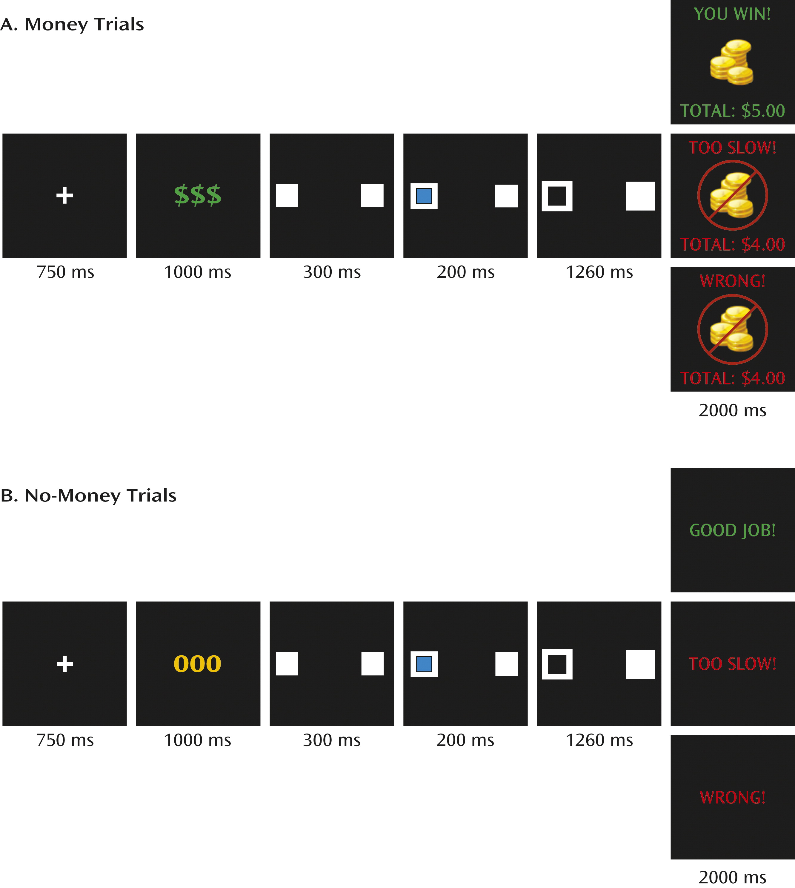
Participants completed an adapted affective Posner task (710). Trials consisted of 1) a fixation cross, 2) a symbol indicating trial type (i.e., money or no money), 3) two white squares, 4) a blue cue appearing in one of the white squares, 5) a target appearing in one of the white squares, and 6) feedback (Figure 1). Participants identified the target’s location as quickly and accurately as possible using a response button box. The blue cue predicted target location on 75% of trials (valid trials) and was in the opposite location on 25% of trials (invalid trials).
FIGURE 1. Schematic of Trial Structure During the Frustration Condition (Game 3)a
a The blue cue and the black target could appear in either the left or the right box. On valid trials, the cue and target were in the same location. On invalid trials, the cue and target were in opposite locations. Participants were instructed to press a button corresponding to the target location. During game 3, which was the frustration condition, participants viewed money trials and no-money trials that were distinguished by different trial type indicators (green “$$$” versus yellow “000”) and the possibility of winning and losing money. During money trials, participants won or lost money depending on performance. During no-money trials, no money was won or lost. For both money and no-money trials in the frustration condition, 40% of correct responses were followed by positive feedback (either “You win” or “Good job”), and 60% of correct responses were followed by negative feedback (“Too slow”). All incorrect responses received negative feedback (“Wrong”).
Participants completed the task as three “games.” During game 1 (50 trials), participants received accurate performance feedback and did not win or lose money. During game 2 (100 trials), participants received accurate feedback and won or lost 50¢ a trial, depending on performance. The frustration manipulation occurred during game 3 (260 trials). During game 3, participants were told that they must respond both quickly and accurately to win money, and that adequate speed was based on a complicated formula that considered performance on prior trials. Frustration was induced by giving participants the feedback that they were “too slow” on 60% of accurate trials, irrespective of the subject’s reaction time (referred to here as “negative feedback trials”).
Game 3 consisted of three trial types: money trials (N=100), no-money trials (N=100), and fixation trials (N=60), randomly presented across three runs (Figure 1). Money trials were identified by three green dollar signs (trial type symbol), indicating that participants could win or lose 50¢ a trial. Feedback consisted of a coin image with green text on positive feedback trials and red text on negative feedback or error trials; the participant’s cumulative winnings were displayed at the bottom of the screen. No-money trials were identified by three yellow circles, and participants did not win or lose money. Thus, on each money or no-money trial, participants received one of three kinds of feedback: error (“wrong” on inaccurate or missed trials), negative (“too slow” on 60% of accurate trials), or positive (“you win” [money trials] or “good job” [no-money trials] on 40% of accurate trials). Fixation trials (N=60) consisted of a fixation cross centrally presented. All trials were followed by a variable intertrial interval (range=850–1150 ms, mean=1000 ms).

Procedure

Participants completed game 1 and 60 trials of game 2 outside the scanner. Next, participants completed 40 trials of game 2 in the scanner while anatomical brain images were collected. This allowed us to determine whether behavioral or affective changes were related to the scanning environment as opposed to the frustration manipulation. Participants then completed three blocks of game 3 during functional imaging. Thus, scanning time was focused on the frustration condition.
Participants self-reported valence, arousal, and frustration at six time points (after game 1 and after each block of games 2 and 3) using 9-point Likert scales (17). After scanning, participants’ deception was assessed via self-report questionnaire and follow-up interview. Participants who knew that the feedback had been manipulated were excluded. No participant reported marked distress associated with the frustration manipulation or deception.

Data Analysis

Demographic characteristics.

Unpaired t tests or chi-square tests were used to compare age, IQ, and sex distribution between groups. Sex distribution differed between groups and was included as a covariate in all analyses.

Self-reported valence, arousal, and frustration ratings.

Group differences on valence, arousal, and frustration were assessed using a time-by-group repeated-measures analysis of covariance (ANCOVA) in which time had six categories: game 1, game 2–out of scanner, game 2–in scanner, game 3–run 1, game 3–run 2, and game 3–run 3.

Behavioral data.

Group differences in response time and accuracy were evaluated using two repeated-measures ANCOVAs. First, we conducted a group-by-time-by-validity (valid, invalid) ANCOVA. Next, to examine whether trial type (money or no-money) influenced group differences in response time, we conducted a group-by-validity-by-trial type ANCOVA that included response time averaged across the three runs of game 3. Two separate ANOVAs were required because game 3, but not games 1 and 2, contained both money and no-money trials. Feedback was not included as a factor in either ANOVA because responses were made before feedback was received. We report interactions and main effects not qualified by interactions (for all findings, see Table S1 in the data supplement that accompanies the online edition of this article).

Imaging data.

Neuroimaging data were acquired on a 1.5-T GE scanner using an eight-channel head coil and including a high-resolution anatomical scan (1.5-mm slices, three-dimensional fast spoiled gradient echo, 20° flip angle, 256×192 matrix, 24 cm field of view). Gradient echo-planar imaging images were collected during game 3 (TR=2900 ms, TE=27 ms, field of view=24 cm, flip angle=90°, voxel size=2.5×3.75×3.75 mm).
Data were analyzed using AFNI (Analysis of Functional Neuroimages) (18). Preprocessing included temporal alignment to the first acquired slice, coregistration, smoothing (kernel full width at half maximum=6), masking, intensity scaling, and transformation into Talairach space. Repetitions with motion >2 mm or >2 degrees relative to the preceding repetition were removed from the analysis.
Event types included three categories: trial type (money, no-money), validity (valid, invalid), and feedback (positive, negative, error). All combinations of these three factors were modeled using individual linear regression with a fixed-shape, gamma-variate response function, convolved with a boxcar function of the stimulus duration. The model included each of the event type regressors, the six motion parameters, and baseline drift for each of the three runs. Beta coefficients and t-statistics were calculated for each voxel and regressor. Because of an insufficient number of invalid and error trials, group analyses focused on the remaining four event types: money–valid–positive feedback; money–valid–negative feedback; no-money–valid–positive feedback; and no-money–valid–negative feedback.
Group-level analyses occurred on two levels. Region-of-interest analyses were conducted on the left and right amygdala and striatum (caudate, putamen, and nucleus accumbens), as defined by the Talairach-Tournoux Daemon. Mean signal intensity was extracted from each region of interest for each of the four event types. Amygdala values were submitted to a group-by-trial type-by-feedback-by hemisphere ANCOVA in SPSS. Striatum values were submitted to a group-by-trial type-by-feedback-by-hemisphere-by-region (regions were caudate, putamen, and nucleus accumbens) ANCOVA in SPSS.
Next, we computed a whole-brain analysis using a group-by-trial type-by-feedback ANCOVA with participant as a random-effects factor. 3dClustSim using group blur estimates indicated that a cluster-extent threshold of k≥37 at p<0.001 resulted in a whole-brain false positive probability of p<0.05. Average signal change values were extracted and post hoc ANCOVAs were performed in SPSS for clusters meeting identified thresholds. Because our primary interest was in group differences in response to positive and negative feedback, we only discuss the group-by-feedback interaction (for all findings, see Table S2 in the online data supplement). Greenhouse-Geisser correction was used when analyses violated sphericity assumptions.
Post hoc analyses examined the effects of medication, comorbid attention deficit hyperactivity disorder (ADHD), and affect ratings (i.e., self-reported frustration during game 3 and irritability over a 6-month period) on neural activation patterns. Exploratory post hoc t tests compared neural activation on negative feedback trials between unmedicated children with severe mood dysregulation (N=7) and healthy comparison children, and between children with severe mood dysregulation without comorbid ADHD (N=4) and healthy comparison children in the brain regions identified in the primary analysis. Pearson correlations conducted separately for each group examined associations between affect measures, behavioral variables, and neural response to negative feedback.

Results

Participants

A total of 61 children enrolled, 19 of whom were excluded for various reasons (inability to complete the task because of frustration or anxiety, five children with severe mood dysregulation; not deceived, two healthy children; technical difficulties, one child with severe mood dysregulation and one healthy child; structural brain abnormality, one healthy child; insufficient numbers of trials/condition (<35% of trials) because of poor behavioral performance or motion >2 mm, five children with severe mood dysregulation and three healthy children; and poor coregistration, one healthy child). The final sample included 19 children with severe mood dysregulation (seven of them unmedicated) and 23 healthy comparison children (Table 1). The groups did not differ significantly in age or full-scale IQ. The severe mood dysregulation group had a greater proportion of boys than the healthy comparison group (χ2=4.27, p<0.04), so sex was a covariate in all analyses.
TABLE 1. Demographic and Clinical Characteristics of Children With Severe Mood Dysregulation and Healthy Comparison Children
CharacteristicSevere Mood Dysregulation Group (N=19)Healthy Comparison Group (N=23)
 MeanSDMeanSD
Age (years)13.62.214.32.2
IQa104.313.8110.412.2
Children’s Depression Rating Scale score26.15.1  
Number of medications1.71.6  
Number of comorbid diagnoses3.31.5  
 N%N%
Maleb1578.91147.8
Comorbid diagnoses    
ADHDc1578.9  
Major depression421.1  
Oppositional defiant disorder1684.2  
Anxiety1263.2  
Conduct disorder15.3  
Medication    
 Unmedicated736.8  
 Atypical antipsychotic736.8  
 Lithium15.3  
 Antiepileptic526.3  
 Antidepressant736.8  
 Stimulant736.8  
a
Wechsler Abbreviated Scale of Intelligence (two-scale IQ).
b
Significant difference between groups, p<0.04.
c
ADHD=attention deficit hyperactivity disorder.

Affect Ratings

A group-by-time interaction (F=3.42, df=5, 190, p<0.05) revealed that children with severe mood dysregulation reported more frustration than healthy children after the last two runs of game 3, but not at earlier assessment points. All participants felt more unhappy during the frustration condition (game 3) than during nonfrustration conditions (games 1 and 2) (time, F=12.76, df=5, 190, p<0.001), but there were no main effects of group or interactions. No findings emerged from the arousal ratings.

Behavioral Results

The first behavioral analysis (group-by-time-by-validity) revealed a time-by-validity interaction for response time (F=7.67, df=5, 195, p<0.001) and accuracy (F=30.05, df=5, 195, p<0.001). In general, participants were faster but less accurate during frustration relative to nonfrustration trials, with no differences between groups.
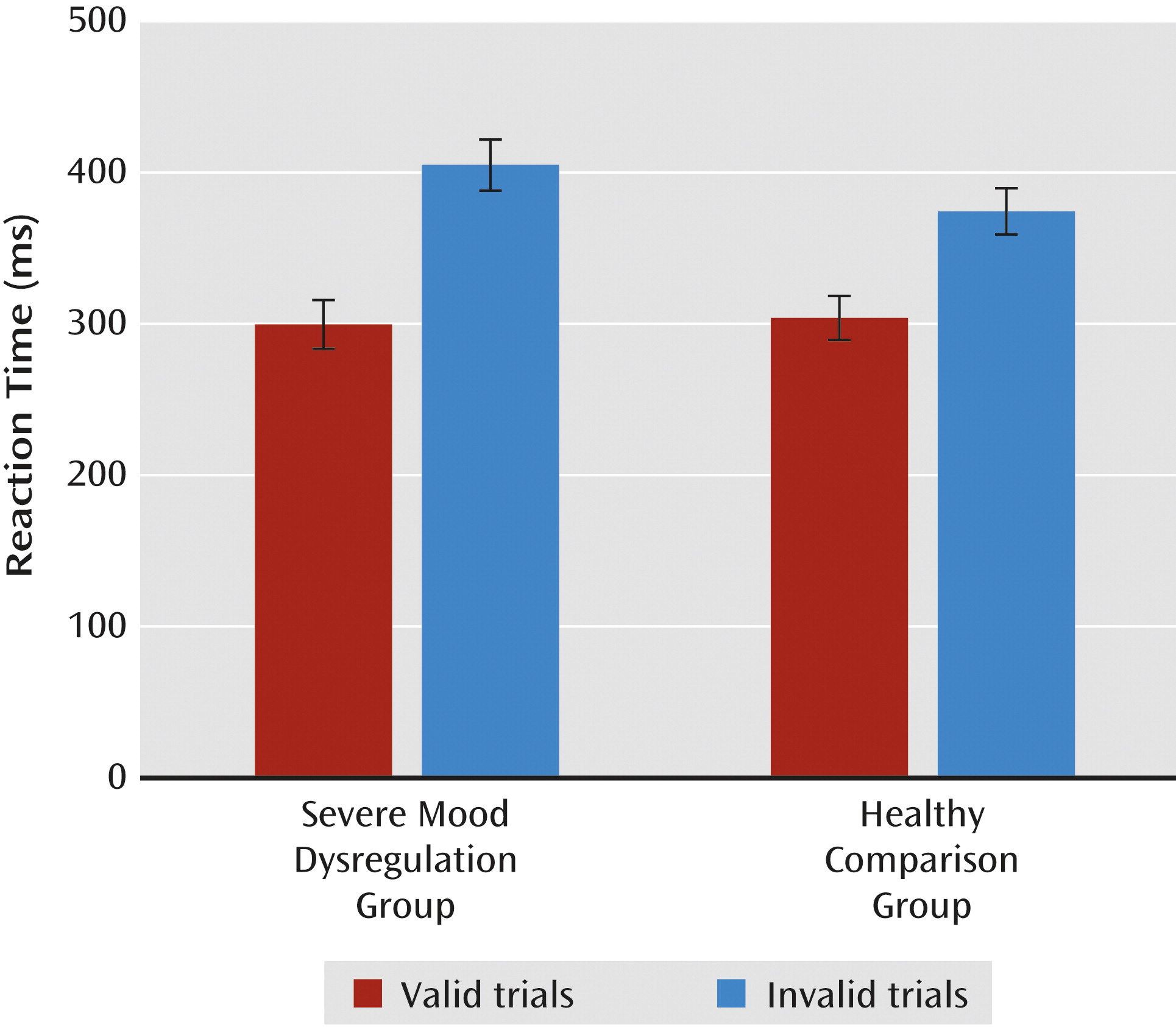
The second behavioral analysis (group-by-validity-by-trial type), was restricted to the frustration condition (game 3), since only game 3 included money and no-money trials. On response time, there was a group-by-validity interaction (F=5.41, df=1, 39, p<0.03). During frustration, all participants responded more slowly during invalid than valid trials; however, children with severe mood dysregulation were slower than healthy children only on invalid trials (Figure 2). A main effect of trial type also emerged (F=8.24, df=1, 39, p<0.01), indicating that participants responded more quickly during money than no-money trials. No group differences emerged for accuracy.
FIGURE 2. Reaction Time to Identify a Target on Valid and Invalid Trials During the Frustration Condition (Game 3)a
a All participants responded more slowly on invalid compared with valid trials; however, children with severe mood dysregulation were slower on invalid trials than were healthy comparison children (p<0.05).

fMRI Activation Results

The amygdala region-of-interest analysis revealed a group-by-feedback-by-hemisphere interaction (F=4.70, df=1, 39, p<0.04). Compared with healthy children, children with severe mood dysregulation exhibited less activation in the left amygdala on negative feedback trials (between-group difference). There was also a within-group difference in the severe mood dysregulation group only, with participants exhibiting less activation in the left amygdala on negative relative to positive feedback trials. Activation in healthy children did not differ between positive and negative feedback trials, and activation did not differ between groups on positive feedback trials.
In the striatum, the group-by-trial type-by-feedback-by-hemisphere-by-region ANCOVA revealed a group-by-feedback interaction (F=5.54, df=1, 39, p<0.03) but no higher-order interactions. The pattern in the striatum was similar to that in the amygdala: children with severe mood dysregulation exhibited less activation in the left and right striatum than healthy children during negative feedback trials (between-group difference). Children with severe mood dysregulation also exhibited less activation in the striatum during negative relative to positive feedback trials. This within-group difference was present in the severe mood dysregulation group only; in healthy children, striatal activation did not differ between positive and negative feedback trials. As in the amygdala, children with severe mood dysregulation did not differ from healthy children on striatal activation during positive feedback trials.
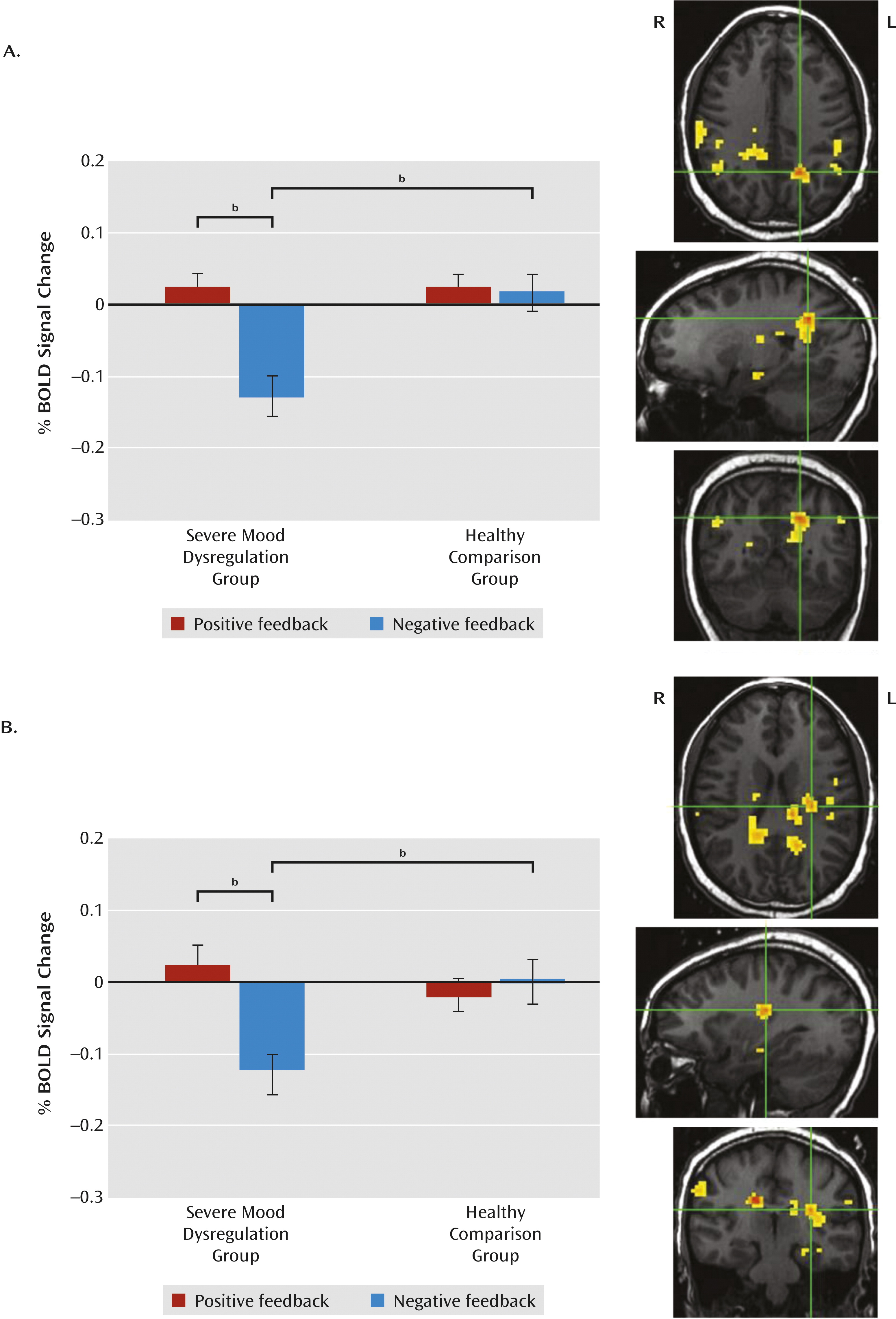
In the whole-brain analysis, no regions showed a group-by-trial type-by-feedback interaction. However, there was a group-by-feedback interaction in 11 regions (Table 2; Figure 3). Activation patterns were identical in each region and similar to the region-of-interest results—that is, we observed a between-group difference during negative feedback trials, no group differences during positive feedback trials, and, in the severe mood dysregulation group only, a within-group difference on negative relative to positive-feedback trials. Specifically, relative to healthy children, children with severe mood dysregulation exhibited less activation in response to negative feedback in parietal, parahippocampal, and thalamic/cingulate/striatal regions (F values, >10.00, p values, <0.005). Activation did not differ between groups on positive feedback trials. In these same regions, children with severe mood dysregulation exhibited less activation in response to negative relative to positive feedback (F values, >7.50, p values, <0.02), whereas healthy children did not show a within-group difference.
TABLE 2. Significant Findings From the Group-by-Feedback Interaction Observed in the Whole-Brain fMRI Analysis
   Talairach Coordinatesb Analysisc
Area of ActivationSideCluster SizeaxyzBrodmann’s AreaFp
Posterior cingulate cortexRight29617–40142920.52<0.001
Posterior cingulate cortexLeft137–19–553217/3114.62<0.001
Postcentral gyrus and inferior parietal lobuleRight8362–2535210.02<0.005
Supramarginal gyrus and inferior parietal lobuleLeft82–37–31261312.07<0.002
InsulaLeft74–28–22201316.61<0.001
Supramarginal gyrusRight6547–52293910.05<0.005
InsulaLeft60–40521321.46<0.001
Parahippocampal gyrusLeft49–13–13–1328/3420.72<0.001
Post-/precentral gyrusLeft45–40–1623614.70<0.002
PrecuneusRight3914–5838710.42<0.005
Cingulate/thalamus/caudateLeft38–13–2823 16.01<0.001
a
Cluster size was determined using a significance threshold of p<0.05, corrected for the number of comparisons.
b
Coordinates refer to the voxel with maximum signal intensity.
c
Statistics refer to the analysis of the extracted clusters in SPSS; df=1, 39.
FIGURE 3. Brain Activation in Children With Severe Mood Dysregulation and Healthy Comparison Children on Positive and Negative Feedback Trialsa
a Panel A shows activation in the left posterior cingulate and precuneus (Talairach coordinates, x=−19, y=−55, z=32). Panel B shows activation in the left insula (Talairach coordinates, x=−28, y=−22, z=20). Images are displayed according to radiologic convention (left=right). BOLD=blood-oxygen-level-dependent.
b Significantly different (p<0.05).

Exploratory Analyses

Post hoc analyses (see Table S3 in the online data supplement) indicated that unmedicated children with severe irritability, as well as children with severe mood dysregulation without comorbid ADHD, exhibited hypoactivation relative to healthy children in the left amygdala, in the left and right striatum, and in 10 of the 11 regions identified by the whole-brain analyses (F values, >4.10, p values, <0.05).
In both children with severe mood dysregulation and healthy children, greater self-reported frustration during the task was associated with reduced activation in response to negative feedback trials in several regions identified in primary analyses (severe mood dysregulation: left inferior parietal lobule/supramarginal gyrus; right supramarginal gyrus; and left parahippocampal gyrus [r values, <−0.47, p values, <0.05]; healthy comparison children: left insula, left parahippocampal gyrus, left pre-/postcentral gyrus, right precuneus, and left cingulate/thalamus/caudate; left amygdala [r values, >−0.42, p values, <0.05]).
In healthy children, greater parent-reported irritability was associated with decreased accuracy on valid trials during frustration (r=−0.46, p<0.05), and greater self-reported irritability was negatively associated with activation to negative feedback trials in the left parahippocampal gyrus (r=−0.78, p<0.001). Self-reported irritability was not associated with any behavioral or neural measure in children with severe mood dysregulation.

Discussion

To our knowledge, this is the first fMRI study to compare the affective, behavioral, and neural correlates of frustration in children with severe and chronic irritability and healthy subjects. During the frustrating game, chronically irritable children exhibited behavioral deficits relative to healthy comparison children, in that children with severe mood dysregulation responded more slowly than healthy comparison children on invalid trials. Also during the frustrating game, irritable children differed from healthy children by showing marked deactivation of neural regions associated with spatial attention, reward processing, and emotional salience on the contrast of negative versus positive feedback trials. Groups did not differ in their neural response to positive feedback trials.
Both groups showed approximately 30% lower accuracy on invalid trials in the frustration versus nonfrustration condition. Therefore, frustration decreased spatial attention flexibility in both groups, perhaps by inhibiting disengagement from the cue. Whereas the groups did not differ on accuracy during frustration, there were between-group differences on reaction time. Specifically, children with severe mood dysregulation were slower than healthy comparison children on invalid trials during frustration, suggesting they had particular difficulty shifting spatial attention away from the cue. Such behavioral deficits may be associated with the parietal hypoactivation observed in children with severe mood dysregulation in response to negative feedback, given the prominent role of parietal regions in mediating spatial attention processes (19). Attention allocation skills are important for successful emotion regulation (20), and deficits in attention control have been associated with increased negative affect and aggression in children (2124). When frustrated and confronted with a negative event, children with severe mood dysregulation may have difficulty disengaging attention from the blocked goal and attending instead to stimuli that might serve as helpful distractors and improve their emotional state (20). Alternatively, an inability to shift attention flexibly may reduce the number of emotion regulation strategies that can be identified and employed. Either mechanism might contribute to the irritable outbursts characteristic of children with severe mood dysregulation.
Alternatively, the behavioral impairments and frustration experienced by children with severe mood dysregulation may stem from deficits in bottom-up processes mediated by the amygdala/insula or striatum. Our observation of left amygdala hypoactivation in children with severe mood dysregulation during negative feedback is surprising, given the amygdala’s role in responding to emotionally salient stimuli (12) and prior evidence of amygdala hyperactivation to frustrating stimuli in adults with high trait anger (25, 26). However, an independent sample of children with severe mood dysregulation exhibited amygdala hypoactivation in response to emotional stimuli on another task (15), perhaps reflecting generalized amygdala dysregulation, with a tendency toward hypoactivation, in chronically irritable children. Additional studies are needed to replicate and clarify these findings.
Decreased striatal response in children with severe mood dysregulation during negative feedback trials may reflect abnormalities in regions supporting reward processing. For example, negative prediction errors, which occur when an outcome is worse than expected, are associated with ventral striatal deactivation (13, 14). Therefore, our striatal finding in children with severe mood dysregulation may indicate that these children experienced the frustrating event as more unexpected and aversive than did healthy children. This response may contribute to their exaggerated and inappropriate responses to frustrating events.
In contrast to two adult studies using similar frustration tasks (25, 26), we did not observe group differences in prefrontal regions. Null findings are difficult to interpret, as they may reflect type II error. Indeed, children with severe mood dysregulation have been found to exhibit abnormal anterior cingulate activation (measured by magnetoencephalography) in response to frustrating feedback (7) and inferior frontal gyrus activation deficits during an fMRI reward study (16). Thus, deficits in prefrontal regions supporting top-down processing in response to frustration may exist in this population even though they were not observed in this study.
In interpreting the deactivation that we observed, frustration tasks probably induce longer-lasting emotional responses than those elicited by the presentation of, for example, emotional faces or pictures. Thus, it is possible that feelings of frustration remained “on” throughout all of game 3, in which case fixation trials may not have been cognitively or affectively neutral. This may complicate the interpretation of neural responses to task-related activity, since these are calculated relative to fixation trials. However, neural response to both positive and negative feedback trials were calculated with respect to fixation trials, and deactivation occurred only during negative trials. We are currently comparing neural responses to feedback under nonfrustration and frustration conditions to examine whether participants’ “baseline” activation changes during frustration.
Our findings in children with severe mood dysregulation are likely to be informative about neural dysfunction in children with disruptive mood dysregulation disorder during frustration. Like those with severe mood dysregulation, children meeting criteria for disruptive mood dysregulation disorder experience recurrent, excessive, and developmentally inappropriate temper outbursts, as well as chronic negative affect between outbursts. Unlike in severe mood dysregulation, the diagnostic criteria for disruptive mood dysregulation disorder do not include hyperarousal symptoms. Therefore, severe mood dysregulation can be viewed as a subset of disruptive mood dysregulation disorder. Since we do not know the degree to which irritability, rather than hyperarousal, is responsible for the abnormalities observed in children with severe mood dysregulation, it is unclear whether our findings would generalize to those with disruptive mood dysregulation disorder who do not have hyperarousal or ADHD symptoms.
Indeed, given high rates of co-occurring ADHD in children with severe mood dysregulation, behavioral and neural deficits related to attention are not surprising and may be related to ADHD symptoms rather than irritability. Post hoc analyses of the deactivation patterns observed in the irritable children in this study suggest that this is not the case. However, a stricter test would involve comparing children with severe mood dysregulation to nonirritable children with ADHD. We are currently conducting such a study. Similar research is necessary to evaluate the contribution of other clinical factors related to severe mood dysregulation (e.g., other co-occurring disorders, psychotropic medications) to confirm the specific effects of irritability. In addition, whether the chronic irritability of children with severe mood dysregulation affects neural and behavioral responses to frustration differently than the episodic irritability characteristic of children with bipolar disorder is unknown and deserves further study.
Our study has some limitation. First, previous studies using the affective Posner paradigm employed methods that isolated neural responses to feedback. In this study, the paradigm modeled the entire trial and thus limited our ability to detect neural activation specific to feedback. Second, behavioral findings, collected across all three games, suggest that attention flexibility differed between nonfrustration and frustration conditions. However, because of time constraints, fMRI data were collected only during the frustration condition (i.e., game 3). An ongoing study includes scanning during frustration and nonfrustration conditions. Third, high rates of psychotropic medication use and of comorbid diagnoses in children with severe mood dysregulation complicate attributions of study findings to irritability specifically. Exploratory post hoc analyses suggest that deficits in irritable children were not attributable to co-occurring ADHD or to psychotropic medications, but additional research is needed to identify the role of irritability independent of other clinical factors. Similarly, an important question for further research is the degree to which state frustration and trait irritability influence behavioral and neural responses to frustration. Finally, the task we used assessed only participants’ ability to shift spatial attention. Frustration likely influences other types of attention and perhaps cognitive control more broadly, and these are important issues to pursue in future research.
Conclusions

Given that irritability is both common and impairing in psychiatric patients, a better understanding of its pathophysiology could inform the broad psychiatric literature. Our findings suggest that frustration impairs attention flexibility and reduces activation in neural regions supporting spatial attention, emotion salience, and reward processing in response to negative feedback in severely irritable children. Research utilizing such paradigms and populations may facilitate the development of novel interventions.

Acknowledgments

The authors gratefully acknowledge the efforts of members of the Emotion and Development Branch and suggestions by R.J.R. Blair.

Supplementary Material

Supplementary Material (1186_ds001.pdf)

References

1.
Leibenluft E, Charney DS, Towbin KE, Bhangoo RK, Pine DS: Defining clinical phenotypes of juvenile mania. Am J Psychiatry 2003; 160:430–437
2.
Leibenluft E: Severe mood dysregulation, irritability, and the diagnostic boundaries of bipolar disorder in youths. Am J Psychiatry 2011; 168:129–142
3.
Safer DJ: Irritable mood and the Diagnostic and Statistical Manual of Mental Disorders. Child Adolesc Psychiatry Ment Health 2009; 3:35
4.
Lewis MD, Lamm C, Segalowitz SJ, Stieben J, Zelazo PD: Neurophysiological correlates of emotion regulation in children and adolescents. J Cogn Neurosci 2006; 18:430–443
5.
Lewis MD, Stieben J: Emotion regulation in the brain: conceptual issues and directions for developmental research. Child Dev 2004; 75:371–376
6.
Moadab I, Gilbert T, Dishion TJ, Tucker DM: Frontolimbic activity in a frustrating task: covariation between patterns of coping and individual differences in externalizing and internalizing symptoms. Dev Psychopathol 2010; 22:391–404
7.
Rich BA, Carver FW, Holroyd T, Rosen HR, Mendoza JK, Cornwell BR, Fox NA, Pine DS, Coppola R, Leibenluft E: Different neural pathways to negative affect in youth with pediatric bipolar disorder and severe mood dysregulation. J Psychiatr Res 2011; 45:1283–1294
8.
Rich BA, Holroyd T, Carver FW, Onelio LM, Mendoza JK, Cornwell BR, Fox NA, Pine DS, Coppola R, Leibenluft E: A preliminary study of the neural mechanisms of frustration in pediatric bipolar disorder using magnetoencephalography. Depress Anxiety 2010; 27:276–286
9.
Rich BA, Schmajuk M, Perez-Edgar KE, Fox NA, Pine DS, Leibenluft E: Different psychophysiological and behavioral responses elicited by frustration in pediatric bipolar disorder and severe mood dysregulation. Am J Psychiatry 2007; 164:309–317
10.
Rich BA, Schmajuk M, Perez-Edgar KE, Pine DS, Fox NA, Leibenluft E: The impact of reward, punishment, and frustration on attention in pediatric bipolar disorder. Biol Psychiatry 2005; 58:532–539
11.
Klöppel S, Stonnington CM, Petrovic P, Mobbs D, Tüscher O, Craufurd D, Tabrizi SJ, Frackowiak RS: Irritability in pre-clinical Huntington’s disease. Neuropsychologia 2010; 48:549–557
12.
LeDoux JE: The Emotional Brain. New York, Simon and Shuster, 1996
13.
Schultz W: Dopamine signals for reward value and risk: basic and recent data. Behav Brain Funct 2010; 6:24
14.
Schultz W, Dayan P, Montague PR: A neural substrate of prediction and reward. Science 1997; 275:1593–1599
15.
Brotman MA, Rich BA, Guyer AE, Lunsford JR, Horsey SE, Reising MM, Thomas LA, Fromm SJ, Towbin K, Pine DS, Leibenluft E: Amygdala activation during emotion processing of neutral faces in children with severe mood dysregulation versus ADHD or bipolar disorder. Am J Psychiatry 2010; 167:61–69
16.
Adleman NE, Kayser R, Dickstein D, Blair RJ, Pine D, Leibenluft E: Neural correlates of reversal learning in severe mood dysregulation and pediatric bipolar disorder. J Am Acad Child Adolesc Psychiatry 2011; 50:1173–1185.e2
17.
McManis MH, Bradley MM, Berg WK, Cuthbert BN, Lang PJ: Emotional reactions in children: verbal, physiological, and behavioral responses to affective pictures. Psychophysiology 2001; 38:222–231
18.
Cox RW: AFNI: software for analysis and visualization of functional magnetic resonance neuroimages. Comput Biomed Res 1996; 29:162–173
19.
Corbetta M, Kincade JM, Ollinger JM, McAvoy MP, Shulman GL: Voluntary orienting is dissociated from target detection in human posterior parietal cortex. Nat Neurosci 2000; 3:292–297
20.
Mischel W, Shoda Y, Rodriguez MI: Delay of gratification in children. Science 1989; 244:933–938
21.
Morasch KC, Bell MA: Self-regulation of negative affect at 5 and 10 months. Dev Psychobiol 2012; 54:215–221
22.
Morris AS, Silk JS, Morris MD, Steinberg L, Aucoin KJ, Keyes AW: The influence of mother-child emotion regulation strategies on children’s expression of anger and sadness. Dev Psychol 2011; 47:213–225
23.
Reijntjes A, Stegge H, Terwogt MM, Kamphuis JH, Telch MJ: Emotion regulation and its effects on mood improvement in response to an in vivo peer rejection challenge. Emotion 2006; 6:543–552
24.
Rothbart MK, Ahadi SA, Hershey KL, Fisher P: Investigations of temperament at three to seven years: the Children’s Behavior Questionnaire. Child Dev 2001; 72:1394–1408
25.
Alia-Klein N, Goldstein RZ, Tomasi D, Woicik PA, Moeller SJ, Williams B, Craig IW, Telang F, Biegon A, Wang GJ, Fowler JS, Volkow ND: Neural mechanisms of anger regulation as a function of genetic risk for violence. Emotion 2009; 9:385–396
26.
Alia-Klein N, Goldstein RZ, Tomasi D, Zhang L, Fagin-Jones S, Telang F, Wang GJ, Fowler JS, Volkow ND: What is in a word? No versus Yes differentially engage the lateral orbitofrontal cortex. Emotion 2007; 7:649–659

Information & Authors

Information

Published In

Go to American Journal of Psychiatry
American Journal of Psychiatry
Pages: 1186 - 1194
PubMed: 23732841

History

Received: 14 July 2012
Revision received: 11 January 2013
Accepted: 14 February 2013
Published online: 1 October 2013
Published in print: October 2013

Authors

Details

Christen M. Deveney, Ph.D.
From the Emotion and Development Branch and the Scientific and Statistical Computing Core, NIMH, Bethesda, Md.
Megan E. Connolly, B.A.
From the Emotion and Development Branch and the Scientific and Statistical Computing Core, NIMH, Bethesda, Md.
Catherine T. Haring, B.A.
From the Emotion and Development Branch and the Scientific and Statistical Computing Core, NIMH, Bethesda, Md.
Brian L. Bones, B.A.
From the Emotion and Development Branch and the Scientific and Statistical Computing Core, NIMH, Bethesda, Md.
Richard C. Reynolds, M.S.
From the Emotion and Development Branch and the Scientific and Statistical Computing Core, NIMH, Bethesda, Md.
Pilyoung Kim, Ph.D.
From the Emotion and Development Branch and the Scientific and Statistical Computing Core, NIMH, Bethesda, Md.
Daniel S. Pine, M.D.
From the Emotion and Development Branch and the Scientific and Statistical Computing Core, NIMH, Bethesda, Md.
Ellen Leibenluft, M.D.
From the Emotion and Development Branch and the Scientific and Statistical Computing Core, NIMH, Bethesda, Md.

Notes

Address correspondence to Dr. Deveney ([email protected]).

Funding Information

The authors report no financial relationships with commercial interests.
Supplementary Material
Supported by the NIMH Intramural Research Program.

Metrics & Citations

Metrics

Citations

Export Citations

If you have the appropriate software installed, you can download article citation data to the citation manager of your choice. Simply select your manager software from the list below and click Download.

For more information or tips please see 'Downloading to a citation manager' in the Help menu.

Format
Citation style
Style
Copy to clipboard

View Options

View options

PDF/EPUB

View PDF/EPUB

Login options

Already a subscriber? Access your subscription through your login credentials or your institution for full access to this article.

Personal login Institutional Login Open Athens login
Purchase Options

Purchase this article to access the full text.

PPV Articles - American Journal of Psychiatry

PPV Articles - American Journal of Psychiatry

Not a subscriber?

Subscribe Now / Learn More

PsychiatryOnline subscription options offer access to the DSM-5-TR® library, books, journals, CME, and patient resources. This all-in-one virtual library provides psychiatrists and mental health professionals with key resources for diagnosis, treatment, research, and professional development.

Need more help? PsychiatryOnline Customer Service may be reached by emailing [email protected] or by calling 800-368-5777 (in the U.S.) or 703-907-7322 (outside the U.S.).

Media

Figures

Other

Tables

Share

Share

Share article link

Share