Abstract

Objective:

Exposure therapy is an effective treatment for posttraumatic stress disorder (PTSD), but a comprehensive, emotion-focused perspective on how psychotherapy affects brain function is lacking. The authors assessed changes in brain function after prolonged exposure therapy across three emotional reactivity and regulation paradigms.

Method:

Individuals with PTSD underwent functional MRI (fMRI) at rest and while completing three tasks assessing emotional reactivity and regulation. Individuals were then randomly assigned to immediate prolonged exposure treatment (N=36) or a waiting list condition (N=30) and underwent a second scan approximately 4 weeks after the last treatment session or a comparable waiting period, respectively.

Results:

Treatment-specific changes were observed only during cognitive reappraisal of negative images. Psychotherapy increased lateral frontopolar cortex activity and connectivity with the ventromedial prefrontal cortex/ventral striatum. Greater increases in frontopolar activation were associated with improvement in hyperarousal symptoms and psychological well-being. The frontopolar cortex also displayed a greater variety of temporal resting-state signal pattern changes after treatment. Concurrent transcranial magnetic stimulation and fMRI in healthy participants demonstrated that the lateral frontopolar cortex exerts downstream influence on the ventromedial prefrontal cortex/ventral striatum.

Conclusions:

Changes in frontopolar function during deliberate regulation of negative affect is one key mechanism of adaptive psychotherapeutic change in PTSD. Given that frontopolar connectivity with ventromedial regions during emotion regulation is enhanced by psychotherapy and that the frontopolar cortex exerts downstream influence on ventromedial regions in healthy individuals, these findings inform a novel conceptualization of how psychotherapy works, and they identify a promising target for stimulation-based therapeutics.
Posttraumatic stress disorder (PTSD) is persistent (1) and impairing (2) but can be treated with psychotherapy (3). One such effective treatment is prolonged exposure (4), which utilizes therapeutic exposure as its primary technique for promoting recovery (5). Formulated from emotional processing theory (6), prolonged exposure helps the patient confront the trauma memory and real-life situations that provoke symptoms. This allows the patient to integrate new, adaptive information regarding safety from threat. Repeated exposure usually results in a reduced fear response and promotes corrective learning, whereby the likelihood and intensity of a future fear response to that stimulus is lessened (6).
This framework suggests that treatment may alter a range of emotional behaviors, from initial detection and orienting toward emotional cues (emotional reactivity) through control of emotional responses (emotion regulation). Although exposure therapies for PTSD have been utilized for decades (4), little is known about how these treatments alter brain function. The literature is largely composed of imaging studies in small samples that assess changes in brain function during a single task before and after treatment (see Table S1 in the data supplement that accompanies the online edition of this article), often without comparison to a control intervention. While this work has provided valuable insight regarding brain changes in the particular process examined, the role of these changes within the larger context of potential mechanisms conveying treatment efficacy has been unclear. As these studies often lacked patient control arms, it is also unclear whether changes reflected the intervention per se or other confounding factors, such as the passage of time or repeated assessments.
Here, we provide a comprehensive assessment of functional brain changes following prolonged exposure therapy for PTSD across three experimental paradigms assessing emotional reactivity and regulation. We used a randomized patient waiting list condition as a control condition and analyzed results using voxel-wise linear mixed-effects modeling in line with the intent-to-treat principle. To identify whether treatment-related changes might reflect one brain region’s direct influence over another, we combined single-pulse transcranial magnetic stimulation (TMS) with functional MRI (fMRI) in a separate sample of healthy individuals. We assessed whether single TMS pulses to one brain region influenced activation in another, thereby demonstrating direct downstream influence. TMS is a noninvasive brain stimulation technique shown to produce elevated activity in the cortical area stimulated by the magnetic field as well as in downstream targets (7), thus mimicking endogenous volitional activation of the targeted region and allowing for experimental manipulation of neural circuitry. Finally, we examined resting-state fMRI in a focused manner to follow up on how task-related neural dynamics altered by treatment might generalize beyond an emotional reactivity and regulation context.
Previous PTSD psychotherapy imaging studies have observed increased prefrontal recruitment during recall of the trauma memory after treatment (810), although decreased recruitment has also been observed during trauma memory recall (11), processing of negative (12) and trauma-related pictures (13), and conflict processing (14). Similarly, increased recruitment of the anterior cingulate has been noted during processing of fearful versus neutral faces (15) and during anticipation of negative versus positive pictures (12), while activation in limbic structures such as the amygdala and anterior insula have shown posttreatment attenuations during recall of the trauma memory (16), a classic (14) and an affective Stroop task (17), and anticipation of affective pictures (12). Thus, the predominant pattern of experimental results is consistent with proposed mechanisms of psychotherapy—increased prefrontal recruitment and control over limbic structures involved in threat detection and emotion induction (18). On the basis of these findings, we expected individuals assigned to receive prolonged exposure therapy to display a greater attenuation of amygdala and anterior insula activation during the processing and detection of emotional stimuli. We also expected to see increased prefrontal recruitment during all phases of emotion processing, from initial detection and processing of stimuli to deployment of automatic and effortful regulatory strategies.

Method

The study methods are presented here in brief, with additional detail available in the Supplemental Methods section in the online data supplement.

Participants, Assessments, and Inclusion Criteria

Individuals 18–60 years of age were recruited to participate in a psychotherapy treatment study for PTSD. Participants provided written informed consent after receiving a complete description of the study. A baseline structured clinical interview was conducted to assess PTSD symptoms and other diagnoses.

Behavioral Paradigms

Emotional reactivity task.

This task (19) probes goal-irrelevant emotional reactivity via conscious and nonconscious presentation of color-tinted fearful and neutral faces. Participants were instructed to identify the color tint of the face stimulus.

Emotional conflict task.

This task (20) induces emotional conflict and conflict adaptation through pairing fearful and happy faces with congruent or incongruent emotion words. Participants were instructed to identify the facial emotion and ignore the emotion word.

Reappraisal task.

Participants viewed negative and neutral pictures from the International Affective Picture System under two conditions: “look” (for negative and neutral) and “decrease” (negative only). During “look” trials, participants were instructed to experience their natural emotional response, and during “decrease” trials, they were instructed to reduce their level of emotional distress by interpreting or seeing the picture differently (21).

Resting state.

Participants completed an 8-minute eyes-open resting-state scan in which they were told to lie still, stay awake, focus on a fixation cross, and allow their mind to wander.

MRI Data Acquisition

See the Supplemental Methods section in the data supplement.

Concurrent TMS-fMRI Mapping in Healthy Participants

To investigate normative patterns of downstream influence in neural circuits demonstrating treatment-related change, we used a separate cohort of 14 healthy individuals who underwent a concurrent TMS-fMRI scanning session conducted according to established protocols (22). See the Supplemental Methods section in the data supplement for further details.

Randomization

After initial assessments and fMRI scanning, participants were randomly assigned either to immediate treatment with prolonged exposure (N=36) or to a waiting list condition (N=30) (see the CONSORT chart in Figure S2 in the data supplement).

Prolonged Exposure Treatment

Treatment sessions occurred either once or twice a week, for a total of nine to 12 sessions, 90 minutes each, that followed manualized procedures (5).

Posttreatment Assessment

Approximately 4 weeks after the final treatment session, participants completed a posttreatment clinical assessment and repeated the imaging protocol. This duration was chosen in order to allow treatment changes to consolidate and symptom levels to equilibrate before the posttreatment assessment.

Image Preprocessing

See the Supplemental Methods section in the data supplement.

Individual-Level Analysis of Task Data

See the Supplemental Methods section in the data supplement.

Individual-Level Analysis of Resting Brain Entropy

To test a hypothesis regarding flexibility of brain states at rest as a follow-up to the primary activation analyses, we investigated regional brain entropy during resting-state fMRI and whether this changed with psychotherapy in regions showing task-related changes. Entropy is a measure of the variety of change patterns in a time-series signal (23), which could index shifts in the way the range of potential brain states available are manifested moment to moment (24). Entropy is also closely related to flexibility, that is, the ability to shift among different states (25).

Assessing Treatment Effects

We analyzed brain activation and connectivity on a voxel-wise level by employing the MacArthur approach (26) embedded in our longitudinal linear mixed-effects models to identify changes over time that were specific to the treatment group. We used a voxel-wise false discovery rate correction within a prespecified anatomical mask to control for type I error (see Figure S1 in the data supplement). In addition to voxel-wise analyses, we conducted region-of-interest analyses using extracted average activation beta weights from the left and right amygdala and anterior insula for each task and contrast of interest. These complement primary voxel-wise analyses by facilitating detection of limbic effects that may be larger in spatial extent but smaller in magnitude, which are unlikely to survive voxel-wise false-discovery-rate correction.

Results

Sample Characteristics

The final sample included 36 individuals assigned to receive immediate prolonged exposure treatment and 30 assigned to the waiting list condition. The groups were well matched on clinical and demographic variables (see Table S2 in the online data supplement).

Treatment Outcome

Compared with the waiting list group, the immediate treatment group showed significantly greater reductions in PTSD and depressive symptom scores as well as greater improvements in certain quality-of-life domains (see Table S3 in the online data supplement).

Baseline Task Effects

See the Supplemental Results section and Table S4 in the data supplement.

Treatment Effects on Task Behavior

During reappraisal, patients in the immediate treatment group compared with those in the waiting list group showed a significantly greater reduction in distress ratings in response to picture presentation, regardless of experimental condition or picture valence. No other significant treatment effects were detected on task behavior.

Treatment Effects on Task Brain Function in Limbic Regions of Interest

There were no significant treatment arm-by-time interaction effects on activation for any task contrast examined in the left and right amygdala or anterior insula (see Table S5 in the data supplement).

Voxel-Wise Analyses for Treatment Effects on Task Brain Function: Regions of Interest

Emotional reactivity task.

We observed no significant time-by-treatment arm effects for unmasked fear versus neutral faces or for masked fear versus neutral faces.

Emotional conflict task.

We observed no significant time-by-treatment arm effects for emotional conflict, conflict regulation, or emotional reactivity (congruent fear versus happy) contrasts.

Reappraisal task.

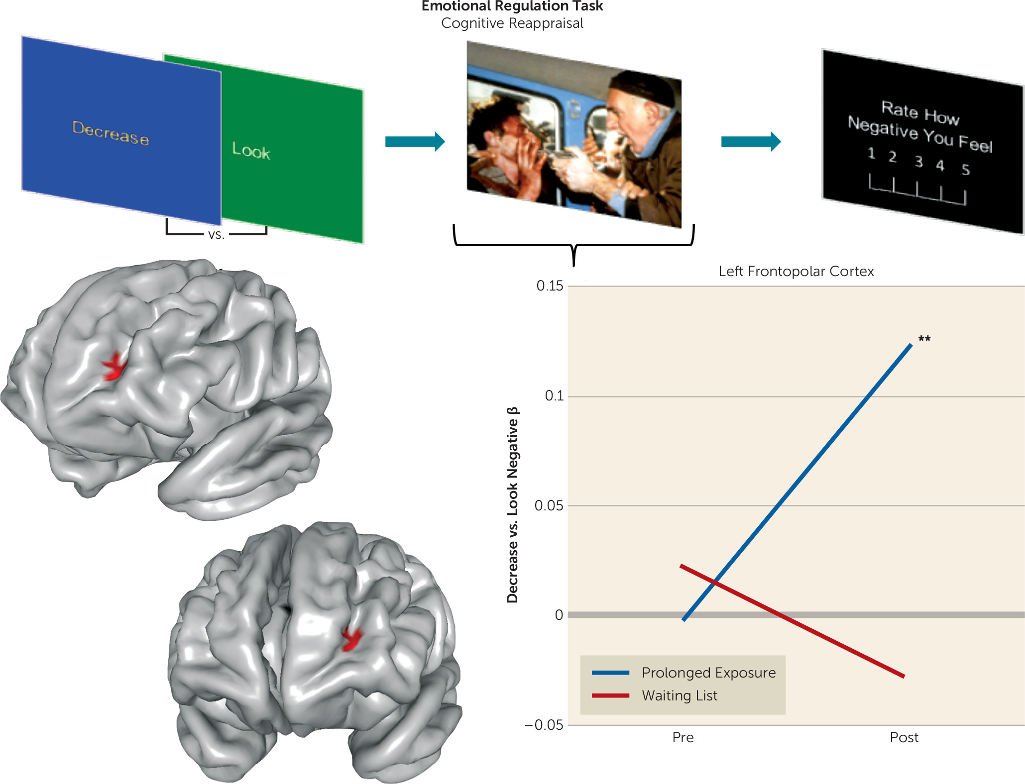
In the regulation contrast from the reappraisal paradigm (“decrease” negative versus “look” negative), we observed a significant time-by-treatment arm effect on left lateral frontopolar cortex activation (middle frontal gyrus; Brodmann’s area [BA] 10) (see Table S6 in the data supplement). Post hoc extractions revealed an increase in activation over time in the immediate treatment group (t=3.32, p=0.002) (Figure 1) and no change in the waiting list group. We observed no other significant effects in regions of interest. We detected no significant time-by-treatment arm interactions for the “look” negative versus “look” neutral contrast. Follow-up analyses across tasks demonstrated that change in left frontopolar activation was selective to reappraisal (see the Supplemental Results section in the data supplement).
FIGURE 1. Increased Left Frontopolar Cortex Activation During Cognitive Reappraisal After Prolonged Exposure Therapya
a A schematic of the task contrast is displayed at the top of the figure, which compares brain activation while individuals deliberately and consciously reduce negative emotion in response to an affectively charged picture relative to when they simply look at the picture and experience their natural emotional response. The line graph depicts the mean individual mixed model-derived predicted values for activation within each treatment arm and at each time point. The interaction effect of time and treatment arm is rendered on a template surface, in which individuals in the prolonged exposure group (N=36) displayed significantly greater increase in activation over time relative to those in the waiting list group (N=30). Pre=pretreatment; Post=posttreatment.
**p<0.01.

Voxel-Wise Treatment Effects on Task Brain Function: Whole Brain Analyses

Across all of the tasks and contrasts tested, no significant effects were detected in the exploratory whole brain analyses.

Exploratory Analyses: Differential Brain Changes as a Function of Remission Status

We also examined whether there were additional brain changes as a function of remission status at end of treatment (see the Supplemental Results section in the data supplement). We observed no additional effects of remission status on differential change in brain activation.

Frontopolar Context-Dependent Connectivity During Reappraisal

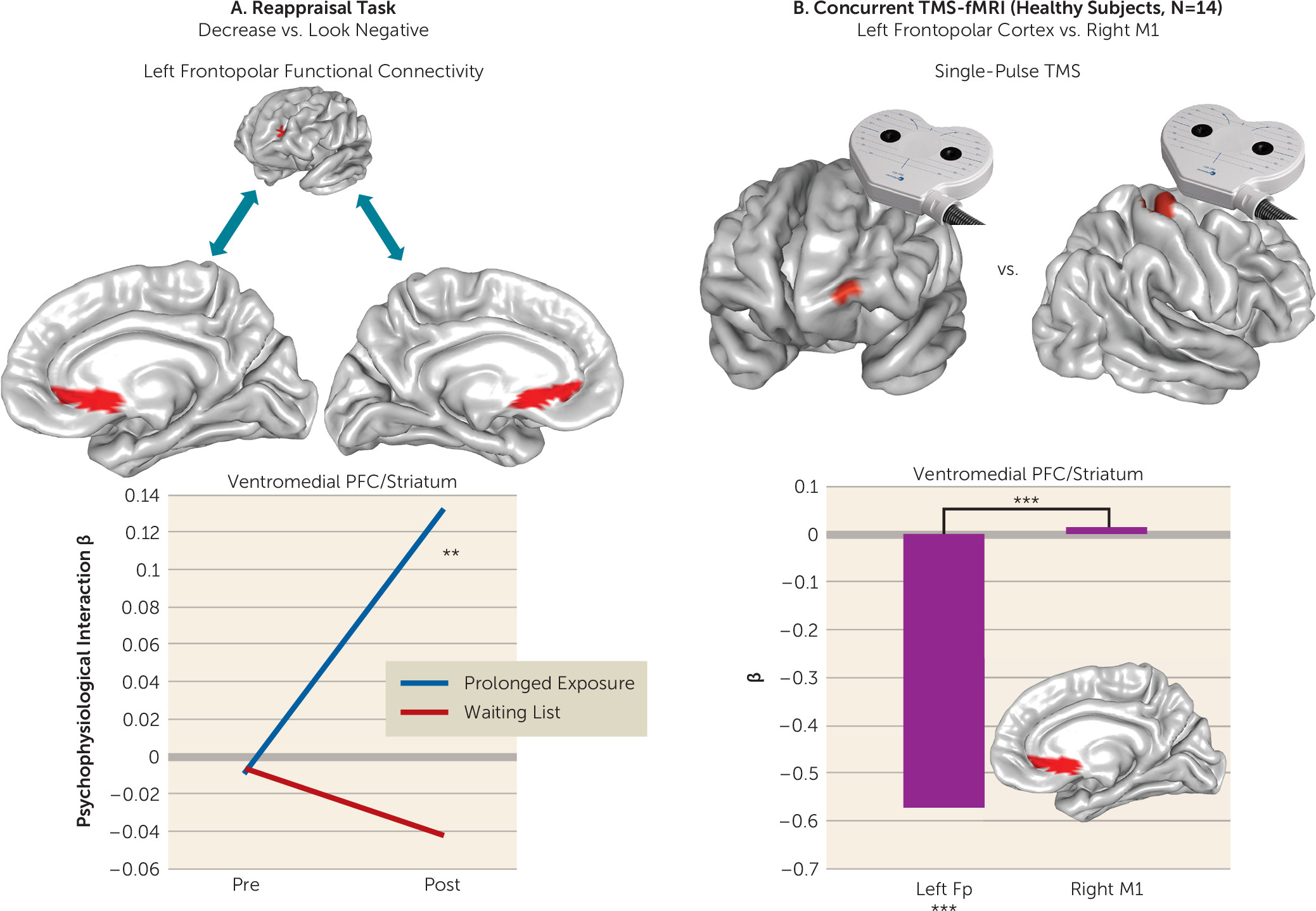
To deepen our mechanistic understanding of the reappraisal effect, we tested left lateral frontopolar cortex context-dependent functional connectivity for treatment-related changes using a generalized psychophysiological interaction analysis (27). We observed a significant time-by-treatment arm interaction effect in the ventromedial prefrontal cortex (olfactory cortex/anterior cingulate/mid-orbital gyrus; BA 25 and 32) extending into the adjacent ventral striatum (nucleus accumbens/caudate nucleus) (see Table S7 in the data supplement). Post hoc extractions revealed increased connectivity between this region and the lateral frontopolar cortex in the immediate treatment arm after treatment (t=3.09, p=0.003) (Figure 2A), with nonsignificantly decreased connectivity in the waiting list arm (t=−1.73, p=0.087). No additional effects were detected in the whole brain analysis.
FIGURE 2. Increased Connectivity Between Left Frontopolar Cortex and Ventromedial Prefrontal Cortex/Ventral Striatum After Prolonged Exposure Therapy: A Pathway of Direct Influence in Healthy Individualsa
a Panel A depicts the treatment arm-by-time interaction effect in which individuals in the prolonged exposure group (N=36) displayed significantly greater increases in connectivity from the left frontopolar cortex to the ventromedial prefrontal cortex/ventral striatum during cognitive reappraisal relative to individuals in the waiting list group (N=30). The line graph depicts the mean individual mixed model–derived predicted values for connectivity within each treatment arm and at each time point. The activation change and its connectivity target are rendered on average surfaces. Panel B depicts the downstream effect of left frontopolar stimulation in healthy individuals (N=14) on blood-oxygen-level-dependent signal change in this same ventromedial prefrontal/striatal region relative to right motor cortex stimulation, which was used as a comparison site. The bar graph depicts the mean individual ventromedial prefrontal/ventral striatal activation values for each stimulation site. The top left brain image depicts the frontopolar stimulation site, and the top right brain image depicts the right motor cortex stimulation site, both rendered on an average surface. fMRI=functional MRI; Fp=frontopolar cortex; M1=primary motor cortex; PFC=prefrontal cortex; Pre=pretreatment; Post=posttreatment; TMS=transcranial magnetic stimulation.
**p<0.01. ***p<0.001.

Brain-Behavior Relationships: Change in Frontopolar Activation

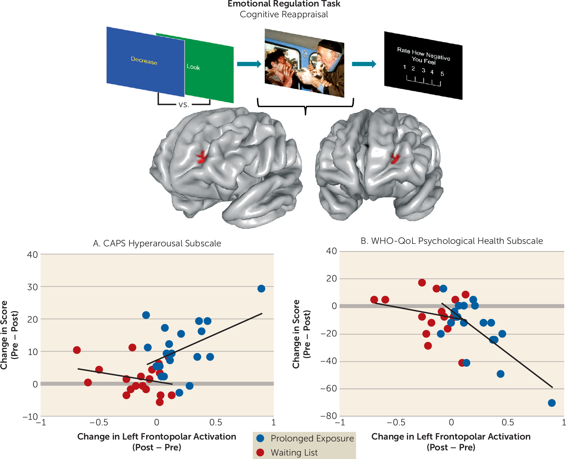
Next, we assessed whether change in left lateral frontopolar reappraisal activation was clinically meaningful by examining its relationship to change in measures of PTSD symptoms and quality of life. These measures were selected to represent outcomes that are disorder specific, symptom focused, and proximal treatment targets as well as those that are transdiagnostic, life-functioning focused, and more distal indicators of treatment success, respectively. Controlling for baseline symptoms and activation in a generalized linear model (with separate models for scores on the Clinician-Administered PTSD Scale for DSM-IV [CAPS], its three subscales, and the three subscales of the World Health Organization Quality of Life Scale–Brief Version [WHO-QoL-BREF]) and using Bonferroni correction for multiple comparisons (p<0.007), we found that greater increases in activation were associated with greater improvements in CAPS hyperarousal symptoms in the immediate treatment group (Wald χ2=7.71, p=0.005). This relationship was significantly different (Wald χ2=8.28, p=0.004) from the relationship between these two measures in the waiting list group (Figure 3A), which was nonsignificant. We also observed that increases in left frontopolar activation were associated with improvement in psychological well-being (the psychological health subscale of the WHO-QoL-BREF) in the immediate treatment group (Wald χ2=95.07, p<0.001). Again, this relationship was significantly different (Wald χ2=7.93, p=0.005) from the relationship between these two measures in the waiting list group (Figure 3B), which was also nonsignificant.
FIGURE 3. Association of Treatment-Related Changes in Left Frontopolar Activation During Reappraisal With Improvements in PTSD Hyperarousal Symptoms and Psychological Well-Beinga
a The diagram at the top depicts the reappraisal contrast from the task, and the treatment arm-by-time interaction effect in the left frontopolar cortex is rendered on an average brain surface below. Scatterplots depict relationships between average increases in within-subject activation over time in the left frontopolar cortex during reappraisal (pretreatment subtracted from posttreatment) and within-subject changes (posttreatment subtracted from pretreatment) in hyperarousal symptoms assessed by the CAPS (panel A) and psychological health assessed by the WHO-QoL (panel B). CAPS=Clinician-Administered PTSD Scale for DSM-IV; WHO-QoL=WHO Quality of Life Scale–Brief Version.

Assessing Treatment-Related Changes in Frontopolar Resting Entropy and Connectivity

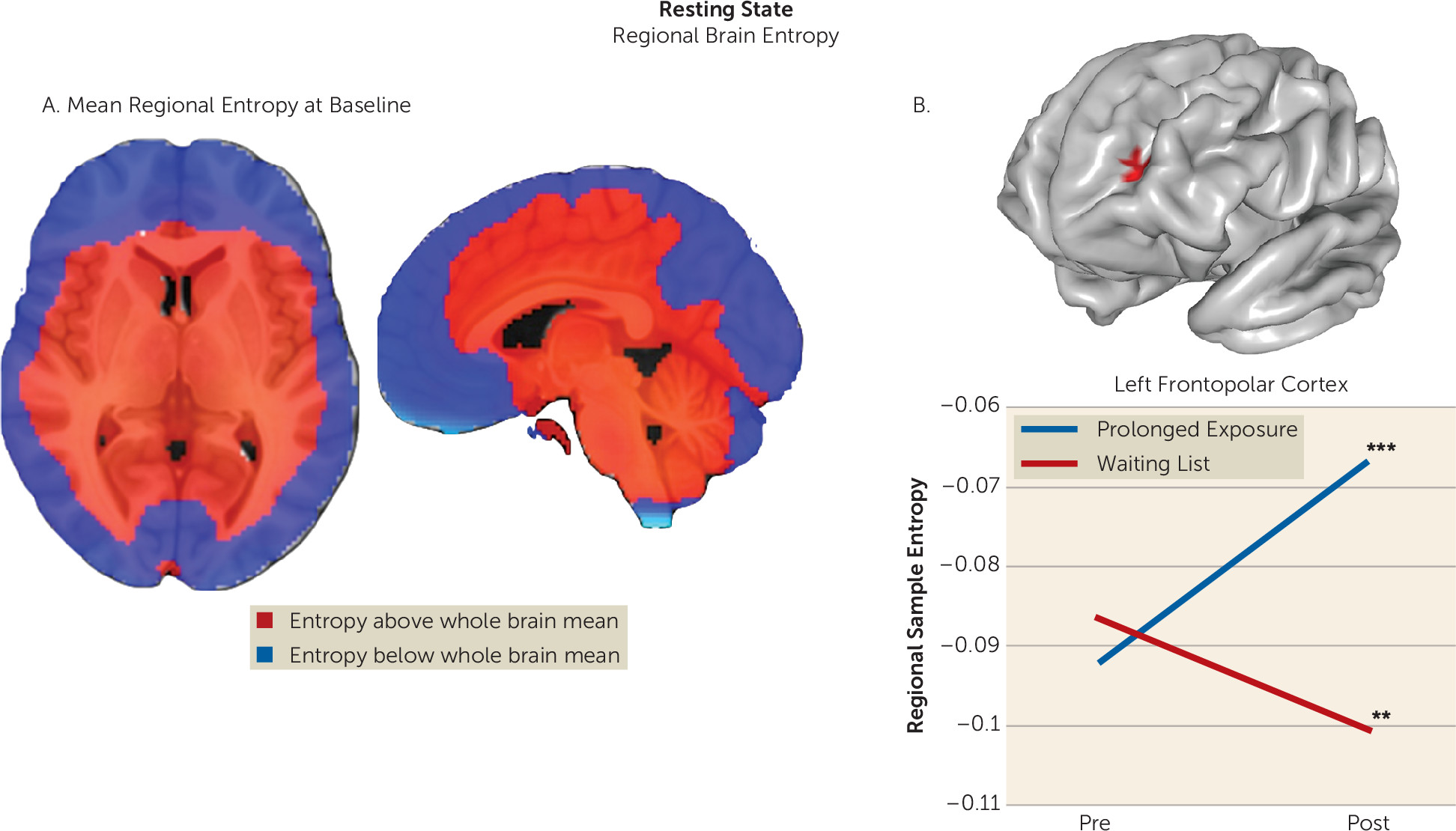
Because previous work has implicated the lateral frontopolar cortex in cognitive flexibility and switching between stimulus-dependent and stimulus-independent mental states (28, 29), we reasoned that frontopolar cortex change may be of a more general relevance and extend beyond emotional reactivity and regulation per se. Thus, we tested whether the blood-oxygen-level-dependent (BOLD) signal at rest in the lateral frontopolar cortex and its ventromedial connectivity target displayed properties of greater flexibility after psychotherapy, one potential brain marker of a more varied repertoire of mental states. We therefore calculated BOLD sample entropy (24) (Figure 4A), which provides a quantitative measure of the variety of change patterns over time. Using entropy values extracted from the clusters identified above, we observed a significant time-by-treatment arm interaction on entropy change in the lateral frontopolar cortex (F=26.57, p<0.001) but not in the ventromedial prefrontal cortex/striatum. In this frontopolar region, the effect was due to an increase in entropy in the treatment group (t=3.968, p<0.001) as well as a decrease in entropy in the waiting list group (t=−3.080, p=0.003) (Figure 4B). We also examined resting connectivity between these regions. We seeded the left frontopolar cortex region that showed change during reappraisal and examined whether intrinsic connectivity with the ventromedial prefrontal cortex/ventral striatum context-dependent connectivity target at rest changed with treatment. There was no significant treatment arm-by-time interaction effect on resting-state connectivity between the two regions demonstrating context-dependent connectivity change during reappraisal (see the Supplemental Results section in the data supplement). Thus, only the resting dynamics of the lateral frontopolar cortex displayed changes in patients after treatment. Specifically, this region showed no changes in intrinsic connectivity with the ventromedial prefrontal cortex/ventral striatum, but rather showed a time course of activity that was more entropic at rest, that is, varied and changed in a greater number of ways than prior to treatment.
FIGURE 4. Increased Resting Regional Brain Entropy After Prolonged Exposure of the Lateral Frontopolar Region Displaying Treatment-Related Change During Cognitive Reappraisala
a Panel A depicts a brain map of the mean regional brain entropy distribution across the entire PTSD sample at baseline, with regions displaying regional entropy values greater than the whole brain mean displayed in red and those displaying regional entropy values lower than the whole brain mean displayed in blue. The image is overlaid on the Montreal Neurological Institute 152-person average T1 structural template. Panel B depicts the reappraisal frontopolar activation effect rendered on an average brain surface. The line graph depicts the mean individual mixed model–derived predicted entropy values within each treatment arm at each time point. Pre=pretreatment; Post=posttreatment.
**p<0.01. ***p<0.001.

Follow-Up Experiment in Healthy Individuals: Testing Frontopolar Influence on the Ventromedial Prefrontal Cortex/Ventral Striatum Using Single-Pulse TMS With fMRI

Given evidence for functional and structural connections of the frontopolar and ventromedial prefrontal cortex in humans (28, 30, 31), we hypothesized that their interactions arise from a direct downstream influence of the frontopolar cortex on the ventromedial prefrontal cortex/ventral striatum. To test this hypothesis, we applied single TMS pulses to the left lateral frontopolar cortex in a separate sample of healthy participants undergoing concurrent TMS-fMRI (N=14). Single TMS pulses to the right hand knob of the primary motor cortex were used as an active comparison stimulation control site. We then compared average within-subject BOLD signal in the region defined by the ventromedial prefrontal/ventral striatal connectivity change during reappraisal (see Table S7 in the data supplement) for each stimulation site. In healthy individuals, TMS stimulation to the left frontopolar cortex induced significant deactivation in this ventromedial prefrontal cortex/ventral striatal region (t=−3.89, p=0.002), and this was significantly different relative to right motor cortex stimulation (t=−2.80, p=0.016) (Figure 2B), which itself did not have an effect. This effect was replicated in a voxel-wise analysis, and additional effects were seen in a whole brain analysis (see the Supplemental Results section and Table S8 in the data supplement).

Discussion

We assessed brain function in individuals with PTSD during emotional reactivity and regulation to better understand how prolonged exposure therapy conveys therapeutic benefit. No treatment-related changes were observed in reactivity to emotional cues or when regulating interference from emotional conflict. However, the left lateral frontopolar cortex displayed increased activation and increased connectivity with the ventromedial prefrontal cortex/ventral striatum during cognitive reappraisal after treatment. Concurrent TMS-fMRI in healthy participants demonstrated that frontopolar cortex stimulation modulates downstream activity in this connectivity target. Increases in frontopolar activation were related to improvement in hyperarousal symptoms and psychological well-being. Finally, the lateral frontopolar region showing activation change during cognitive reappraisal also demonstrated a wider variety of resting-state signal fluctuation patterns over time. Taken together, these findings indicate that 1) the most prominent therapeutic brain change following prolonged exposure is prefrontal rather than limbic and manifests during deliberate emotion regulation; 2) this change is clinically relevant and is related to improvement in symptoms and psychological well-being; 3) this change manifests in the lateral frontopolar cortex and its interactions with the ventromedial prefrontal cortex/ventral striatum, a recipient of frontopolar downstream influence; and 4) this change is evident during both regulation of emotion and at rest and may therefore reflect a generalized shift in frontopolar function.
These results inform a novel view of the brain mechanism of exposure therapy. In contrast to existing accounts of psychotherapy mechanisms (18, 32), we observed no limbic attenuation during emotional reactivity, no increased recruitment of posterior lateral prefrontal substrates implicated in top-down control (33), and no prefrontal change during emotional reactivity, emotional conflict, or emotional conflict regulation. Notably, this contrasts with treatment moderation results in this sample, wherein emotional reactivity and emotional conflict regulation–related brain function predicted treatment outcome, as reported in the companion article (34). Instead, we demonstrate that exposure therapy alters functioning of the most anterior portion of the prefrontal cortex (BA 10) during deliberate emotion regulation, as well as its connectivity with a ventromedial corticostriatal target that is a target of its downstream influence. Together, these findings point toward a prominent, selective effect of exposure therapy on a cortical substrate that is anatomically and functionally distinct (31) from other prefrontal cortical regions widely held to convey the efficacy of psychotherapy (18, 35).
In contrast to prefrontal cortical regions implicated in executive control or salience (36), the frontopolar cortex (also referred to as the anterior prefrontal cortex [31] or the rostral prefrontal cortex [29]) is believed to control the balance of stimulus-dependent attention (e.g., to the external environment) and stimulus-independent attention (e.g., attention toward the internal milieu) (29). The lateral frontopolar region identified here has been implicated in higher-order processes requiring a continual integration of inner mental phenomena with outward attention to “keep something in mind” while performing concurrent tasks (29). The frontopolar cortex is composed primarily of BA 10, a substrate with unique cytoarchitecture (31). Substantially enlarged in humans, it is one of the last regions to mature developmentally and is almost exclusively interconnected with higher-order associative cortices involved in cross-modal information integration (31). Hemodynamic changes in this region occur across many paradigms (29), consistent with its proposed role as a coordinator of multiple component cognitive functions processed by more posterior prefrontal areas (31). Meta-analytic data indicate that this region is activated by reappraisal (37), particularly in the later temporal phases (38), and is hypoactive in PTSD (39), suggesting that the effects observed here may indicate normalization of an abnormality.
Increased activation in this region was concomitant with increased ventromedial prefrontal (BA 25 and 32)/ventral striatal connectivity. Activation of this ventromedial (BA 25)/ventral striatal region moderated treatment response during emotional conflict regulation at baseline (34), illustrating a potential connection between these processes and a common substrate. BA 25, the subgenual cingulate, has been implicated in parasympathetic modulation of internal state (40), while the nucleus accumbens/ventral striatum has been shown to mediate relationships between successful reappraisal and both ventromedial prefrontal and frontopolar function (41). That greater activation in this region at baseline predicted more favorable psychotherapy outcomes in this sample was interpreted in the context of emotional conflict regulation as an enhanced capacity to attenuate arousal/vigilance after perturbation by a salient stimulus (34). Consistent with the proposed role of the lateral frontopolar cortex in switching between stimulus-dependent and stimulus-independent attention (42), this convergence suggests that psychotherapy may train the lateral frontopolar cortex to better evoke, amplify, or integrate attention toward an internal regulatory process that mediates successful emotion regulation and marks cessation of reappraisal (41). Clinically, this may manifest as less engagement in avoidance strategies to regulate emotional state and more moderate, less excessive responses to emotionally salient stimuli.
It is noteworthy that BA 10 has demonstrated psychotherapy-related changes in two PTSD imaging studies, one showing increased left hemisphere recruitment during script-driven imagery in police officers (10), and the other showing attenuated right hemisphere activation during anticipation of negative versus positive images in assaulted women (12). Thus, our findings add to accumulating evidence that frontopolar cortical function conveys at least some of the beneficial effects of PTSD psychotherapy. We expand on initial findings by demonstrating change in lateral frontopolar reappraisal-related activation, connectivity with the ventromedial prefrontal cortex/ventral striatum, and frontopolar resting entropy, as well as by demonstrating that lateral frontopolar cortex stimulation can directly modulate ventromedial prefrontal/ventral striatal function. Studies in social anxiety disorder have also demonstrated functional changes in BA 10 after treatment, for example, during social evaluation after treatment with nefazodone (43) and during threat processing after cognitive-behavioral therapy (44). The frontopolar cortex is also an efficacious TMS site for the treatment of major depression (45), and frontopolar cerebral blood flow indexes treatment response after exposure with response prevention for obsessive-compulsive disorder (46). Thus, the frontopolar cortex may be a transdiagnostic therapeutic target across disorders that are characterized by diminished positive affect and exaggerated fear, anxiety, and threat reactivity.
We demonstrate the capacity for lateral frontopolar stimulation to influence ventromedial prefrontal/striatal signal in healthy individuals, which provides initial evidence for an integrated communication pathway operating in multiple contexts. This communication may therefore reflect a process of general relevance, consistent with the interactions of these regions during a range of behaviors (28, 30) and with the proposed role of the frontopolar cortex as an attentional gate (29). Specifically, transient lateral frontopolar activations are also thought to support bidirectional switching between stimulus-dependent and stimulus-independent processing modes (29), which may underlie TMS-induced deactivation of the ventromedial prefrontal cortex. As this region is implicated in control and awareness of one’s internal state (40), attenuation of regional activity here by frontopolar stimulation may signal a shift away from a stimulus-independent state of rest (47). Likewise, increased resting entropy in the lateral frontopolar cortex after psychotherapy suggests that this region is able to function more flexibly and assume a more varied repertoire of configurations, which may reflect a wider range of mental states. Here, we utilized TMS-fMRI and resting-state data only to follow up on primary analyses of task findings, and we did not undertake an extensive investigation of these metrics. Therefore, the findings should be considered initial supporting evidence to better contextualize the results of task effects, while future investigations focusing specifically on TMS-fMRI and resting-state metrics in PTSD will provide further insights.
This study has several limitations. The first is the lack of a traumatized healthy comparison sample, which might have allowed us to determine whether functional changes reflect normalization of abnormalities or compensatory adaptations. Second, we did not collect frontopolar TMS-fMRI data in patients, which would have been most informative for this investigation. We note that the TMS-fMRI findings reported here may not necessarily apply to individuals with PTSD. Third, we did not counterbalance task order across participants, as it was not possible to ensure balanced administrations across randomized groups. However, this could also reduce generalizability of brain change effects if the task administration order exerted habituation effects on the brain dynamics in a given task that showed a differential change over time between treatment arms. It is notable that we did not detect hypothesized treatment-related changes in limbic regions (e.g., the amygdala and insula) previously demonstrated to be hyperactive in PTSD and to display changes after therapy (1416). This may reflect a lack of power to detect effects of smaller magnitude. However, it is also noteworthy that among randomized controlled PTSD imaging studies, changes in prefrontal function in the absence of limbic changes have been observed with a frequency (10, 11) equivalent to that of limbic changes (14, 16), which may be related to variation in the experimental task, study sample characteristics, or other factors. Further studies are needed to understand these sources of variation. Additionally, that effects were observed only during the reappraisal task could be related to differences in evoked arousal related to the complex affective picture stimuli utilized in that task, as opposed to the emotional faces utilized in other tasks. However, the fact that lateral frontopolar entropy changes were observed in the immediate treatment group at rest, a low-arousal state, suggests that this is not the case. Studies examining peripheral arousal measures during task completion (e.g., skin conductance response) will be helpful in delineating whether selective effects during one task are related to evoked arousal, the mental process engaged, or both.
Despite the limitations, our findings have import for understanding the mechanism of exposure therapy by demonstrating that the most prominent functional brain change during the processing and regulation of nontraumatic emotional stimuli occurs in an anatomically distinct, higher-order frontal structure (31). This region may also be responsible for the instantiation of a conceptually distinct process—a gating mechanism dictating the balance of awareness of the internal and external world (29). Although additional studies are needed to further elaborate on the functional significance of the frontopolar cortex in PTSD and its change after exposure therapy, the present findings identify an underexplored anatomical brain target and pathway of influence to the ventromedial prefrontal cortex with promise for stimulation-based therapeutics and augmentation of psychotherapy effects.
Footnote

ClinicalTrials.gov identifier: NCT01507948.

Supplementary Material

File (appi.ajp.2017.16091073.ds001.pdf)

References

1.
Marmar CR, Schlenger W, Henn-Haase C, et al: Course of posttraumatic stress disorder 40 years after the Vietnam War: findings from the National Vietnam Veterans Longitudinal Study. JAMA Psychiatry 2015; 72:875–881
2.
Schnurr PP, Lunney CA, Bovin MJ, et al: Posttraumatic stress disorder and quality of life: extension of findings to veterans of the wars in Iraq and Afghanistan. Clin Psychol Rev 2009; 29:727–735
3.
Cusack K, Jonas DE, Forneris CA, et al: Psychological treatments for adults with posttraumatic stress disorder: a systematic review and meta-analysis. Clin Psychol Rev 2016; 43:128–141
4.
Foa EB, Gillihan SJ, Bryant RA: Challenges and successes in dissemination of evidence-based treatments for posttraumatic stress: lessons learned from prolonged exposure therapy for PTSD. Psychol Sci Public Interest 2013; 14:65–111
5.
Foa EB, Hembree EA, Rothbaum BO: Prolonged Exposure Therapy for PTSD. Oxford, UK, Oxford University Press, 2007
6.
Foa EB, Kozak MJ: Emotional processing of fear: exposure to corrective information. Psychol Bull 1986; 99:20–35
7.
Bohning DE, Shastri A, McGavin L, et al: Motor cortex brain activity induced by 1-Hz transcranial magnetic stimulation is similar in location and level to that for volitional movement. Invest Radiol 2000; 35:676–683
8.
Levin P, Lazrove S, van der Kolk B: What psychological testing and neuroimaging tell us about the treatment of posttraumatic stress disorder by eye movement desensitization and reprocessing. J Anxiety Disord 1999; 13:159–172
9.
Lansing K, Amen DG, Hanks C, et al: High-resolution brain SPECT imaging and eye movement desensitization and reprocessing in police officers with PTSD. J Neuropsychiatry Clin Neurosci 2005; 17:526–532
10.
Peres JF, Newberg AB, Mercante JP, et al: Cerebral blood flow changes during retrieval of traumatic memories before and after psychotherapy: a SPECT study. Psychol Med 2007; 37:1481–1491
11.
Lindauer RJ, Booij J, Habraken JB, et al: Effects of psychotherapy on regional cerebral blood flow during trauma imagery in patients with post-traumatic stress disorder: a randomized clinical trial. Psychol Med 2008; 38:543–554
12.
Aupperle RL, Allard CB, Simmons AN, et al: Neural responses during emotional processing before and after cognitive trauma therapy for battered women. Psychiatry Res 2013; 214:48–55
13.
Rabe S, Zoellner T, Beauducel A, et al: Changes in brain electrical activity after cognitive behavioral therapy for posttraumatic stress disorder in patients injured in motor vehicle accidents. Psychosom Med 2008; 70:13–19
14.
Thomaes K, Dorrepaal E, Draijer N, et al: Treatment effects on insular and anterior cingulate cortex activation during classic and emotional Stroop interference in child abuse-related complex post-traumatic stress disorder. Psychol Med 2012; 42:2337–2349
15.
Felmingham K, Kemp A, Williams L, et al: Changes in anterior cingulate and amygdala after cognitive behavior therapy of posttraumatic stress disorder. Psychol Sci 2007; 18:127–129
16.
Peres JF, Foerster B, Santana LG, et al: Police officers under attack: resilience implications of an fMRI study. J Psychiatr Res 2011; 45:727–734
17.
Roy MJ, Francis J, Friedlander J, et al: Improvement in cerebral function with treatment of posttraumatic stress disorder. Ann N Y Acad Sci 2010; 1208:142–149
18.
Brooks SJ, Stein DJ: A systematic review of the neural bases of psychotherapy for anxiety and related disorders. Dialogues Clin Neurosci 2015; 17:261–279
19.
Etkin A, Klemenhagen KC, Dudman JT, et al: Individual differences in trait anxiety predict the response of the basolateral amygdala to unconsciously processed fearful faces. Neuron 2004; 44:1043–1055
20.
Etkin A, Schatzberg AF: Common abnormalities and disorder-specific compensation during implicit regulation of emotional processing in generalized anxiety and major depressive disorders. Am J Psychiatry 2011; 168:968–978
21.
Minkel JD, McNealy K, Gianaros PJ, et al: Sleep quality and neural circuit function supporting emotion regulation. Biol Mood Anxiety Disord 2012; 2:22
22.
Chen AC, Oathes DJ, Chang C, et al: Causal interactions between fronto-parietal central executive and default-mode networks in humans. Proc Natl Acad Sci USA 2013; 110:19944–19949
23.
Shannon CE: A mathematical theory of communication. Bell System Technical Journal 1948; 27:379–423
24.
Wang Z, Li Y, Childress AR, et al: Brain entropy mapping using fMRI. PLoS One 2014; 9:e89948
25.
Shuiabi E, Thomson V, Bhuiyan N: Entropy as a measure of operational flexibility. Eur J Oper Res 2005; 165:696–707
26.
Kraemer HC, Wilson GT, Fairburn CG, et al: Mediators and moderators of treatment effects in randomized clinical trials. Arch Gen Psychiatry 2002; 59:877–883
27.
McLaren DG, Ries ML, Xu G, et al: A generalized form of context-dependent psychophysiological interactions (gPPI): a comparison to standard approaches. Neuroimage 2012; 61:1277–1286
28.
Daw ND, O’Doherty JP, Dayan P, et al: Cortical substrates for exploratory decisions in humans. Nature 2006; 441:876–879
29.
Burgess PW, Dumontheil I, Gilbert SJ: The gateway hypothesis of rostral prefrontal cortex (area 10) function. Trends Cogn Sci 2007; 11:290–298
30.
Turner MS, Simons JS, Gilbert SJ, et al: Distinct roles for lateral and medial rostral prefrontal cortex in source monitoring of perceived and imagined events. Neuropsychologia 2008; 46:1442–1453
31.
Ramnani N, Owen AM: Anterior prefrontal cortex: insights into function from anatomy and neuroimaging. Nat Rev Neurosci 2004; 5:184–194
32.
Milad MR, Rosenbaum BL, Simon NM: Neuroscience of fear extinction: implications for assessment and treatment of fear-based and anxiety related disorders. Behav Res Ther 2014; 62:17–23
33.
Buhle JT, Silvers JA, Wager TD, et al: Cognitive reappraisal of emotion: a meta-analysis of human neuroimaging studies. Cereb Cortex 2014; 24:2981–2990
34.
Fonzo GA, Goodkind MS, Oathes DJ, et al: PTSD psychotherapy outcome predicted by brain activation during emotional reactivity and regulation. Am J Psychiatry 2017; 174:1163–1174
35.
Messina I, Sambin M, Palmieri A, et al: Neural correlates of psychotherapy in anxiety and depression: a meta-analysis. PLoS One 2013; 8:e74657
36.
Seeley WW, Menon V, Schatzberg AF, et al: Dissociable intrinsic connectivity networks for salience processing and executive control. J Neurosci 2007; 27:2349–2356
37.
Yarkoni T, Poldrack RA, Nichols TE, et al: Large-scale automated synthesis of human functional neuroimaging data. Nat Methods 2011; 8:665–670
38.
Kalisch R: The functional neuroanatomy of reappraisal: time matters. Neurosci Biobehav Rev 2009; 33:1215–1226
39.
Patel R, Spreng RN, Shin LM, et al: Neurocircuitry models of posttraumatic stress disorder and beyond: a meta-analysis of functional neuroimaging studies. Neurosci Biobehav Rev 2012; 36:2130–2142
40.
Critchley HD, Nagai Y, Gray MA, et al: Dissecting axes of autonomic control in humans: insights from neuroimaging. Auton Neurosci 2011; 161:34–42
41.
Wager TD, Davidson ML, Hughes BL, et al: Prefrontal-subcortical pathways mediating successful emotion regulation. Neuron 2008; 59:1037–1050
42.
Burgess PW, Gilbert SJ, Dumontheil I: Function and localization within rostral prefrontal cortex (area 10). Philos Trans R Soc Lond B Biol Sci 2007; 362:887–899
43.
Kilts CD, Kelsey JE, Knight B, et al: The neural correlates of social anxiety disorder and response to pharmacotherapy. Neuropsychopharmacology 2006; 31:2243–2253
44.
Klumpp H, Fitzgerald DA, Phan KL: Neural predictors and mechanisms of cognitive behavioral therapy on threat processing in social anxiety disorder. Prog Neuropsychopharmacol Biol Psychiatry 2013; 45:83–91
45.
Nahas Z, Anderson BS, Borckardt J, et al: Bilateral epidural prefrontal cortical stimulation for treatment-resistant depression. Biol Psychiatry 2010; 67:101–109
46.
Yamanishi T, Nakaaki S, Omori IM, et al: Changes after behavior therapy among responsive and nonresponsive patients with obsessive-compulsive disorder. Psychiatry Res 2009; 172:242–250
47.
Nagai Y, Critchley HD, Featherstone E, et al: Activity in ventromedial prefrontal cortex covaries with sympathetic skin conductance level: a physiological account of a “default mode” of brain function. Neuroimage 2004; 22:243–251

Information & Authors

Information

Published In

Go to American Journal of Psychiatry
American Journal of Psychiatry
Pages: 1175 - 1184
PubMed: 28715907

History

Received: 25 September 2016
Revision received: 31 March 2017
Accepted: 20 April 2017
Published online: 18 July 2017
Published in print: December 01, 2017

Keywords

  1. Posttraumatic Stress Disorder
  2. Brain Imaging Techniques
  3. Psychotherapy
  4. Emotion Regulation
  5. Transcranial Magnetic Stimulation
  6. Frontopolar Cortex

Authors

Details

Gregory A. Fonzo, Ph.D.
From the Department of Psychiatry and Behavioral Sciences, Stanford University School of Medicine, Stanford, Calif.; the Stanford Neurosciences Institute, Stanford University, Stanford; the Veterans Affairs Palo Alto Healthcare System and the Sierra Pacific Mental Illness, Research, Education, and Clinical Center (MIRECC), Palo Alto, Calif.; the New Mexico Veterans Affairs Healthcare System, Albuquerque; the Center for Neuromodulation in Depression and Stress, Department of Psychiatry, University of Pennsylvania Perelman School of Medicine, Philadelphia; and the Department of Psychiatry, Emory University School of Medicine, Atlanta.
Madeleine S. Goodkind, Ph.D.
From the Department of Psychiatry and Behavioral Sciences, Stanford University School of Medicine, Stanford, Calif.; the Stanford Neurosciences Institute, Stanford University, Stanford; the Veterans Affairs Palo Alto Healthcare System and the Sierra Pacific Mental Illness, Research, Education, and Clinical Center (MIRECC), Palo Alto, Calif.; the New Mexico Veterans Affairs Healthcare System, Albuquerque; the Center for Neuromodulation in Depression and Stress, Department of Psychiatry, University of Pennsylvania Perelman School of Medicine, Philadelphia; and the Department of Psychiatry, Emory University School of Medicine, Atlanta.
Desmond J. Oathes, Ph.D.
From the Department of Psychiatry and Behavioral Sciences, Stanford University School of Medicine, Stanford, Calif.; the Stanford Neurosciences Institute, Stanford University, Stanford; the Veterans Affairs Palo Alto Healthcare System and the Sierra Pacific Mental Illness, Research, Education, and Clinical Center (MIRECC), Palo Alto, Calif.; the New Mexico Veterans Affairs Healthcare System, Albuquerque; the Center for Neuromodulation in Depression and Stress, Department of Psychiatry, University of Pennsylvania Perelman School of Medicine, Philadelphia; and the Department of Psychiatry, Emory University School of Medicine, Atlanta.
Yevgeniya V. Zaiko, B.A.
From the Department of Psychiatry and Behavioral Sciences, Stanford University School of Medicine, Stanford, Calif.; the Stanford Neurosciences Institute, Stanford University, Stanford; the Veterans Affairs Palo Alto Healthcare System and the Sierra Pacific Mental Illness, Research, Education, and Clinical Center (MIRECC), Palo Alto, Calif.; the New Mexico Veterans Affairs Healthcare System, Albuquerque; the Center for Neuromodulation in Depression and Stress, Department of Psychiatry, University of Pennsylvania Perelman School of Medicine, Philadelphia; and the Department of Psychiatry, Emory University School of Medicine, Atlanta.
Meredith Harvey, B.A.
From the Department of Psychiatry and Behavioral Sciences, Stanford University School of Medicine, Stanford, Calif.; the Stanford Neurosciences Institute, Stanford University, Stanford; the Veterans Affairs Palo Alto Healthcare System and the Sierra Pacific Mental Illness, Research, Education, and Clinical Center (MIRECC), Palo Alto, Calif.; the New Mexico Veterans Affairs Healthcare System, Albuquerque; the Center for Neuromodulation in Depression and Stress, Department of Psychiatry, University of Pennsylvania Perelman School of Medicine, Philadelphia; and the Department of Psychiatry, Emory University School of Medicine, Atlanta.
Kathy K. Peng, M.A.
From the Department of Psychiatry and Behavioral Sciences, Stanford University School of Medicine, Stanford, Calif.; the Stanford Neurosciences Institute, Stanford University, Stanford; the Veterans Affairs Palo Alto Healthcare System and the Sierra Pacific Mental Illness, Research, Education, and Clinical Center (MIRECC), Palo Alto, Calif.; the New Mexico Veterans Affairs Healthcare System, Albuquerque; the Center for Neuromodulation in Depression and Stress, Department of Psychiatry, University of Pennsylvania Perelman School of Medicine, Philadelphia; and the Department of Psychiatry, Emory University School of Medicine, Atlanta.
M. Elizabeth Weiss, Ph.D.
From the Department of Psychiatry and Behavioral Sciences, Stanford University School of Medicine, Stanford, Calif.; the Stanford Neurosciences Institute, Stanford University, Stanford; the Veterans Affairs Palo Alto Healthcare System and the Sierra Pacific Mental Illness, Research, Education, and Clinical Center (MIRECC), Palo Alto, Calif.; the New Mexico Veterans Affairs Healthcare System, Albuquerque; the Center for Neuromodulation in Depression and Stress, Department of Psychiatry, University of Pennsylvania Perelman School of Medicine, Philadelphia; and the Department of Psychiatry, Emory University School of Medicine, Atlanta.
Allison L. Thompson, Ph.D.
From the Department of Psychiatry and Behavioral Sciences, Stanford University School of Medicine, Stanford, Calif.; the Stanford Neurosciences Institute, Stanford University, Stanford; the Veterans Affairs Palo Alto Healthcare System and the Sierra Pacific Mental Illness, Research, Education, and Clinical Center (MIRECC), Palo Alto, Calif.; the New Mexico Veterans Affairs Healthcare System, Albuquerque; the Center for Neuromodulation in Depression and Stress, Department of Psychiatry, University of Pennsylvania Perelman School of Medicine, Philadelphia; and the Department of Psychiatry, Emory University School of Medicine, Atlanta.
Sanno E. Zack, Ph.D.
From the Department of Psychiatry and Behavioral Sciences, Stanford University School of Medicine, Stanford, Calif.; the Stanford Neurosciences Institute, Stanford University, Stanford; the Veterans Affairs Palo Alto Healthcare System and the Sierra Pacific Mental Illness, Research, Education, and Clinical Center (MIRECC), Palo Alto, Calif.; the New Mexico Veterans Affairs Healthcare System, Albuquerque; the Center for Neuromodulation in Depression and Stress, Department of Psychiatry, University of Pennsylvania Perelman School of Medicine, Philadelphia; and the Department of Psychiatry, Emory University School of Medicine, Atlanta.
Colleen E. Mills-Finnerty, Ph.D.
From the Department of Psychiatry and Behavioral Sciences, Stanford University School of Medicine, Stanford, Calif.; the Stanford Neurosciences Institute, Stanford University, Stanford; the Veterans Affairs Palo Alto Healthcare System and the Sierra Pacific Mental Illness, Research, Education, and Clinical Center (MIRECC), Palo Alto, Calif.; the New Mexico Veterans Affairs Healthcare System, Albuquerque; the Center for Neuromodulation in Depression and Stress, Department of Psychiatry, University of Pennsylvania Perelman School of Medicine, Philadelphia; and the Department of Psychiatry, Emory University School of Medicine, Atlanta.
Benjamin M. Rosenberg, B.A.
From the Department of Psychiatry and Behavioral Sciences, Stanford University School of Medicine, Stanford, Calif.; the Stanford Neurosciences Institute, Stanford University, Stanford; the Veterans Affairs Palo Alto Healthcare System and the Sierra Pacific Mental Illness, Research, Education, and Clinical Center (MIRECC), Palo Alto, Calif.; the New Mexico Veterans Affairs Healthcare System, Albuquerque; the Center for Neuromodulation in Depression and Stress, Department of Psychiatry, University of Pennsylvania Perelman School of Medicine, Philadelphia; and the Department of Psychiatry, Emory University School of Medicine, Atlanta.
Raleigh Edelstein, B.A.
From the Department of Psychiatry and Behavioral Sciences, Stanford University School of Medicine, Stanford, Calif.; the Stanford Neurosciences Institute, Stanford University, Stanford; the Veterans Affairs Palo Alto Healthcare System and the Sierra Pacific Mental Illness, Research, Education, and Clinical Center (MIRECC), Palo Alto, Calif.; the New Mexico Veterans Affairs Healthcare System, Albuquerque; the Center for Neuromodulation in Depression and Stress, Department of Psychiatry, University of Pennsylvania Perelman School of Medicine, Philadelphia; and the Department of Psychiatry, Emory University School of Medicine, Atlanta.
Rachael N. Wright, B.S.
From the Department of Psychiatry and Behavioral Sciences, Stanford University School of Medicine, Stanford, Calif.; the Stanford Neurosciences Institute, Stanford University, Stanford; the Veterans Affairs Palo Alto Healthcare System and the Sierra Pacific Mental Illness, Research, Education, and Clinical Center (MIRECC), Palo Alto, Calif.; the New Mexico Veterans Affairs Healthcare System, Albuquerque; the Center for Neuromodulation in Depression and Stress, Department of Psychiatry, University of Pennsylvania Perelman School of Medicine, Philadelphia; and the Department of Psychiatry, Emory University School of Medicine, Atlanta.
Carena A. Kole, B.S.
From the Department of Psychiatry and Behavioral Sciences, Stanford University School of Medicine, Stanford, Calif.; the Stanford Neurosciences Institute, Stanford University, Stanford; the Veterans Affairs Palo Alto Healthcare System and the Sierra Pacific Mental Illness, Research, Education, and Clinical Center (MIRECC), Palo Alto, Calif.; the New Mexico Veterans Affairs Healthcare System, Albuquerque; the Center for Neuromodulation in Depression and Stress, Department of Psychiatry, University of Pennsylvania Perelman School of Medicine, Philadelphia; and the Department of Psychiatry, Emory University School of Medicine, Atlanta.
Steven E. Lindley, M.D., Ph.D.
From the Department of Psychiatry and Behavioral Sciences, Stanford University School of Medicine, Stanford, Calif.; the Stanford Neurosciences Institute, Stanford University, Stanford; the Veterans Affairs Palo Alto Healthcare System and the Sierra Pacific Mental Illness, Research, Education, and Clinical Center (MIRECC), Palo Alto, Calif.; the New Mexico Veterans Affairs Healthcare System, Albuquerque; the Center for Neuromodulation in Depression and Stress, Department of Psychiatry, University of Pennsylvania Perelman School of Medicine, Philadelphia; and the Department of Psychiatry, Emory University School of Medicine, Atlanta.
Bruce A. Arnow, Ph.D.
From the Department of Psychiatry and Behavioral Sciences, Stanford University School of Medicine, Stanford, Calif.; the Stanford Neurosciences Institute, Stanford University, Stanford; the Veterans Affairs Palo Alto Healthcare System and the Sierra Pacific Mental Illness, Research, Education, and Clinical Center (MIRECC), Palo Alto, Calif.; the New Mexico Veterans Affairs Healthcare System, Albuquerque; the Center for Neuromodulation in Depression and Stress, Department of Psychiatry, University of Pennsylvania Perelman School of Medicine, Philadelphia; and the Department of Psychiatry, Emory University School of Medicine, Atlanta.
Booil Jo, Ph.D.
From the Department of Psychiatry and Behavioral Sciences, Stanford University School of Medicine, Stanford, Calif.; the Stanford Neurosciences Institute, Stanford University, Stanford; the Veterans Affairs Palo Alto Healthcare System and the Sierra Pacific Mental Illness, Research, Education, and Clinical Center (MIRECC), Palo Alto, Calif.; the New Mexico Veterans Affairs Healthcare System, Albuquerque; the Center for Neuromodulation in Depression and Stress, Department of Psychiatry, University of Pennsylvania Perelman School of Medicine, Philadelphia; and the Department of Psychiatry, Emory University School of Medicine, Atlanta.
James J. Gross, Ph.D.
From the Department of Psychiatry and Behavioral Sciences, Stanford University School of Medicine, Stanford, Calif.; the Stanford Neurosciences Institute, Stanford University, Stanford; the Veterans Affairs Palo Alto Healthcare System and the Sierra Pacific Mental Illness, Research, Education, and Clinical Center (MIRECC), Palo Alto, Calif.; the New Mexico Veterans Affairs Healthcare System, Albuquerque; the Center for Neuromodulation in Depression and Stress, Department of Psychiatry, University of Pennsylvania Perelman School of Medicine, Philadelphia; and the Department of Psychiatry, Emory University School of Medicine, Atlanta.
Barbara O. Rothbaum, Ph.D.
From the Department of Psychiatry and Behavioral Sciences, Stanford University School of Medicine, Stanford, Calif.; the Stanford Neurosciences Institute, Stanford University, Stanford; the Veterans Affairs Palo Alto Healthcare System and the Sierra Pacific Mental Illness, Research, Education, and Clinical Center (MIRECC), Palo Alto, Calif.; the New Mexico Veterans Affairs Healthcare System, Albuquerque; the Center for Neuromodulation in Depression and Stress, Department of Psychiatry, University of Pennsylvania Perelman School of Medicine, Philadelphia; and the Department of Psychiatry, Emory University School of Medicine, Atlanta.
Amit Etkin, M.D., Ph.D. [email protected]
From the Department of Psychiatry and Behavioral Sciences, Stanford University School of Medicine, Stanford, Calif.; the Stanford Neurosciences Institute, Stanford University, Stanford; the Veterans Affairs Palo Alto Healthcare System and the Sierra Pacific Mental Illness, Research, Education, and Clinical Center (MIRECC), Palo Alto, Calif.; the New Mexico Veterans Affairs Healthcare System, Albuquerque; the Center for Neuromodulation in Depression and Stress, Department of Psychiatry, University of Pennsylvania Perelman School of Medicine, Philadelphia; and the Department of Psychiatry, Emory University School of Medicine, Atlanta.

Notes

Address correspondence to Dr. Etkin ([email protected]).
Presented at the 54th annual meeting of the American College of Neuropsychopharmacology, Hollywood, Fla., Dec. 6–10, 2015.

Author Contributions

Drs. Fonzo and Goodkind contributed equally as first authors.

Funding Information

National Institute of Mental Health10.13039/100000025: R01 MH091860, T32 MH019938
Supported by NIMH grant R01 MH091860 to Dr. Etkin. Dr. Fonzo was partially supported by NIMH grant T32 MH019938.Dr. Rothbaum has received funding from the Wounded Warrior Project, the Department of Defense (Clinical Trial Grant W81XWH-10-1-1045), NIMH (grant 1R01MH094757-01), a Brain and Behavior Research Foundation (NARSAD) Distinguished Investigator Grant, the McCormick Foundation, and Transcept Pharmaceuticals; she has served on an advisory board for Genentech, owns equity in Virtually Better, and has received royalties from Oxford University Press, Guilford, American Psychiatric Publishing, and Emory University. Dr. Etkin has served as a consultant for Takeda, Otsuka, and Acadia, has received a research grant from Brain Resource, Inc., and owns equity in Akili Interactive. The other authors report no financial relationships with commercial interests.

Metrics & Citations

Metrics

Citations

Export Citations

If you have the appropriate software installed, you can download article citation data to the citation manager of your choice. Simply select your manager software from the list below and click Download.

For more information or tips please see 'Downloading to a citation manager' in the Help menu.

Format
Citation style
Style
Copy to clipboard

View Options

View options

PDF/EPUB

View PDF/EPUB

Get Access

Login options

Already a subscriber? Access your subscription through your login credentials or your institution for full access to this article.

Personal login Institutional Login Open Athens login
Purchase Options

Purchase this article to access the full text.

PPV Articles - American Journal of Psychiatry

PPV Articles - American Journal of Psychiatry

Not a subscriber?

Subscribe Now / Learn More

PsychiatryOnline subscription options offer access to the DSM-5-TR® library, books, journals, CME, and patient resources. This all-in-one virtual library provides psychiatrists and mental health professionals with key resources for diagnosis, treatment, research, and professional development.

Need more help? PsychiatryOnline Customer Service may be reached by emailing [email protected] or by calling 800-368-5777 (in the U.S.) or 703-907-7322 (outside the U.S.).

Media

Figures

Other

Tables

Share

Share

Share article link

Share