Abstract

Objective:

This study tested for differences of white matter integrity between treated and never-treated long-term schizophrenia patients, matched on illness duration, and for differential changes in relation to age in these two groups relative to healthy comparison subjects.

Method:

This cross-sectional diffusion tensor imaging study included 31 never-treated and 46 matched antipsychotic-treated patients with long-term schizophrenia and 58 healthy comparison subjects. Fractional anisotropy measures of white matter tracts were extracted and compared. Linear regression analysis was used to explore the association between age and fractional anisotropy among the three groups.

Results:

Fractional anisotropy significantly differed among the three groups in 14 of 20 white matter tracts defined in the Johns Hopkins University white matter template. Never-treated patients displayed greater reduction of fractional anisotropy than antipsychotic-treated patients in the left anterior thalamic radiation, the left cingulum-hippocampus pathway, the splenium and genu of the corpus callosum, and the left superior longitudinal fasciculus, and greater fractional anisotropy in the right uncinate fasciculus. Both patient groups showed multiple reductions relative to healthy comparison subjects. Never-treated patients showed an accelerated and clinically relevant age-related reduction of fractional anisotropy in the genu of the corpus callosum.

Conclusions:

These psychoradiological findings provide insight into the regional distribution of white matter deficits in the years after illness onset in long-term schizophrenia. Findings of greater impairments in never-treated patients, and a greater age-related reduction in the genu of the corpus callosum in these patients, suggest that long-term antipsychotic treatment does not adversely affect white matter tracts over the longer-term course of illness and may confer benefits.
Psychosocial deterioration over the course of illness in schizophrenia is well established (13). Several studies have reported progressive cerebral alterations as well (4, 5). However, the degree to which these latter changes are progressive over the longer-term course of illness and the degree to which they are secondary to antipsychotic treatment remain unclear. Separating influences of these two mechanisms remains challenging because nearly all patients are treated following diagnosis, and antipsychotic drugs are known to have robust effects on brain anatomy (611). In this context, a cross-sectional comparison of duration-matched never-treated and treated long-term schizophrenia patients may shed light on progression and treatment effects on brain anatomy over the longer-term course of illness.
Recent advances in neuroimaging methodology have led to a large increase in diseased brain studies. In particular, the evolving field of psychoradiology (https://radiopaedia.org/articles/psychoradiology) provides not only insights into abnormal brain function and underlying neuropsychopathologies but also potential clinical utility in patients with psychiatric disorders, from diagnosis and prognosis to planning and monitoring therapeutic interventions (12).
Multiple longitudinal psychoradiological studies of treated patients document decreases in whole brain, frontal, and temporal lobe volumes, as well as increases in ventricle size, from 1 to 7 years following illness onset (5, 9, 13, 14). Another line of work has reported both gray and white matter changes after antipsychotic treatment (6, 7, 11, 1518). However, fewer studies have reviewed white matter changes in longitudinal studies (14) and after antipsychotic treatment (8, 1924), and most involve shorter-term follow-up. Psychoradiological studies of white matter change during short-term antipsychotic treatment have reported both increased (23) and decreased fractional anisotropy (8, 25) after 6 to 12 weeks of antipsychotic treatment. Over a 5-year follow-up of early-course patients, increased white matter alterations have been reported (26). Cross-sectional studies suggest accelerated loss of white matter integrity with age in schizophrenia, but results are inconsistent, and again the potential influences of antipsychotic treatment remain uncertain (4, 2737). Generally, while some clinical and preclinical data suggest that antipsychotic medications may have beneficial effects on white matter with chronic treatment (36, 37), other data, especially after acute treatment in early-course patients, suggest adverse effects (19, 20, 22).
A direct comparison of never-treated and treated long-term schizophrenia patients with a wide range of illness duration provides a tractable strategy for contrasting brain alterations in relation to illness duration and to test for differences related to treatment status. Although a cross-sectional study has limitations, such a study can provide information relative to important questions about course of illness and long-term drug effects on brain alterations in schizophrenia that is not otherwise available.
In developing countries such as China and India, there have been reports of neuroimaging findings in rare cases of never-treated long-term schizophrenia identified by community outreach programs in less affluent and rural areas (3840). In a study of patients who were never treated for more than 5 years after illness onset, we demonstrated greater age-related cortical thinning in prefrontal and temporal gyri than in healthy comparison subjects (40). In a study of 17 long-term never-treated schizophrenia patients with a mean illness duration of 15 years (SD=6) (39), a limited reduction of fractional anisotropy in the left temporal lobe was observed relative to healthy comparison subjects. In this latter study with a relatively limited age range, no significant association was observed between white matter deficits and age or illness duration, and there was no comparison with treated patients.
In the present study, we conducted diffusion tensor imaging (DTI) studies with long-term never-treated schizophrenia patients, antipsychotic-treated patients matched on illness duration, and healthy comparison subjects. We hypothesized that more severe white matter deficits would be found in never-treated long-term schizophrenia patients relative to treated patients and that age-related decline in white matter tract integrity would be accelerated in never-treated patients relative to treated patients and healthy comparison subjects, especially in the corpus callosum.

Method

Participants

Thirty-one never-treated long-term schizophrenia patients with illness duration ranging from 5 to 47 years, 46 illness duration–matched schizophrenia patients who had received long-term antipsychotic treatment, and 58 healthy comparison subjects underwent DTI studies. Participants were all of Han ancestry and thus were ethnically homogeneous. Illness onset was determined using the Nottingham Onset Schedule (41) with information provided by patients, family members, and medical records. Diagnosis of schizophrenia was confirmed using the Structured Clinical Interview for DSM-IV. Psychiatric symptoms were assessed using the Positive and Negative Syndrome Scale (PANSS) and were more severe in the never-treated patients (Table 1). All participants were right handed. The study was approved by the West China Hospital research ethics committee of Sichuan University, and written informed consent was obtained from all study participants.
TABLE 1. Demographic and Clinical Characteristics of Never-Treated Long-Term Schizophrenia Patients, Antipsychotic-Treated Schizophrenia Patients of Similar Illness Duration, and Healthy Comparison Subjects
CharacteristicNever-Treated Long-Term Schizophrenia Patients (N=31)Antipsychotic-Treated Schizophrenia Patients (N=46)Healthy Comparison Subjects (N=58)p
 MeanSDMeanSDMeanSD 
Age (years)46.3513.1946.229.5445.0310.300.80
Education (years)7.733.958.673.379.292.880.06
Duration of illness (years)21.2912.0621.6510.32 0.89
Age at onset25.086.6724.675.98 0.78
Positive and Negative Syndrome Scale scores    
 Total score92.6118.1452.4410.79 <0.001
 Positive symptom score23.296.4810.943.08 <0.001
 Negative symptom score24.829.0617.786.70 <0.001
 General psychopathology symptom score43.898.0024.724.62 <0.001
Antipsychotic dosage (chlorpromazine equivalents, mg/day) 823.90617.70  
 N%N%N%p
Gender      0.76
 Female1548.42350.03356.9 
 Male1651.62350.02543.1 
Never-treated patients were identified from a community mental health screening program designed to identify and provide psychiatric care to individuals with serious but untreated mental illness. The never-treated schizophrenia patients, most in their 40s or older, were typically recruited from within 35 km of downtown Chengdu, a large city in western China. Twenty lived in rural areas, mostly in small villages on the outskirts of Chengdu, and the others in urban or suburban areas. Based on available retrospective information, onset of symptoms was gradual and insidious for 22 of the patients, and the other nine developed acute psychosis after some significant life trauma, such as the loss of a close relative or parental marital difficulties. Nineteen had no history of employment. Twelve were able to care for themselves in many activities of daily living. Most patients and their family members had 9 years of education, including 6 years in primary school and 3 years in junior high school, as was common in China when they were in school. Patients had not received treatment with psychiatric medications for various reasons, primarily because of parental concern about family stigma (prominent in 17 cases); a lack of family understanding of the severity of mental illness in a family member (noteworthy in seven cases); poor socioeconomic conditions that limited travel and funds for medical care (four cases); and conflicts with physicians when the patient was first brought to medical attention near the time of illness onset (three cases). As a result of these factors, these patients had been cared for and sheltered in the parents’ home without medical care through the course of their illness.
The treated patient group was recruited from the same or nearby communities and had received antipsychotic treatment relatively consistently beginning early in their course of illness, according to medical records and reports from family members and patients. Both never-treated and treated patients had no known neurological disorder or comorbidity of other psychiatric disorders, including alcohol or drug abuse. None had significant medical conditions, or treatments for such conditions, with known impact on CNS function or anatomy. Because of the long history of treatment by different providers, details of antipsychotic drug treatment for each treated patient are not available, but we estimated typical medication dosages for the most recent 10 years using all available data (Table 1).

Data Acquisition

MRI examinations were performed using a 3-T scanner (EXCITE, General Electric, Milwaukee) with an eight-channel phase array head coil. DTI data were acquired using a bipolar diffusion weighted spin-echo echo planar imaging sequence (TR=10,000 ms, TE=70 ms) with a 128×128 matrix over a field of view of 240×240 mm, with 50 axial slices of 3-mm thickness covering the whole brain without gap. Each DTI data set included 15 images of unique diffusion directions (B=1,000) and a nondiffusion image (B=0). High-resolution T1-weighted anatomical images were acquired for registration purposes using a three-dimensional spoiled gradient sequence (TR=8.5 ms, TE=3.5 ms, TI=400 ms, flip angle=12°) with a 240×240 matrix over a field of view of 240×240 mm and 156 axial slices of 1-mm thickness. All scans were reviewed by an experienced neuroradiologist to exclude individuals with gross brain abnormalities.

Imaging Processing

Routine DTI preprocessing, with head motion and eddy current correction, brain extraction, and tensor model fitting, was performed using FSL (FMRIB Software Library, http://www.fmrib.ox.ac.uk/fsl). We then used automated fiber quantification software (42) (https://github.com/jyeatman/AFQ) to identify 20 white matter tracts in individual subjects. The identification procedure involved three primary steps: whole brain deterministic fiber tractography, waypoint region of interest–based tract segmentation, and probability map–based fiber refinement using the 20-tract Johns Hopkins University white matter template (http://neuro.debian.net/pkgs/fsl-jhu-dti-whitematter-atlas.html). Tracts included the left and right anterior thalamic radiation, corticospinal tract, cingulum-cingulate, cingulum-hippocampus pathway, inferior fronto-occipital fasciculus, inferior longitudinal fasciculus, superior longitudinal fasciculus, uncinate fasciculus and arcuate fasciculus, and the forceps major of the splenium and the forceps minor of the genu of the corpus callosum. After tract identification, the diffusion measurement along the tract core, defined as the tract profile, was extracted from each fiber tract. Tracts were smoothed using a 10-point moving average filter to reduce local variation caused by imaging noise.

Statistical Analysis

We used analysis of variance (ANOVA) (three groups by 20 regions) to compare average fractional anisotropy values among the three groups across fiber tracts, followed by post hoc one-way ANOVAs comparing groups on tracts separately, and pairwise post hoc tests in tracts with significant group differences using Tukey’s multiple comparison procedure. In exploratory descriptive analyses, we examined each tract separately divided into 100 equal segments to characterize group differences along fiber tracts using pointwise comparisons (see Figure S1 in the online supplement).
Pearson correlation analysis between fractional anisotropy of fiber tracts that showed significant group differences and PANSS scores was performed to evaluate relationships between fiber tract integrity and clinical symptoms. Linear regression analysis was used to explore associations between age and fractional anisotropy among the three groups; regression coefficients were compared across groups to test for differential rates of age-related change across groups.

Results

Fractional Anisotropy Alterations in Never-Treated and Treated Long-Term Schizophrenia Patients

The two-way ANOVA demonstrated a significant main effect for group (F=60.35, df=2, 2640, p<0.0001), fiber tract (F=225.6, df=19, 2640, p<0.0001), and their interaction (F=1.57, df=38, 2640, p=0.015) (p values corrected for multiple comparisons). Fractional anisotropy of white matter microstructure for the three participant groups differed significantly in the left and right anterior radiation, the left cingulum-hippocampus pathway, the splenium and genu of the corpus callosum, the left and right inferior fronto-occipital fasciculus, the left and right inferior frontal fasciculus, the left and right superior longitudinal fasciculus, the left and right uncinate fasciculus, and the left arcuate fasciculus (Table 2, p<0.05, corrected).
TABLE 2. Fractional Anisotropy Among Never-Treated Long-Term Schizophrenia Patients, Antipsychotic-Treated Schizophrenia Patients of Similar Illness Duration, and Healthy Comparison Subjects
Fractional Anisotropy LocationNever-Treated Long-Term Schizophrenia Patients (NTSZ)Antipsychotic-Treated Schizophrenia Patients of Similar Illness Duration (TSZ)Healthy Comparison Subjects (HC)AnalysisPost Hoc Pairwise Comparisons
 MeanSDMeanSDMeanSDFpNTSZ versus TSZNTSZ versus HCTSZ versus HC
Left anterior thalamic radiation0.4480.0440.4700.0370.4750.0355.130.007** 
Right anterior thalamic radiation0.4600.0390.4730.0370.4790.0283.170.045 * 
Left corticospinal tract0.6050.0360.6190.0440.6070.0281.950.147   
Right corticospinal tract0.5890.0300.6090.0460.6010.0292.970.055   
Left cingulum-cingulate0.4520.0610.4670.0770.4840.0682.380.097   
Right cingulum-cingulate0.4260.0580.4370.0680.4530.0452.670.073   
Left cingulum-hippocampus pathway0.4140.0540.4360.0430.4430.0394.530.013** 
Right cingulum-hippocampus pathway0.4180.0370.4300.0540.4360.0561.140.322   
Splenium of corpus callosum0.5760.0760.6050.0730.6150.0602.450.043**** 
Genu of corpus callosum0.5460.0530.5660.0360.5990.03213.83<0.001******
Left inferior fronto-occipital fasciculus0.4890.0430.4870.0430.5190.03011.50<0.001 ***
Right inferior fronto-occipital fasciculus0.4820.0370.4800.0450.5090.0309.25<0.001 ***
Left inferior longitudinal fasciculus0.4530.0360.4610.0320.4780.0307.86<0.001 * 
Right inferior longitudinal fasciculus0.4240.0310.4200.0400.4380.0304.060.020  *
Left superior longitudinal fasciculus0.4290.0530.4570.0520.4540.0453.550.031** 
Right superior longitudinal fasciculus0.4290.0440.4370.0570.4630.0426.290.003 ***
Left uncinate fasciculus0.4600.0510.4550.0430.4770.0473.070.049 **
Right uncinate fasciculus0.4210.0350.3940.0550.4360.03212.72<0.001**
Left arcuate fasciculus0.4800.0540.5000.0520.5070.0353.610.030 * 
Right arcuate fasciculus0.4620.0460.4660.0500.4830.0422.980.054   
*
p<0.05.
**
p<0.01.
***
p<0.001.
p<0.0001.
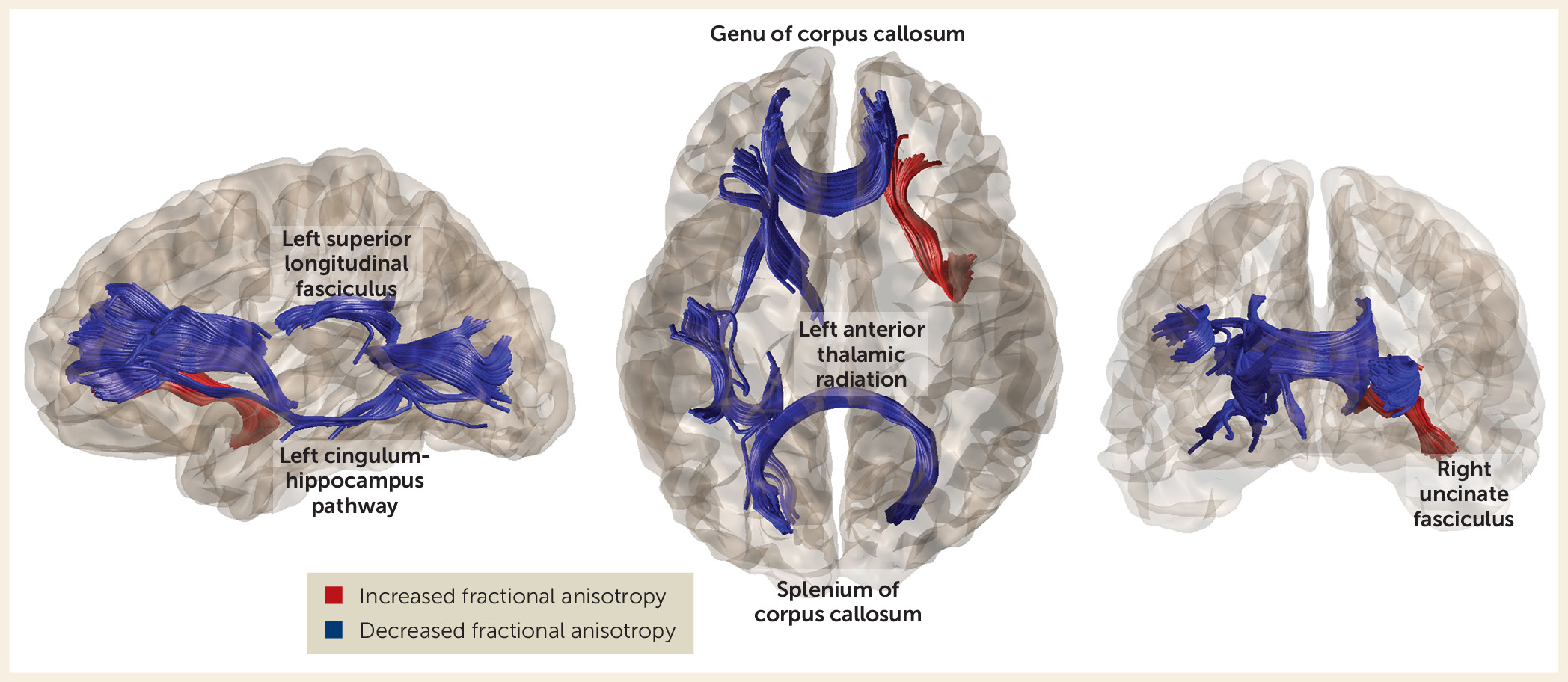
Comparisons of the two schizophrenia groups revealed significantly reduced fractional anisotropy in the never-treated group in the left anterior thalamic radiation, the left cingulum-hippocampus pathway, the splenium and genu of the corpus callosum, and the left superior longitudinal fasciculus, as well as greater fractional anisotropy in the right uncinate fasciculus (Figure 1). Pairwise comparisons showed multiple abnormalities relative to comparison subjects in both patient groups (Table 2).
FIGURE 1. Comparison of Mean Fractional Anisotropy in Brain White Matter Tracts Between Never-Treated Long-Term Schizophrenia Patients and Antipsychotic-Treated Schizophrenia Patients of Similar Illness Durationa
a Red and blue indicate increased and decreased fractional anisotropy, respectively, in fibers in never-treated relative to antipsychotic-treated schizophrenia patients.

Correlations Between Changes in Fractional Anisotropy and Clinical Symptoms

Within the never-treated schizophrenia patient group, among tracts with decreased fractional anisotropy relative to healthy comparison subjects, reductions in fractional anisotropy of the genu of the corpus callosum (p<0.05, r=−0.42) and the right superior longitudinal fasciculus (p<0.05, r=−0.44) were associated with greater total PANSS score. No correlations were significant in the antipsychotic-treated schizophrenia patients.

Relationship of Age and White Matter Microstructure

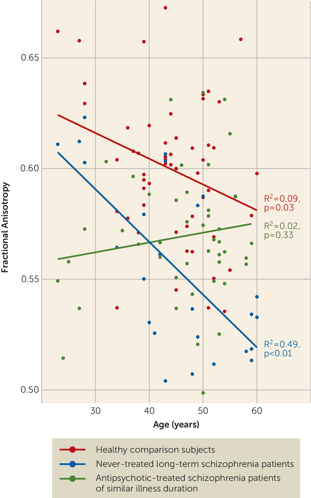
We examined relationships between age and fractional anisotropy for white matter fibers that differed among the three groups. The slopes of the three regression lines were significantly different (F=10.0617, p<0.0001) in the genu of the corpus callosum. Post hoc analyses compared regression coefficients (slopes) between each pair of participant groups, with p values false-discovery-rate-corrected for multiple comparisons. The age-related decline in fractional anisotropy of the genu of the corpus callosum in never-treated patients was significantly greater than that in healthy comparison subjects (F=4.60, p=0.035) and treated patients (F=16.26, p<0.001) (Figure 2). Treated patients showed limited change with age, with significantly less age-related change than even healthy comparison subjects (F=5.27, p=0.024). Treated patients also demonstrated somewhat greater reduction in fractional anisotropy closer to illness onset and treatment initiation, with little decline in relation to age (Figure 2).
FIGURE 2. Relationships Between Age and Mean Fractional Anisotropy of the Genu of the Corpus Callosum in Treated and Never-Treated Schizophrenia Patients, and Healthy Comparison Subjectsa
a Outliers were excluded when age effects were modeled to better track the potential trajectory in later life of individuals with never-treated long-term schizophrenia and healthy comparison subjects.

Discussion

By comparing a rare group of never-treated long-term schizophrenia patients with both treated illness duration–matched patients and healthy comparison subjects, the present study demonstrated altered fractional anisotropy in both patient groups but greater fractional anisotropy alterations in never-treated patients. These alterations were predominantly in pathways of the frontal cortex (Figure 1, Table 2). In addition to extending previous reports of fractional anisotropy alteration in both treated and never-treated schizophrenia (29, 39, 43, 44), we demonstrated a greater age-related reduction of fractional anisotropy in the genu of the corpus callosum in never-treated long-term schizophrenia patients relative to both healthy comparison subjects and antipsychotic-treated patients. Fractional anisotropy of the genu of the corpus callosum was related to symptom severity among never-treated patients. These findings provide novel insights into white matter abnormalities over the long-term course of illness without the influences of antipsychotic medication, and the findings regarding the anterior corpus callosum carry clinical relevance.
The greater reductions of fractional anisotropy in several fiber bundles throughout the brain in never-treated patients relative to treated patients suggest that long-term antipsychotic treatment may not only not cause adverse effects but may confer some beneficial effects on brain white matter via cumulative pharmacological effects or indirect benefits from treated psychosis, as has been argued previously (43, 45, 46). Animal studies have shown that antipsychotics can facilitate oligodendrocyte regeneration and myelin repair following injury (47). Because the oligodendrocyte abnormalities widely observed in postmortem studies of schizophrenia are thought to be one reason for decreased fractional anisotropy in white matter (48, 49), this is one possible mechanism by which antipsychotic treatment might have beneficial effects on brain white matter. There are multiple direct and indirect mechanisms by which antipsychotic treatment may confer beneficial effects on white matter. Although the specific mechanisms remain to be identified, our findings of greater reduction of fractional anisotropy in never-treated patients relative to treated patients are consistent with a possible beneficial role of long-term antipsychotic treatment on brain white matter over the course of schizophrenia.
It is noteworthy that findings from the few previous short-term studies of antipsychotic medications on white matter integrity in schizophrenia are varied, with reports of increased fractional anisotropy (23), decreased fractional anisotropy (8, 25), and no changes (24) after 6–12 weeks of acute antipsychotic treatment. Because our findings indicate greater fractional anisotropy reductions in never-treated patients than in treated patients across multiple tracts, it is possible that there are differences between early and longer-term antipsychotic treatment effects on white matter integrity. Acute treatment effects may be adverse, as suggested by some first-episode data (8, 25) and by the somewhat lower fractional anisotropy values in younger treated patients in the present study. Longer-term treatment effects may be positive via cumulative pharmacologic effects, which may reduce adverse effects of persistent acute psychosis and improve multiple factors in general medical health over the illness course.
There are several significant challenges to studying very long-term effects of antipsychotic drugs on brain systems in patients. Besides the decades of follow-up required for longitudinal studies to examine such effects directly, there are problems of participant attrition, changes in MRI systems over time, difficulty controlling drug treatment plans, variable treatment adherence over a lifespan, and the fact that illness course and drug treatment are confounded because nearly all schizophrenia patients are treated once diagnosed, and dosage is related to illness severity. In the face of these challenges, the field lacks direct evidence about long-term effects of antipsychotic drugs on the brain in schizophrenia patients.
In this context, the present cross-sectional study of never-treated patients with long-term illness failed to detect evidence for an adverse effect of longer-term antipsychotic treatment. In addition, our data provide suggestive evidence supporting preclinical data indicating a potential beneficial effect of antipsychotic treatment—specifically, that it may help preserve white matter microstructure over the course of illness (50, 51). Such an effect may in part explain the better overall clinical outcomes in antipsychotic-treated patients than in never-treated patients over the course of longer-term follow-up (38).
The greater age-related reduction of fractional anisotropy in the genu of the corpus callosum in never-treated patients than in treated patients and healthy comparison subjects suggests that in this circuitry, antipsychotic treatment effects might be particularly beneficial. The association of genu alterations with clinical symptoms in the present study and in previous reports (52) strengthens this argument. Abnormalities in the genu have been reported previously (27, 29, 43) and have been considered an important neurobiological observation because the tract provides most interhemispheric connectivity of the frontal lobes. This connectivity has long been known to influence cognitive and affective processes that are disrupted in schizophrenia (43, 53). Previous observations of greater callosal alterations in chronic patients relative to first-episode patients (54) are also consistent with the decline over the illness course seen in our never-treated patients.
Patterns observed in the genu of treated patients were notably distinct, as shown in Figure 2. This group demonstrated little change with age, with fractional anisotropy appearing to be more reduced in the earlier years after illness onset compared with never-treated patients. This observation parallels that of a recent study in which healthy controls showed a modest fractional anisotropy decline with age while treated patients showed little age-related change. In that study, younger treated patients had lower fractional anisotropy than controls, but the difference disappeared with age (27). One possible interpretation of these findings in relation to our own data is that any beneficial effects of antipsychotic treatment may be cumulative over the years of illness course or more pronounced later in the course of illness.
In our previous study with a partially overlapping sample of never-treated long-term patients, we observed an increased rate of age-related decline in cortical thickness in the ventral frontal cortex and superior temporal gyrus (40). In the present study, while tracts in the temporal lobe had abnormal fractional anisotropy, the only differential age-related fiber tract effect in the never-treated patients was in the genu of the corpus callosum. This tract connects ventral prefrontal cortex regions across the hemispheres and thus may be related to previously reported accelerated age-related changes in ventral prefrontal cortical thickness.
Although findings of fractional anisotropy reduction were consistent across most tracts, an exception was the observation that the fractional anisotropy of the right uncinate fasciculus was higher in untreated patients than in treated patients. The uncinate fasciculus is a late-maturing pathway connecting the hippocampus and amygdala with the orbitofrontal cortex and thus is important for emotion and cognitive processing (55). This finding may suggest a specific and different effect of antipsychotic treatment on this pathway, leading to decreased diffusion signaling along this fiber tract. While short-term effects on social cognition do not appear to be adverse (56, 57), longer-term effects remain to be determined.
Several limitations should be considered when interpreting our findings. First, inferences that differences between the two patient groups are related to their treatment history are constrained by the lack of an experimental control (i.e., random assignment of patients to the two patient groups). Our approach may be perhaps the only feasible immediate-term strategy for addressing questions about the long-term course of schizophrenia and antipsychotic drug effects on brain changes. However, the validity of our inferences depends on the observed group differences representing treatment effects rather than some systematic difference in our patient groups only indirectly related to their treatment history. In the developed world, patients who refuse treatment can be atypical. In the circumstances of the developing world, where access to services has been historically limited, economic and cultural factors and proximity to care often determine treatment access. Furthermore, the high level of psychopathology in the never-treated patient group does not suggest that they were higher functioning or had less severe psychotic disorders, allowing them to function well in the community. These considerations, together with the fact that our patient groups were recruited from the same community and did not differ in demographic factors, age at onset, or illness duration, limit to some degree concerns that our data reflect intrinsic patient group differences rather than treatment history effects. Nonetheless, these factors need to be considered when interpreting our findings.
Second, given limitations associated with our cross-sectional design, age-related modeling provides only a preliminary test for progressive brain changes. Third, as there were robust group differences across multiple tracts but differential aging effects limited to the genu, much of the change in DTI parameters appears to occur earlier than was evaluated in our study and appears not to be significantly and differentially progressive with age (5 years after onset and later). Fourth, we used a DTI protocol with 15 directions long used in our schizophrenia studies. Fifteen diffusion gradients could limit sensitivity to detect changes in the microstructural integrity of white matter fibers, especially in superficial areas. However, we note that we had sufficient power to detect significant effects in most tracts with the DTI protocol employed. Fifth, given variable drug choice and dosage over time, we were not able to link these treatment parameters to white matter alterations observed in the treated patient group. Last, to clarify mechanisms of effects, animal model work will be needed to examine longer-term antipsychotic effects on white matter to experimentally confirm a relationship between antipsychotic drugs and white matter changes, and to establish the neurobiological mechanisms underlying the relationship.
The present study revealed more widespread alteration of white matter microstructure in never-treated long-term schizophrenia patients than in those who received long-term antipsychotic treatment. These psychoradiological findings provide insights into white matter deficits over the course of schizophrenia without the confounding effects of antipsychotic medication. The results also suggest that long-term antipsychotic treatment not only does not appear to confer adverse effects on brain white matter but also may provide some benefits for white matter over the course of illness.
Acknowledgments

Supported by the National Natural Science Foundation (grants 81621003, 81671664, 81371527, 81030027, and 81761128023), by the National Youth Top-Notch Talent Support Program, by the China Medical Board Distinguished Professorship Award (F510000/G16916411), by the National Key Technologies Research and Development Program (2012BAI01B03), by the Program for Changjiang Scholars and Innovative Research Team in University (grant IRT16R52) of China, and by a grant from the Humboldt Foundation to Dr. Sweeney.

Supplementary Material

File (appi.ajp.2018.17121402.ds001.pdf)

References

1.
Austin SF, Mors O, Budtz-Jørgensen E, et al: Long-term trajectories of positive and negative symptoms in first episode psychosis: a 10-year follow-up study in the OPUS cohort. Schizophr Res 2015; 168:84–91
2.
Kotov R, Fochtmann L, Li K, et al: Declining clinical course of psychotic disorders over the two decades following first hospitalization: evidence from the Suffolk County Mental Health Project. Am J Psychiatry 2017; 174:1064–1074
3.
Velthorst E, Fett AJ, Reichenberg A, et al: The 20-year longitudinal trajectories of social functioning in individuals with psychotic disorders. Am J Psychiatry 2017; 174:1075–1085
4.
Cropley VL, Klauser P, Lenroot RK, et al: Accelerated gray and white matter deterioration with age in schizophrenia. Am J Psychiatry 2017; 174:286–295
5.
Fusar-Poli P, Smieskova R, Kempton MJ, et al: Progressive brain changes in schizophrenia related to antipsychotic treatment? A meta-analysis of longitudinal MRI studies. Neurosci Biobehav Rev 2013; 37:1680–1691
6.
Konopaske GT, Dorph-Petersen KA, Pierri JN, et al: Effect of chronic exposure to antipsychotic medication on cell numbers in the parietal cortex of macaque monkeys. Neuropsychopharmacology 2007; 32:1216–1223
7.
Konopaske GT, Dorph-Petersen KA, Sweet RA, et al: Effect of chronic antipsychotic exposure on astrocyte and oligodendrocyte numbers in macaque monkeys. Biol Psychiatry 2008; 63:759–765
8.
Szeszko PR, Robinson DG, Ikuta T, et al: White matter changes associated with antipsychotic treatment in first-episode psychosis. Neuropsychopharmacology 2014; 39:1324–1331
9.
Ho BC, Andreasen NC, Ziebell S, et al: Long-term antipsychotic treatment and brain volumes: a longitudinal study of first-episode schizophrenia. Arch Gen Psychiatry 2011; 68:128–137
10.
Keshavan MS, Bagwell WW, Haas GL, et al: Changes in caudate volume with neuroleptic treatment. Lancet 1994; 344:1434
11.
Vita A, De Peri L, Deste G, et al: The effect of antipsychotic treatment on cortical gray matter changes in schizophrenia: does the class matter? A meta-analysis and meta-regression of longitudinal magnetic resonance imaging studies. Biol Psychiatry 2015; 78:403–412
12.
Lui S, Zhou XJ, Sweeney JA, et al: Psychoradiology: the frontier of neuroimaging in psychiatry. Radiology 2016; 281:357–372
13.
Vita A, De Peri L, Deste G, et al: Progressive loss of cortical gray matter in schizophrenia: a meta-analysis and meta-regression of longitudinal MRI studies. Transl Psychiatry 2012; 2:e190
14.
Dietsche B, Kircher T, Falkenberg I: Structural brain changes in schizophrenia at different stages of the illness: a selective review of longitudinal magnetic resonance imaging studies. Aust N Z J Psychiatry 2017; 51:500–508
15.
Dorph-Petersen KA, Pierri JN, Perel JM, et al: The influence of chronic exposure to antipsychotic medications on brain size before and after tissue fixation: a comparison of haloperidol and olanzapine in macaque monkeys. Neuropsychopharmacology 2005; 30:1649–1661
16.
Lesh TA, Tanase C, Geib BR, et al: A multimodal analysis of antipsychotic effects on brain structure and function in first-episode schizophrenia. JAMA Psychiatry 2015; 72:226–234
17.
Lottman KK, Kraguljac NV, White DM, et al: Risperidone effects on brain dynamic connectivity: a prospective resting-state fMRI study in schizophrenia. Front Psychiatry 2017; 8:14
18.
Torres US, Duran FL, Schaufelberger MS, et al: Patterns of regional gray matter loss at different stages of schizophrenia: a multisite, cross-sectional VBM study in first-episode and chronic illness. Neuroimage Clin 2016; 12:1–15
19.
Bartzokis G, Lu PH, Stewart SB, et al: In vivo evidence of differential impact of typical and atypical antipsychotics on intracortical myelin in adults with schizophrenia. Schizophr Res 2009; 113:322–331
20.
Wang HN, Liu GH, Zhang RG, et al: Quetiapine ameliorates schizophrenia-like behaviors and protects myelin integrity in cuprizone intoxicated mice: the involvement of notch signaling pathway. Int J Neuropsychopharmacol 2015; 19:pyv088
21.
Hu M, Zong X, Zheng J, et al: Risperidone-induced topological alterations of anatomical brain network in first-episode drug-naive schizophrenia patients: a longitudinal diffusion tensor imaging study. Psychol Med 2016; 46:2549–2560
22.
Goghari VM, Lang DJ, Khorram B, et al: Anterior internal capsule volumes increase in patients with schizophrenia switched from typical antipsychotics to olanzapine. J Psychopharmacol 2011; 25:621–629
23.
Reis Marques T, Taylor H, Chaddock C, et al: White matter integrity as a predictor of response to treatment in first episode psychosis. Brain 2014; 137:172–182
24.
Zeng B, Ardekani BA, Tang Y, et al: Abnormal white matter microstructure in drug-naive first episode schizophrenia patients before and after eight weeks of antipsychotic treatment. Schizophr Res 2016; 172:1–8
25.
Wang Q, Cheung C, Deng W, et al: White-matter microstructure in previously drug-naive patients with schizophrenia after 6 weeks of treatment. Psychol Med 2013; 43:2301–2309
26.
Sun Y, Chen Y, Lee R, et al: Disruption of brain anatomical networks in schizophrenia: a longitudinal, diffusion tensor imaging based study. Schizophr Res 2016; 171:149–157
27.
Kanaan RA, Picchioni MM, McDonald C, et al: White matter deficits in schizophrenia are global and don’t progress with age. Aust N Z J Psychiatry 2017; 51:1020–1031
28.
Bijanki KR, Hodis B, Magnotta VA, et al: Effects of age on white matter integrity and negative symptoms in schizophrenia. Schizophr Res 2015; 161:29–35
29.
Kelly S, Jahanshad N, Zalesky A, et al: Widespread white matter microstructural differences in schizophrenia across 4322 individuals: results from the ENIGMA Schizophrenia DTI Working Group. Mol Psychiatry 2018; 23:1261–1269
30.
Ellison-Wright I, Bullmore E: Meta-analysis of diffusion tensor imaging studies in schizophrenia. Schizophr Res 2009; 108:3–10
31.
Kanaan RA, Kim JS, Kaufmann WE, et al: Diffusion tensor imaging in schizophrenia. Biol Psychiatry 2005; 58:921–929
32.
Zalesky A, Fornito A, Seal ML, et al: Disrupted axonal fiber connectivity in schizophrenia. Biol Psychiatry 2011; 69:80–89
33.
Levitt JJ, Nestor PG, Levin L, et al: Reduced structural connectivity in frontostriatal white matter tracts in the associative loop in schizophrenia. Am J Psychiatry 2017; 174:1102–1111
34.
Nelson BG, Bassett DS, Camchong J, et al: Comparison of large-scale human brain functional and anatomical networks in schizophrenia. Neuroimage Clin 2017; 15:439–448
35.
White T, Ehrlich S, Ho BC, et al: Spatial characteristics of white matter abnormalities in schizophrenia. Schizophr Bull 2013; 39:1077–1086
36.
Vos T, Flaxman AD, Naghavi M, et al: Years lived with disability (YLDs) for 1160 sequelae of 289 diseases and injuries 1990–2010: a systematic analysis for the Global Burden of Disease Study 2010. Lancet 2012; 380:2163–2196; correction, 2013; 381:628
37.
Whiteford HA, Ferrari AJ, Degenhardt L, et al: The global burden of mental, neurological and substance use disorders: an analysis from the Global Burden of Disease Study 2010. PLoS One 2015; 10:e0116820
38.
Ran MS, Weng X, Chan CL, et al: Different outcomes of never-treated and treated patients with schizophrenia: 14-year follow-up study in rural China. Br J Psychiatry 2015; 207:495–500
39.
Liu X, Lai Y, Wang X, et al: Reduced white matter integrity and cognitive deficit in never-medicated chronic schizophrenia: a diffusion tensor study using TBSS. Behav Brain Res 2013; 252:157–163
40.
Zhang W, Deng W, Yao L, et al: Brain structural abnormalities in a group of never-medicated patients with long-term schizophrenia. Am J Psychiatry 2015; 172:995–1003
41.
Singh SP, Cooper JE, Fisher HL, et al: Determining the chronology and components of psychosis onset: the Nottingham Onset Schedule (NOS). Schizophr Res 2005; 80:117–130
42.
Yeatman JD, Dougherty RF, Myall NJ, et al: Tract profiles of white matter properties: automating fiber-tract quantification. PLoS One 2012; 7:e49790
43.
Ellison-Wright I, Nathan PJ, Bullmore ET, et al: Distribution of tract deficits in schizophrenia. BMC Psychiatry 2014; 14:99
44.
Kochunov P, Williamson DE, Lancaster J, et al: Fractional anisotropy of water diffusion in cerebral white matter across the lifespan. Neurobiol Aging 2012; 33:9–20
45.
Bartzokis G, Lu PH, Amar CP, et al: Long acting injection versus oral risperidone in first-episode schizophrenia: differential impact on white matter myelination trajectory. Schizophr Res 2011; 132:35–41
46.
Bartzokis G: Neuroglialpharmacology: myelination as a shared mechanism of action of psychotropic treatments. Neuropharmacology 2012; 62:2137–2153
47.
Zhang Y, Zhang H, Wang L, et al: Quetiapine enhances oligodendrocyte regeneration and myelin repair after cuprizone-induced demyelination. Schizophr Res 2012; 138:8–17
48.
Trépanier MO, Hopperton KE, Mizrahi R, et al: Postmortem evidence of cerebral inflammation in schizophrenia: a systematic review. Mol Psychiatry 2016; 21:1009–1026
49.
Vikhreva OV, Rakhmanova VI, Orlovskaya DD, et al: Ultrastructural alterations of oligodendrocytes in prefrontal white matter in schizophrenia: a post-mortem morphometric study. Schizophr Res 2016; 177:28–36
50.
Hu X, Zhou H, Zhang D, et al: Clozapine protects dopaminergic neurons from inflammation-induced damage by inhibiting microglial overactivation. J Neuroimmune Pharmacol 2012; 7:187–201
51.
Jiang L, Wu X, Wang S, et al: Clozapine metabolites protect dopaminergic neurons through inhibition of microglial NADPH oxidase. J Neuroinflammation 2016; 13:110
52.
Saito J, Hori M, Nemoto T, et al: Longitudinal study examining abnormal white matter integrity using a tract-specific analysis in individuals with a high risk for psychosis. Psychiatry Clin Neurosci 2017; 71:530–541
53.
Edwards TJ, Sherr EH, Barkovich AJ, et al: Clinical, genetic and imaging findings identify new causes for corpus callosum development syndromes. Brain 2014; 137:1579–1613
54.
Zhuo C, Liu M, Wang L, et al: Diffusion tensor MR imaging evaluation of callosal abnormalities in schizophrenia: a meta-analysis. PLoS One 2016; 11:e0161406
55.
Singh S, Singh K, Trivedi R, et al: Microstructural abnormalities of uncinate fasciculus as a function of impaired cognition in schizophrenia: a DTI study. J Biosci 2016; 41:419–426
56.
Insel T, Cuthbert B, Garvey M, et al: Research domain criteria (RDoC): toward a new classification framework for research on mental disorders. Am J Psychiatry 2010; 167:748–751
57.
Chekroud AM, Zotti RJ, Shehzad Z, et al: Cross-trial prediction of treatment outcome in depression: a machine learning approach. Lancet Psychiatry 2016; 3:243–250

Information & Authors

Information

Published In

Go to American Journal of Psychiatry
American Journal of Psychiatry
Pages: 1129 - 1136
PubMed: 30068259

History

Received: 29 December 2017
Revision received: 27 March 2018
Accepted: 17 May 2018
Published online: 2 August 2018
Published in print: November 01, 2018

Keywords

  1. Schizophrenia
  2. Psychoradiology
  3. Brain Imaging Techniques

Authors

Details

Yuan Xiao, M.D.
From Huaxi MR Research Center (HMRRC), Department of Radiology, West China Hospital of Sichuan University, Chengdu, Sichuan, China; the Department of Psychiatry, Chengdu Tianfu New District Psychiatric Hospital, Chengdu, Sichuan, China; the Department of Psychiatry, Chengdu First Mental Health Prevention Hospital, Chengdu, Sichuan, China; and the Department of Psychiatry and Behavioral Neuroscience, University of Cincinnati, Cincinnati.
Huaiqiang Sun, Ph.D.
From Huaxi MR Research Center (HMRRC), Department of Radiology, West China Hospital of Sichuan University, Chengdu, Sichuan, China; the Department of Psychiatry, Chengdu Tianfu New District Psychiatric Hospital, Chengdu, Sichuan, China; the Department of Psychiatry, Chengdu First Mental Health Prevention Hospital, Chengdu, Sichuan, China; and the Department of Psychiatry and Behavioral Neuroscience, University of Cincinnati, Cincinnati.
Shulin Shi, M.D.
From Huaxi MR Research Center (HMRRC), Department of Radiology, West China Hospital of Sichuan University, Chengdu, Sichuan, China; the Department of Psychiatry, Chengdu Tianfu New District Psychiatric Hospital, Chengdu, Sichuan, China; the Department of Psychiatry, Chengdu First Mental Health Prevention Hospital, Chengdu, Sichuan, China; and the Department of Psychiatry and Behavioral Neuroscience, University of Cincinnati, Cincinnati.
Dan Jiang, M.D.
From Huaxi MR Research Center (HMRRC), Department of Radiology, West China Hospital of Sichuan University, Chengdu, Sichuan, China; the Department of Psychiatry, Chengdu Tianfu New District Psychiatric Hospital, Chengdu, Sichuan, China; the Department of Psychiatry, Chengdu First Mental Health Prevention Hospital, Chengdu, Sichuan, China; and the Department of Psychiatry and Behavioral Neuroscience, University of Cincinnati, Cincinnati.
Bo Tao, M.D.
From Huaxi MR Research Center (HMRRC), Department of Radiology, West China Hospital of Sichuan University, Chengdu, Sichuan, China; the Department of Psychiatry, Chengdu Tianfu New District Psychiatric Hospital, Chengdu, Sichuan, China; the Department of Psychiatry, Chengdu First Mental Health Prevention Hospital, Chengdu, Sichuan, China; and the Department of Psychiatry and Behavioral Neuroscience, University of Cincinnati, Cincinnati.
Youjin Zhao, Ph.D.
From Huaxi MR Research Center (HMRRC), Department of Radiology, West China Hospital of Sichuan University, Chengdu, Sichuan, China; the Department of Psychiatry, Chengdu Tianfu New District Psychiatric Hospital, Chengdu, Sichuan, China; the Department of Psychiatry, Chengdu First Mental Health Prevention Hospital, Chengdu, Sichuan, China; and the Department of Psychiatry and Behavioral Neuroscience, University of Cincinnati, Cincinnati.
Wenjing Zhang, Ph.D.
From Huaxi MR Research Center (HMRRC), Department of Radiology, West China Hospital of Sichuan University, Chengdu, Sichuan, China; the Department of Psychiatry, Chengdu Tianfu New District Psychiatric Hospital, Chengdu, Sichuan, China; the Department of Psychiatry, Chengdu First Mental Health Prevention Hospital, Chengdu, Sichuan, China; and the Department of Psychiatry and Behavioral Neuroscience, University of Cincinnati, Cincinnati.
Qiyong Gong, M.D., Ph.D. [email protected]
From Huaxi MR Research Center (HMRRC), Department of Radiology, West China Hospital of Sichuan University, Chengdu, Sichuan, China; the Department of Psychiatry, Chengdu Tianfu New District Psychiatric Hospital, Chengdu, Sichuan, China; the Department of Psychiatry, Chengdu First Mental Health Prevention Hospital, Chengdu, Sichuan, China; and the Department of Psychiatry and Behavioral Neuroscience, University of Cincinnati, Cincinnati.
John A. Sweeney, Ph.D.
From Huaxi MR Research Center (HMRRC), Department of Radiology, West China Hospital of Sichuan University, Chengdu, Sichuan, China; the Department of Psychiatry, Chengdu Tianfu New District Psychiatric Hospital, Chengdu, Sichuan, China; the Department of Psychiatry, Chengdu First Mental Health Prevention Hospital, Chengdu, Sichuan, China; and the Department of Psychiatry and Behavioral Neuroscience, University of Cincinnati, Cincinnati.
Su Lui, M.D., Ph.D. [email protected]
From Huaxi MR Research Center (HMRRC), Department of Radiology, West China Hospital of Sichuan University, Chengdu, Sichuan, China; the Department of Psychiatry, Chengdu Tianfu New District Psychiatric Hospital, Chengdu, Sichuan, China; the Department of Psychiatry, Chengdu First Mental Health Prevention Hospital, Chengdu, Sichuan, China; and the Department of Psychiatry and Behavioral Neuroscience, University of Cincinnati, Cincinnati.

Notes

Address correspondence to Dr. Gong ([email protected]) or Dr. Lui ([email protected]).

Competing Interests

The authors report no financial relationships with commercial interests.

Funding Information

Alexander von Humboldt-Stiftung10.13039/100005156
Program for Changjiang Scholars and Innovative Research Team in University
National Natural Science Foundation of China10.13039/501100001809: 81621003, 81671664, 81371527, 81030027, 8122010801
the CMB Distinguished Professorship Award: F510000/ G16916411
National Youth Top-notch Talent Support Program

Metrics & Citations

Metrics

Citations

Export Citations

If you have the appropriate software installed, you can download article citation data to the citation manager of your choice. Simply select your manager software from the list below and click Download.

For more information or tips please see 'Downloading to a citation manager' in the Help menu.

Format
Citation style
Style
Copy to clipboard

View Options

View options

PDF/EPUB

View PDF/EPUB

Login options

Already a subscriber? Access your subscription through your login credentials or your institution for full access to this article.

Personal login Institutional Login Open Athens login
Purchase Options

Purchase this article to access the full text.

PPV Articles - American Journal of Psychiatry

PPV Articles - American Journal of Psychiatry

Not a subscriber?

Subscribe Now / Learn More

PsychiatryOnline subscription options offer access to the DSM-5-TR® library, books, journals, CME, and patient resources. This all-in-one virtual library provides psychiatrists and mental health professionals with key resources for diagnosis, treatment, research, and professional development.

Need more help? PsychiatryOnline Customer Service may be reached by emailing [email protected] or by calling 800-368-5777 (in the U.S.) or 703-907-7322 (outside the U.S.).

Media

Figures

Other

Tables

Share

Share

Share article link

Share