Site maintenance Wednesday, November 13th, 2024. Please note that access to some content and account information will be unavailable on this date.
Skip to main content
Full access
Editor’s Note
Published Online: 3 August 2022

Neurodevelopmental and Neurodegenerative Illnesses: Autism, Fragile X Syndrome, Parkinson’s Disease, and Dementia

Publication: American Journal of Psychiatry
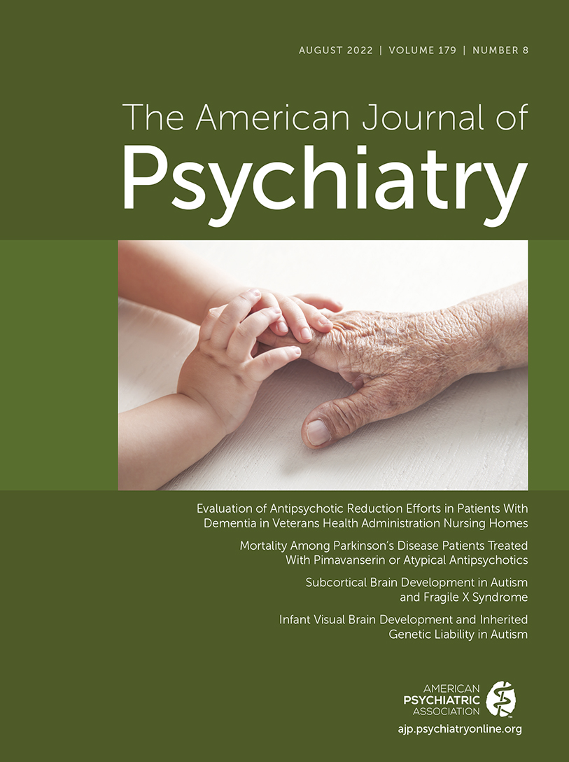
This issue of the Journal is focused on neuropsychiatric illnesses with underlying mechanisms that involve aberrant neurodevelopment—such as autism spectrum disorder (ASD) and fragile X syndrome—or that involve neurodegenerative processes such as dementias and Parkinson’s disease. Included in this issue are three papers relevant to an important treatment concern in dementia patients: the management of behavioral disturbances and psychotic symptoms. We begin with a comprehensive overview authored by Dr. Dilip Jeste from the University of California at San Diego along with Dr. Rajesh R. Tampi (1) that discusses the challenges and importance of effectively treating neuropsychiatric symptoms in patients with dementias, emphasizing the efficacy of behavioral interventions as well as the risks and benefits of using psychopharmacological agents and other modalities like electroconvulsive therapy (ECT) and transcranial magnetic stimulation (TMS). This overview sets the stage for the two original research papers that follow. The first paper reports on changes over time in antipsychotic use in VA nursing home patients with dementia. The second paper reports data comparing mortality rates associated with pimavanserin treatment with mortality rates associated with the administration of other atypical antipsychotics in Parkinson’s disease patients with psychotic symptoms and dementia.
In relation to neurodevelopmental disorders, two papers focus on ASD and fragile X syndrome. The results presented in these papers are derived from data collected from the Infant Brain Imaging Study Network. This network is a highly valuable resource that, as a consortium of five different sites, collects longitudinal imaging and phenotyping data to uncover early-life alterations in brain development that are associated with the risks for developing ASD. Findings from these papers highlight the detection of developmental brain abnormalities during infancy that precede the development of ASD symptoms and involve the amygdala and occipital cortex as well as early-life alterations in the caudate that are associated with fragile X syndrome.

Reductions in Antipsychotic Use in VA Patients With Dementia Living in Nursing Homes

Antipsychotic medications are frequently used for the treatment of behavioral problems and associated psychotic symptoms in individuals with dementia. While this approach can be effective, there is considerable concern, as reflected in the black box warnings issued by the FDA, regarding the association between antipsychotic use and death in elderly dementia patients. A study assessing the risk of antipsychotic-associated death in VA patients with dementia who were 65 years or older found that, depending on the specific antipsychotic used, when followed for 180 days, the increased mortality risk ranged from 2% to 3.8% (2). Gerlach et al. (3) examine the use of antipsychotic medications in veterans suffering from dementia residing in nursing homes. As the authors point out, programs have been instituted in the VA and the Centers of Medicare and Medicaid Services to reduce the use of antipsychotics in patients with dementia living in nursing homes, and the current study focuses on characterizing the extent to which this has occurred over the period from 2009 to 2018. The investigators used data from over 35,000 VA patients with dementia who were living in a nursing home for a minimum of 30 days to assess treatment with antipsychotics as well as the use of antidepressants, anxiolytics, antiepileptics, opiates, and medication for memory impairment. Over the 9-year study period, the percentage of individuals prescribed antipsychotics decreased from 33.7% to 27.5%, and quetiapine was the most prescribed antipsychotic medication. The number of individuals who were prescribed anxiolytics also decreased over this time period from 33.5% to 27.1%. The use of opiates was common in this population and prescriptions were initially found to increase, followed by a decrease to 41.2% of individuals. In addition, prescriptions for memory-related medications declined from 32.4% to 21.8%. In contrast to the reduction in the prescriptions for these drugs, prescriptions for antiepileptic drugs increased from 26.8% to 43.3%, with gabapentin being the most prescribed antiepileptic agent. Sertraline was the most frequently used antidepressant, and overall antidepressant prescriptions increased from 56.8% to 63.4%. Memory-related medications decreased from 32.4% to 21.8%. These findings show that the efforts within the VA to reduce the use of antipsychotics in patients with dementia living in nursing homes have been effective, and at the same time the use of antiepileptics and antidepressants increased. The authors emphasize that while there are concerns regarding the use of antipsychotics in this population, the evidence for treating dementia-related behavioral and psychotic symptoms is strongest for this class of drugs. Of concern, the other drugs that have been substituted for antipsychotics are less likely to be effective and have potential significant risks. In his editorial, Dr. Lon Schneider from the University of Southern California provides a historical perspective on the use of antipsychotics in treating dementia-related psychotic and behavioral disturbances (4), and comments on the Gerlach et al. findings in relation to the relative benefits and risks of using antipsychotics as compared to medications with less of an evidence base supporting their efficacy.

Pimavanserin and Other Atypical Antipsychotic Associated Mortality in Parkinson’s Disease Patients

The Parkinson’s Foundation estimates that 1 million people in the United States and more than 10 million people around the world suffer from PD. In the United States there are approximately 60,000 new PD cases per year (5). In addition to its well-known effects on motor behavior, PD is frequently comorbid with depression, anxiety, psychotic symptoms, and cognitive deficits. In fact, up to 50% of PD patients experience hallucinations or delusions, and when advanced, PD often progresses to dementia. Degeneration of midbrain dopamine neurons, especially those in the substantia nigra, is a critical component of the pathophysiological process that underlies PD. It is thought that this neuronal toxicity stems from alterations in the neurotransmitter-associated protein, alpha-synuclein, which ultimately results in the development of neuronal Lewy bodies. While both atypical antipsychotics and pimavanserin, also an atypical antipsychotic, are used for treating psychotic symptoms in PD, pimavanserin is the only drug that is FDA approved for this indication. Pimavanserin has a unique receptor binding profile acting as an inverse agonist and antagonist at the 5-HT2A receptor, with minimal effects at other monoamine receptors including the D2 receptor (6). Mosholder et al. (7) use data from a Medicare cohort to examine and compare mortality in PD patients receiving pimavanserin (N=3,227) or atypical antipsychotics (N=18,442; 78% of individuals in the atypical antipsychotic group were taking quetiapine). Data from patients were included in the analysis if they began these treatments during the 3-year period from 2016 to 2019. The mean age of the patients in the study was approximately 78 years, and the median length of exposure to the treatments was 78 days. Additionally, it is noteworthy that the atypical antipsychotic group had a greater incidence of medical comorbidities, which were statistically controlled for in the analysis. Dementia was also diagnosed in 55% of the patients in the pimavanserin group and 60% of the patients in the atypical antipsychotic group. The results revealed that pimavanserin treatment was associated with a 35% lower mortality rate compared with atypical antipsychotics when controlling for comorbid medical conditions and other relevant variables. However, this effect was only evident during the first 180 days of treatment and was only observed in the approximately 85% of patients that were not in nursing homes. In his editorial (4), Dr. Lon Schneider also discusses the significance of these findings regarding the relatively decreased mortality rate reported with pimavanserin treatment. He provides background regarding the clinical trial used to gain FDA approval for pimavanserin’s use in PD patients, and he encourages the reader to interpret the reported advantages of pimavanserin over other atypical antipsychotics with caution based on some of the limitations in the present study.

Differences in Amygdala and Caudate Development in Infants With Autism Compared With Fragile X Syndrome

Autism spectrum disorder (ASD) and fragile X syndrome are neurodevelopmental disorders with very different etiologies that are known to have overlapping symptoms, both associated with marked changes in behavior and a range of alterations in intellectual development as well as high levels of comorbid pathological anxiety. In contrast to ASD, which most commonly involves mutations in various risk genes and chromosomal copy number variants (8), fragile X syndrome is due to mutations in the FMR1 gene, which, when expressed in the brain, encodes for a protein thought to be involved in synaptic development. Various abnormalities in brain development are well-known to occur in young children with ASD and fragile X syndrome. For example, previous work has identified altered amygdala development, characterized by increased size during early childhood, to be associated with ASD. The amygdala is of particular interest as it is involved in mediating fear- and anxiety-related behaviors as well as in contributing to social behavior. Differing from ASD, fragile X syndrome has been associated with aberrant development of the caudate. In this issue, Shen and colleagues (9) report data from a longitudinal imaging study examining the early-life development of select brain regions including the amygdala and caudate in infants at risk for developing ASD, infants with fragile X syndrome, and control infants. Infants were studied as participants in the Infant Brain Imaging Study Network and were considered to be at risk for ASD if they had a sibling with confirmed ASD. Infants were initially scanned at 6 months of age and then again at 12 months and 24 months. At 24 months the at-risk ASD participants were assessed as to whether they met criteria for ASD. Results demonstrated that when comparing at-risk infants that did not develop ASD (N=212), fragile X syndrome infants (N=29), and control infants (N=109), the at-risk infants that developed ASD (N=58) had significantly greater amygdala volumes at 12 and 24 months. In contrast, the fragile X syndrome infants demonstrated increased volume of the caudate, putamen, and globus pallidus at 6, 12, and 24 months of age. In addition to the group differences, at the individual level, more rapid amygdala growth from 6 to 12 months predicted the magnitude of social deficits in the infants that developed ASD at 2 years of age. Conversely, individual differences in caudate volume at 12 months of age predicted greater repetitive behaviors in fragile X syndrome infants at 24 months of age. Taken together, these findings lend insights into alterations in different brain regions and developmental time courses that are associated with ASD and fragile X syndrome. Of particular interest is the finding that early-life amygdala overgrowth precedes the onset of social deficit symptoms in infants that later develop ASD. This points to the possibility of using the trajectory of early-life amygdala development as a neural marker of risk as well as a potential early-life treatment target. In their editorial, Drs. David Amaral and Christine Nordahl from the University of California at Davis (10) provide a discussion of the findings from this paper and an in-depth discussion of the amygdala as it may relate to mediating ASD-related social deficits. They also discuss potential mechanisms that may underlie altered amygdala development and comment on data from their own work suggesting that there are different patterns of altered amygdala development in autistic children, which may be important in understanding the heterogeneity of symptoms experienced by ASD individuals.
Altered Development of Visual Pathways Reflect Heritable Risk to Develop Autism

Girault and colleagues (11) also use data from the Infant Brain Imaging Study Network to identify developing brain regions that may be related to the heritable risk to develop ASD. Based on the familial occurrence of ASD, and the assumption that greater symptoms reflect greater genetic liability, the approach taken by the researchers was to examine relations between the ASD symptoms of an affected child with brain development in their at-risk, younger sibling. Data from MRI scans from 384 ASD-sibling pairs collected at 6, 12, and 24 months of age was used. In the 89 sibling pairs that were concordant for ASD, significant associations were found between the symptoms of the older ASD sibling with brain alterations in the younger sibling. In contrast, these associations were not present in the 289 sibling pairs that were discordant for ASD. Specifically, in the concordant ASD pairs, the authors found that symptoms related to sociability and communication in the older affected sibling were associated with total cerebral volume and surface area in the younger sibling that developed ASD. Because of interest in the early development of visual pathways in relation to social deficits, the researchers focused their analyses on occipital cortical regions and the splenium of the corpus callosum. Here, results revealed that sociability-related symptoms in the older ASD sibling predicted right middle occipital gyrus cortical surface area at 6, 12, and 24 months of age in their younger sibling later diagnosed with ASD. Additionally, similar relations in concordant pairs were found between the older sibling’s symptoms and the microstructural integrity of the splenium of the corpus callosum in the younger sibling. However, this association was only significant at 6 months of age and not at 12 and 24 months. Functional connectivity analyses from the data collected at 6 months of age revealed decreased visual network connectivity, a finding that is generally consistent with the structural findings. The results presented demonstrate that structural and functional alterations in visual pathways can be detected prior to the onset of ASD symptoms in ASD children, and importantly, link these early-life brain alterations to symptoms severity in their ASD older sibling. While these effects are considered to be heritable, it is important to keep in mind that shared environmental factors could also contribute to the reported associations. In their editorial (12), Drs. Iska Moxon-Emre and Stephanie Ameis from the University of Toronto discuss the potential importance of these findings, place the findings into the context of other work, and pose questions for the future.

Conclusions

The papers in this issue are focused on neurodegenerative and neurodevelopmental disorders that are relevant to understanding and caring for patients across the lifespan.
Regarding neurodegenerative disorders, the overview by Drs. Tampi and Jeste presents evidence supporting the use of behavioral and psychopharmacological strategies for the management of behavioral disturbances and psychotic symptoms in patients with dementia. While the administration of antipsychotics to these patients confers a small increase in mortality risk, the push to decrease their use in the VA nursing home population has been supplanted by other pharmacological agents that are less likely to be effective. We also learn that pimavanserin treatment in PD patients with psychotic symptoms or dementia may be associated with decreased mortality when compared with other atypical antipsychotics. However, this pimavanserin-related advantage appears to be short lived.
From the papers on ASD and fragile X syndrome, we learn about abnormal patterns of brain development during infancy in patients with these disorders. Of particular interest is the finding that alterations in amygdala development can be detected prior to the onset of ASD symptoms, and are also predictive of individual differences in ASD-related deficits in social functioning. Fragile X syndrome infants do not have amygdala abnormalities but instead have alterations in caudate development.
In psychiatry, we work to help patients overcome their symptoms and disabilities at very different stages of their lives, from early childhood to adolescence into adulthood and old age. The papers in this issue of the Journal are focused on illnesses that present at the two ends of the age spectrum, infancy and old age. The papers involving neurodegenerative disorders have important clinical implications for our readership providing evidence supporting the optimal treatment of behavioral disturbances and psychotic symptoms in patients with dementia and PD. The papers on ASD and fragile X syndrome characterize patterns of aberrant neural development, bringing to our awareness the possibility of identifying early-life neural risk markers for these illnesses, as well as treatment strategies instituted prior to the onset of significant symptoms that could target alterations in aberrant neurodevelopmental processes.

References

1.
Tampi RR, Jeste DV: Dementia is more than memory loss: neuropsychiatric symptoms of dementia and their nonpharmacological and pharmacological management. Am J Psychiatry 2022; 179:528–543
2.
Maust DT, Kim HM, Seyfried LS, et al: Antipsychotics, other psychotropics, and the risk of death in patients with dementia: number needed to harm. JAMA Psychiatry 2015; 72:438–445
3.
Gerlach LB, Maust DT, Kales HC, et al: Evaluation of antipsychotic reduction efforts in patients with dementia in Veterans Health Administration nursing homes. Am J Psychiatry 2022; 179:544–552
4.
Schneider LS: The safety of pimavanserin for Parkinson’s disease and efforts to reduce antipsychotics for people with dementia. Am J Psychiatry 2022; 179:519–521
5.
Parkinson’s Foundation: Statistics. https://www.parkinson.org/Understanding-Parkinsons/Statistics. Accessed June 24, 2022
6.
Vanover KE, Weiner DM, Makhay M, et al: Pharmacological and behavioral profile of N-(4-fluorophenylmethyl)-N-(1-methylpiperidin-4-yl)-N’-(4-(2-methylpropyloxy)phenylmethyl) carbamide (2R, 3R)-dihydroxybutanedioate (2:1) (ACP-103), a novel 5-hydroxytryptamine(2A) receptor inverse agonist.-(4-Fluorophenylmethyl)--(1-methylpiperidin-4-yl)--(4-(2-methylpropyloxy)phenylmethyl) Carbamide (2)-Dihydroxybutanedioate (2:1) (ACP-103), a Novel 5-Hydroxytryptamine2A Receptor Inverse Agonist. J Pharmacol Exp Ther 2006; 317:910–918
7.
Mosholder AD, Ma Y, Akhtar S, et al: Mortality among Parkinson’s disease patients treated with pimavanserin or atypical antipsychotics: an observational study in Medicare beneficiaries. Am J Psychiatry 2022; 179:553–561
8.
Manoli DS, State MW: Autism spectrum disorder genetics and the search for pathological mechanisms. Am J Psychiatry 2021; 178:30–38
9.
Shen MD, Swanson MR, Wolff JJ, et al: Subcortical brain development in autism and fragile X syndrome: evidence for dynamic, age- and disorder-specific trajectories in infancy. Am J Psychiatry 2022; 179:562–572
10.
Amaral DG, Nordahl CW: Amygdala involvement in autism: early postnatal changes but what are the behavioral consequences? Am J Psychiatry 2022; 179:522–524
11.
Girault JB, Donovan K, Hawks Z, et al: Infant visual brain development and inherited genetic liability in autism. Am J Psychiatry 2022; 179:573–585
12.
Moxon-Emre I, Ameis S: Infant brain signatures of genetic liability for autism: the critical need for longitudinal research. Am J Psychiatry 2022; 179:525–527

Information & Authors

Information

Published In

Go to American Journal of Psychiatry
American Journal of Psychiatry
Pages: 515 - 518

History

Published in print: August 2022
Published online: 3 August 2022

Keywords

  1. Neurodevelopmental Disorders
  2. Dementia
  3. Neurodevelopmental Disorders
  4. Autism Spectrum Disorder

Authors

Details

Ned H. Kalin, M.D. [email protected]
Department of Psychiatry, University of Wisconsin School of Medicine and Public Health, Madison.

Notes

Send correspondence to Dr. Kalin ([email protected]).

Funding Information

Disclosures of Editors’ financial relationships appear in the April 2022 issue of the Journal.

Metrics & Citations

Metrics

Citations

Export Citations

If you have the appropriate software installed, you can download article citation data to the citation manager of your choice. Simply select your manager software from the list below and click Download.

For more information or tips please see 'Downloading to a citation manager' in the Help menu.

Format
Citation style
Style
Copy to clipboard

View Options

View options

PDF/EPUB

View PDF/EPUB

Get Access

Login options

Already a subscriber? Access your subscription through your login credentials or your institution for full access to this article.

Personal login Institutional Login Open Athens login

Not a subscriber?

Subscribe Now / Learn More

PsychiatryOnline subscription options offer access to the DSM-5-TR® library, books, journals, CME, and patient resources. This all-in-one virtual library provides psychiatrists and mental health professionals with key resources for diagnosis, treatment, research, and professional development.

Need more help? PsychiatryOnline Customer Service may be reached by emailing [email protected] or by calling 800-368-5777 (in the U.S.) or 703-907-7322 (outside the U.S.).

Media

Figures

Other

Tables

Share

Share

Share article link

Share